A Rare Case of Ovarian Hyperstimulation Syndrome in a Preterm Infant
Asieh Mosallanejad1, Shahrzad Tabatabai2, Marjan Shakiba3, Mohammad Reza Alaei4, Hedieh Saneifard5
1 Assistant Professor, Imam Hosein Medical Center, Shahid Beheshti University of Medical Sciencce, Tehran, Iran.
2 Assistant Professor, Imam Hosein Medical Center, Shahid Beheshti University of Medical Sciencce, Tehran, Iran.
3 Assistant Professor, Mofid Children Hospital, Shahid Beheshti University of Medical Sciencce, Tehran, Iran.
4 Associate Professor, Mofid Children Hospital, Shahid Beheshti University of Medical Sciencce, Tehran, Iran.
5 Assistant Professor, Mofid Children Hospital, Shahid Beheshti University of Medical Sciencce, Tehran, Iran.
NAME, ADDRESS, E-MAIL ID OF THE CORRESPONDING AUTHOR: Dr. Asieh Mosallanejad, Assistant Professor, Imam Hosein Medical Center, Shahid Madani Street, Tehran, Iran.
E-mail: mosalladr@gmail.com
Ovarian hyperstimulation syndrome is a rare disease among preterm infants. This syndrome was first described in 1985 in four infants with a gestational age of <30 weeks. Several explanations for this syndrome have been suggested namely the immaturity of Hypothalamic-Pituitary-Gonadal (HPG) axis, lack of negative feedback, increased sensitivity of Follicle Stimulating Hormone (FSH) receptors due to mutation and high level of estradiol. In this report, a case of hyperstimulation syndrome in a newborn with gestational age of 30 weeks is presented and the probable mechanisms in the literature are discussed.
Genital oedema, Hypothalamic-pituitary-gonadal axis, Ovarian cysts
Case Report
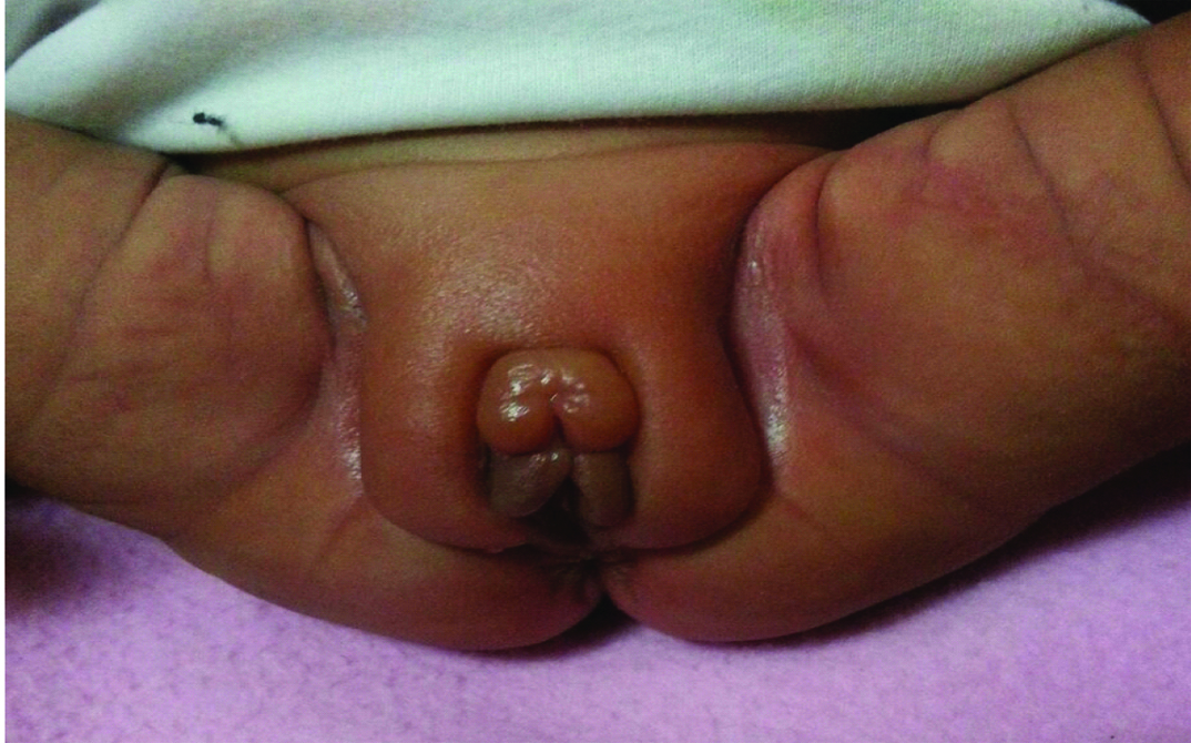
A two-month-old female infant was brought to our clinic due to swelling in external genitals. She was a premature infant with a gestational age of 30 weeks and was delivered by caesarean section because of mother’s appendicitis. Weight of the child was 1500 grams at birth. Apgar score in the first and fifth minute after delivery was 7 and 8 respectively. She had been hospitalized for 32 days in neonatal intensive care unit due to neonatal respiratory distress syndrome and prematurity. The patient had developed gradual swelling of external genitalia starting at post conception age of 35 weeks. On physical examination tense oedema of clitoris, labia major and minor, the lower abdominal wall and upper thighs was observed [Table/Fig-1]. There was no swelling in other places and other physical examinations were normal. On laboratory evaluations serum electrolyte, liver and renal function tests and serum albumin levels were all normal for this age (Na=136mEq/L normal range is 133-145mEq/L, Cr=0.4mg/dl normal range is 0.27-0.5mg/dl, AST=35unit/L normal range is 35-140unit/L, ALT= 28unit/L normal range is 3-35unit/L and serum albumin=3.8g/dl normal range is 3-5.5g/dl) Serum estradiol, Luteinizing Hormone (LH) and FSH were measured using micro chemiluminescence (Abbot Architect, USA) method. Laboratory findings were as follow: serum level of estradiol=34pg/ml (normal range <25pg/ml), FSH=4.6mIU/ml (normal range is <0.2 -4.6mIU/ml), LH=0.8mIU/ml (normal range is <0.5mIU/ml).
Oedema of clitoris, labia major and minor, and upper thighs was observed on physical examination.
On sonographic examination of pelvis and abdominal cavity two 15mm by 18mm and 6mm by 8mm follicular cysts in the left ovary and another 4.5 mm cyst in the right ovary was observed. The size of uterus was 8 ×12 × 39mm.
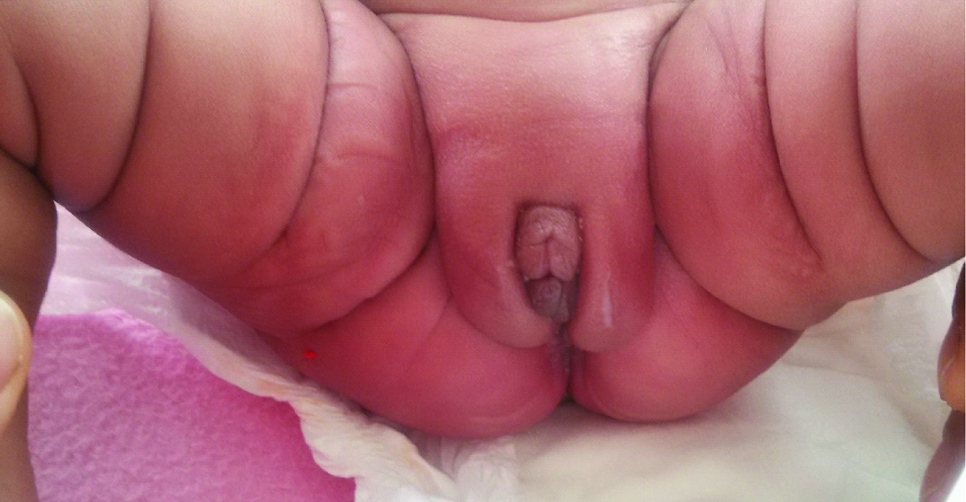
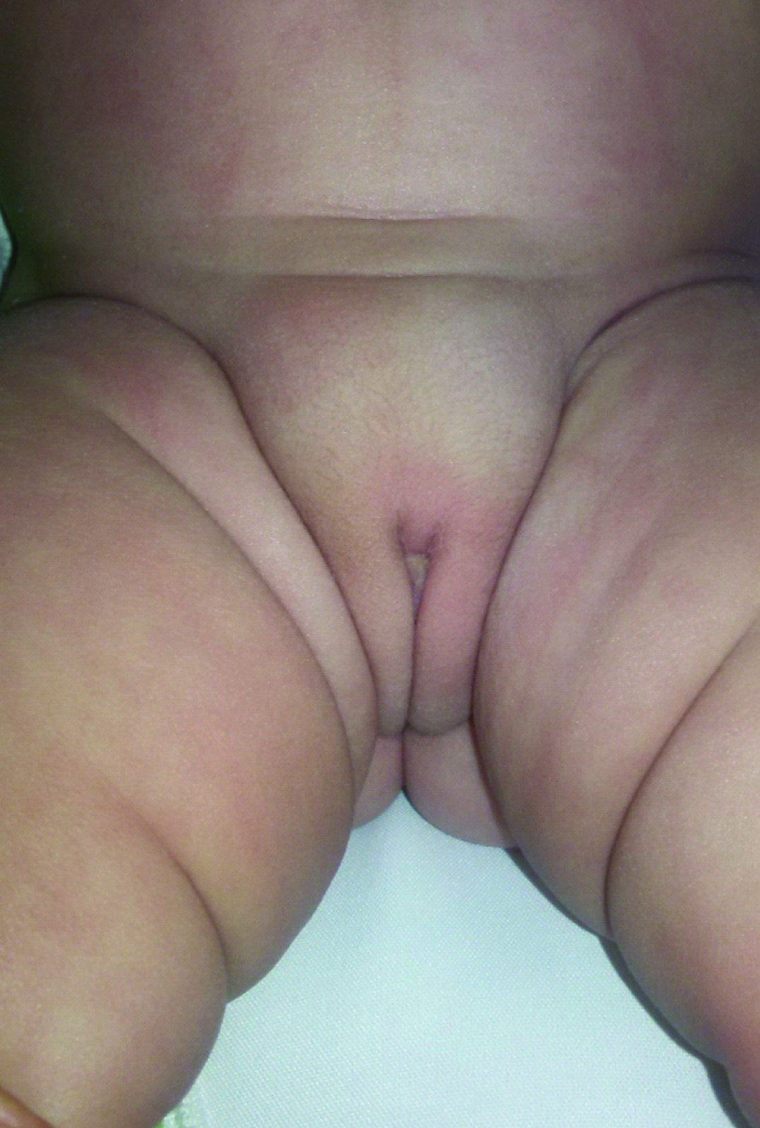
Without any intervention the patient was followed and after three weeks the genital and upper thighs oedema was reduced [Table/Fig-2]. On repeated sonographic examination a follicular cyst in the left ovary with a size of 10mm was observed and the right ovary was normal. After six weeks the genital examination was completely normal [Table/Fig-3] and the sonographic examination revealed normal ovaries.
After three weeks of follow-up the genital and upper thighs oedema was reduced.
After six weeks of follow-up the genital examination was completely normal.
At this time the laboratory findings were as follow: Estradiol=25pg/ml (normal range <25). Serum FSH and LH were in normal range for this sex and age.
Discussion
Although follicular ovarian cysts are the most common abdominal masses among female fetuses and infants (30% to 40%) [1], their occurrence is seldom accompanied by other signs like genital oedema or overgrowth of breast glandular tissue [2–8].
We reported a preterm infant (gestational age 30 weeks) with ovarian hyperstimulation syndrome. Preterm hyperstimulation syndrome was first described by Sedin et al., [4]. This syndrome has been reported in preterm infants with one sided or two sided ovarian cysts and oedema of the external genitalia and upper legs with varying degree of elevation in gonadotropin and estradiol levels [4]. The negative feedback mechanism for Hypothalamic–Pituitary–Gonadal (HPG) axis in preterm infants is immature, thus the increased levels of LH and FSH in response to the withdrawal of placental steroids might cause the appearance of this syndrome in infants [4]. In patients reported by Sedin et al., the serum levels of gonadotropins and estradiol were significantly higher than normal and IV injection of luteinizing hormone-releasing hormone caused post-pubertal type reaction [4].
The coincidence of genital oedema with ovarian cysts and the reduction of oedema with disappearance of cysts might indicate the role of cysts in creating the genital oedema. The molecules secreted from granulosa and theca cells like vascular endothelial growth factor might have a role in this phenomenon [6]. Artini et al., in study of ovarian hyperstimulation among adult females have noticed an increase in the serum levels of vascular endothelial growth factor [8]. The increase of vasogenic molecules which might cause vascular leakage [9] as well as some other inflammatory compounds like Interleukin-6 might play a role in vascular hyperpermeability and oedema formation [8,10].
Sedin et al., in their study emphasized on the role of estradiol in the appearance of this syndrome [4], but this role has been questioned in later studies [3,5]. Similarly in our case the serum level of estradiol was a little higher than normal range indicating that increased levels of estradiol and gonadotropins is not specific for preterm ovarian hyperstimulation syndrome.
The presence of ovarian cysts and severe oedema of external genitalia might be a sign of irregular activation of FSH receptor and suggest that this syndrome might be the result of FSH receptor mutation. Smits et al., have reported the mutation of FSH receptor in an adult patient with ovarian hyperstimulation syndrome and concluded that this mutation has caused increased basal activity of the mutated receptor [9].
Diagnosis: Genitalia swelling are abnormal in neonates. Hyperstimulation syndrome should be considered as a diagnosis in preterm infants with external genitalia oedema which might extend upto the upper legs and hypogastric area [5]. Gonadotropin levels may be increased and multiple cysts with different sizes may be seen in ovarian sonography [5].
The pathognomonic sign of hyperstimulation syndrome is oedema in external genitalia among preterm infants which might extend to the upper legs and hypogastric area [5].
This syndrome is self limited and usually does not necessitate medical or surgical treatment [5]. In our case the ovarian cysts subsided in a 5 to 6 weeks period. In ovarian cysts bigger than 4-5cm in size which do not subside in time there is a risk of Torrtion. In these patients drainage through aspiration might be helpful. Also, the use of progesterone acetate in severe cases that don’t regress might reduce the symptoms [11,12].
Conclusion
In preterm female infants who present with genital oedema hyperstimulation syndrome should be considered. The presence of ovarian cysts in sonography aids in diagnosis of this syndrome and gonadotropin as well as estradiol levels might be increased. This syndrome is usually self limiting and does not need pharmaceutical or surgical treatments.
[1]. Akın MA, Akın L, Özbek S, Tireli G, Kavuncuoglu S, Sander S, Fetal-neonatal ovarian cysts—their monitoring and management: retrospective evaluation of 20 cases and review of the literatureJ Clin Res Pediatr Endocrinol 2010 2(1):28-33. [Google Scholar]
[2]. Bergqvist C, Esscher T, Lindgren PG, Lundkvist K, Sedin G, Cystic ovaries in a pre-term newborn infantZ Kinderchir 1984 39(6):403-04. [Google Scholar]
[3]. Altuntas N, Turkyilmaz C, Yuce O, Kulali F, Hirfanoglu IM, Onal E, Preterm ovarian hyperstimulation syndrome presented with vaginal bleeding: a case reportJ Pediatr Endocrinol Metab 2014 27(3-4):355-58. [Google Scholar]
[4]. Sedin G, Bergquist C, Lindgren PG, Ovarian hyperstimulation syndrome in preterm infantsPediatr Res 1985 19(6):548-52. [Google Scholar]
[5]. Starzyk J, Wójcik M, Wojtyś J, Tomasik P, Mitkowska Z, Pietrzyk JJ, Ovarian hyperstimulation syndrome in newborns—a case presentation and literature reviewHorm Res 2009 71(1):60-64. [Google Scholar]
[6]. Halis H, Memur S, Korkmaz L, Baştuğ O, Güneş T, Kurtoğlu S, Ovarian hyperstimulation syndrome treated by medroxyprogesterone acetateJ Pediatr Endocrinol Metab 2014 27(9-10):997-99. [Google Scholar]
[7]. Esen I, Demirel F, Images in clinical medicine. Preterm ovarian hyperstimulationN Engl J Med 2015 372(24):2336 [Google Scholar]
[8]. Artini PG, Monti M, Fasciani A, Battaglia C, D’Ambrogio G, Genazzani AR, Vascular endothelial growth factor, interleukin-6 and interleukin-2 in serum and follicular fluid of patients with ovarian hyperstimulation syndromeEur J Obstet Gynecol Reprod Biol 2002 101(2):169-74. [Google Scholar]
[9]. Smits G, Olatunbosun O, Delbaere A, Pierson R, Vassart G, Costagliola S, Ovarian hyperstimulation syndrome due to a mutation in the follicle-stimulating hormone receptorN Engl J Med 2003 349(8):760-66. [Google Scholar]
[10]. Revel A, Barak V, Lavy Y, Anteby E, Abramov Y, Schenker JJ, Characterization of intraperitoneal cytokines and nitrites in women with severe ovarian hyperstimulation syndromeFertil Steril 1996 66(1):66-71. [Google Scholar]
[11]. Luzzatto C, Midrio P, Toffolutti T, Suma V, Neonatal ovarian cysts: management and follow-upPediatr Surg Int 2000 16(1-2):56-59. [Google Scholar]
[12]. Kurtoğlu S, Baştuğ O, Mini puberty and its interpretationTurk Pediatri Ars 2014 49(3):186-91. [Google Scholar]