Prosthodontic Approach in Management of Prolonged Neonatal Intubation
Vikas B Kamble1, Shital K Shah2, Vishnu B Rathod3, Priyanka S Ambadkar4, Charudutt N Patil5
1 Head of Department, Department of Prosthodontics, Crown and Bridge, P.M.N.M. Dental College and Hospital, Bagalkot, Karnataka, India.
2 Head, Department of Paediatrics and Neonatology, Navjeevan Children’s Hospital, Pandharpur, Maharashtra, India.
3 Postgraduate Student, Department of Paediatrics and Neonatology, Navjeevan Children’s Hospital, Pandharpur, Maharashtra, India.
4 Postgraduate Student, Department of Prosthodontics, Crown and Bridge, P.M.N.M. Dental College and Hospital, Bagalkot, Karnataka, India.
5 Postgraduate Student, Department of Prosthodontics, Crown and Bridge, P.M.N.M. Dental College and Hospital, Bagalkot, Karnataka, India.
NAME, ADDRESS, E-MAIL ID OF THE CORRESPONDING AUTHOR: Dr. Vikas B Kamble, Head of Department, Department of Prosthodontics, Crown and Bridge, P.M.N.M. Dental College and Hospital, Bagalkot, Karnataka, India.
E-mail: drvikaskamble2002@yahoo.co.in
Intubation is a routine intervention in the Neonatal Intensive Care Unit (NICU) for preterm neonates with respiratory distress, inadequate gag reflex, poor sucking and swallowing. Prolonged intubation in neonates can be done by nasal or oral route. Although naso-tracheal intubation may reduce movement of the tube, it may contribute to airway obstruction, possible hypoxia, and occlusion of the nasal aperture during a crucial period of development further contributing to laboured breathing. Being obligate nasal breathers, oro-tracheal route is the preferred method of intubation in premature infants as oral mucosa is less susceptible to damage than nasal mucosa. Ineffective stabilization of the tubes is a frequent problem often resulting in accidental extubation and displacement of orotracheal and orogastric tube. Hence, these tubes must be stabilized against displacement from tongue and jaw movements to prevent discomfort and subsequent tissue trauma. Complications of prolonged endotracheal intubation include palatal groove formation by pressure against the hard palate, infection, accidental extubation, malposition, laryngeal or tracheal edema and ulceration, tracheal stenosis, vocal cord injury. Various oral appliances are used for infants to stabilize the tubes and prevent complications associated with long term intubation. This case report describes a prosthodontic approach in management of prolonged neonatal intubation.
Endotracheal intubation, Neonate, Orogastric Intubation, Prolonged intubation
Case Report
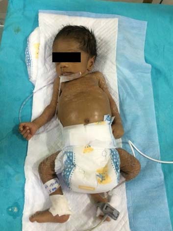
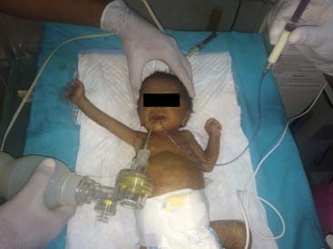
A four day-old-male neonate was referred to Navjeevan Hospital, Pandharpur, Maharashtra to improve his upper respiratory distress [Table/Fig-1]. He was diagnosed with respiratory distress, intra-uterine growth restriction, and hypoglycaemia and was advised prolonged orotracheal and orogastric intubation. He was intubated and the tube was stabilized using adhesive tape. Because of improper stabilization of tubes, accidental extubation was being experienced. After consultation with the paediatrician, it was planned to improve the stabilization of both the tubes and also eliminate chances of groove formation over the palate because of prolonged intubation. The procedure was explained to the parents of the infant and written consent was obtained.
Pre-operative extra oral view.
Technique
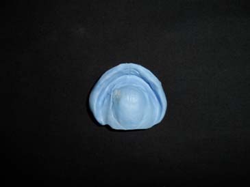
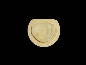
A polysiloxane putty (Virtual Putty Base Regular Set, Ivoclar Vivadent, Germany Lot No.- 5795) impression of the maxillary arch of the neonate was made using a preformed spoon like acrylic tray [Table/Fig-2] and a plaster (Gypsum Plaster, Asian Chemicals, Rajkot, Batch No. 5872/398) cast was prepared [Table/Fig-3].
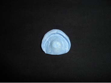
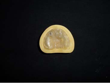
An impression tray was fabricated on this plaster cast using clear autopolymerizing acrylic resin (DPI-RR, Mumbai, Batch No. 4269) which acts as a special tray. This tray was used to make another polysiloxane putty (Virtual Putty Base Regular Set, Ivoclar Vivadent, Germany Lot No. 5795) impression to accurately record the maxillary arch [Table/Fig-4] and master cast was prepared in dental stone (Gold Stone, Asian Chemical, Gujarat, Batch No. 2468/414) [Table/Fig-5].
A 2mm thickness plate of clear autopolymerizing acrylic resin (DPI-RR, Mumbai, Batch No. 4269) was fabricated on this master cast. Heat polymerizing methyl methacrylate acrylic resin could also be used if the appliance was not required for immediate use.
Following polymerization, a groove was cut in the cameo surface of this appliance to accept the appropriate sized orotracheal and orogastric tube and was modified with autopolymerizing acrylic resin to provide a snap fit for the tubes.
One small stainless steel tag was incorporated anteriorly in the appliance to provide attachment for dental floss which extends 6 inches outside the oral cavity, used to retrieve the appliance if it accidentally dislodges from the palate. Finishing and polishing of the appliance was done followed with cold sterilization in 2% glutaraldehyde for 10-30 minutes [Table/Fig-6].
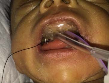
The appliance was inserted in infant’s mouth and when properly secured against the palate, the orotracheal and orogastric tubes were pressed into the appropriate grooves [Table/Fig-7]. Respiratory support was initiated and feeding was performed as required [Table/Fig-8].
Post insertion instructions were given to the nursing staff and the cleaning procedure of the appliance was done every 24 hours and oral hygiene was performed.
After removal of appliance for cleaning, the mucosa of the palate was carefully observed for any tissue trauma and mucositis.
Polysiloxane putty impression made with spoon like acrylic tray.
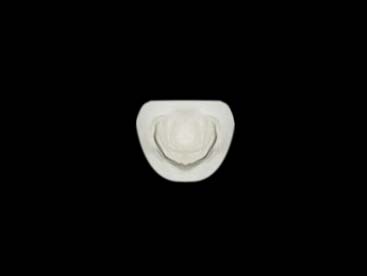
Polysiloxane putty impression made with acrylic impression tray.
Appliance secured with orotracheal and orogastric tube.
Respiration supported by oro-tracheal intubation and feeding performed by oro-gastric tube.
Discussion
The neonatal airway has become a specific area of concern in recent years as modern advances in neonatology have improved the survival rates of preterm and other critically ill newborns [1]. Intubation is the method of choice as incidence of mortality and morbidity for tracheostomy in the neonate is much higher than that of endotracheal intubation. Oral mucosa is less susceptible to damage than nasal mucosa [2]. Oro-tracheal route is the preferred method of intubation in premature infants as they are obligate nasal breathers [2]. Ineffective stabilization of the tubes is a frequent problem often resulting in accidental extubation and displacement of orotracheal and orogastric tube [3]. Complications of prolonged endotracheal intubation include palatal groove formation by pressure against the hard palate, infection, accidental extubation, malposition, laryngeal or tracheal oedema and ulceration, tracheal stenosis, vocal cord injury. Various oral appliances [4–8] to stabilize the tubes are being used for infants who require prolonged intubation as mentioned in [Table/Fig-9].
Various oral appliances to stabilize orotracheal and orogastric tube used for infants who require prolong intubation.
| Year | Author | Appliance |
|---|
| 1981 | Richards SD [7] | A double stick disc was attached to the endotracheal tube connector, further attached to the non-adhesive side of the tape. Disadvantage - Once placed it is difficult to remove the tape and need to be pulled back and re-positioned which even results in rashes on skin. |
| 1981 | Sullivan PG and Haringman H [4] | A palatal appliance was fabricated with anterior and posterior grooves, modified for the snap fit of the intubation tubes with small stainless steel tag. Advantages: 1) Tubes can be inserted into the appliance after they are placed at its location; 2) Snap fit allows for easy placement and retrieval; 3) Appliance need not be removed for replacing either tube. |
| 1984 | Erenberg A and Nowak A [5] | A palatal appliance was fabricated with continuous antero-posterior groove, further modified for the snap fit of the tubes along with a stainless steel tag in the anterior portion of plate. Disadvantages - It covers more area as it covers the tubes antero-posteriorly. |
| 1992 | McCourt JW [8] | A palatal appliance was constructed with visible light cured urethane, stainless steel ball clasp wire, a closed-four-link synthetic elastic latex polymer chain. It was slipped under the taped orotracheal tube. The chain was moved over the intubation tubes to engage its third link. Advantages - Superior strength, complete polymerization, and low bacterial adherence. Disadvantages - Urethane is more brittle than acrylic resins, cumbersome to fabricate. |
| 2002 | Shimoyama T et al., [6] | A palatal appliance was fabricated with an outer guide antero-posteriorly through which the tube could be passed. Disadvantage - It would not hold the tube tight enough as the diameter of the outer tube is greater than the tube and the tube needs to be placed through the outer guide after insertion of the appliance. |
In this case report, the appliance for stabilization of tubes was fabricated as suggested by Sullivan PG and Haringman H [4]. The advantages of this appliance are, along with stabilization of tubes it also prevents the palatal groove formation because of prolonged intubation. This appliance gives effective stabilization as the tubes are more securely held in the anterior and posterior grooves of the appliance. As the area of modification is less in this appliance, there are less chances of tongue irritation. Even after the infant is extubated, the appliance can be used as long as an orogastric tube is required for feeding or continuous gastric suction [3]. It was observed by the consulting paediatrician, that the appliance was well tolerated by the neonate and was easily manipulated by the nursing staff during routine infant care with no complications like accidental extubation or displacement of tubes. Artificial respiration and orogastric feeding was effectively performed. Limitation of this appliance is that a new appliance has to be fabricated if the infant needs intubation for more than four weeks or when head circumference increases by 2.5 cms [5,8]. This appliance is contraindicated if the intubation required is less than 24 hours. This approach is also contraindicated in neonatal care set-ups where nursing staff are not educated and well trained for its use.
Thus, use of this palatal appliance is justified as it is proved to be beneficial in stabilization of orotracheal and orogastric intubation and prevented the complications resulting from prolonged neonatal intubation. Thus, early prosthodontic care is essential by successfully advocating this technique in neonates requiring prolonged intubation.
Conclusion
The low birth weight and ill, term neonate frequently requires the use of orotracheal and orogastric tubes, both of which are associated with iatrogenic complications such as palatal grove formation, tracheal mucosal damage, subglottic stenosis and laryngeal damage. In an effort to reduce these complications, an intra-oral appliance was used to stabilize the orogastric and orotracheal tubes. This case report describes prosthodontic approach in management of prolonged neonatal intubation.
[1]. Dankle SK, Schuller DE, McClead RE, Prolonged intubation of neonatesArch Otolaryngel Head Neck Surg 1987 11:841-43. [Google Scholar]
[2]. Gonten AS, Meyer JB, Kim AK, Dental management of neonates requiring prolonged oral intubationJournal of Prosthodontics 1995 4(4):222-25. [Google Scholar]
[3]. Budreau G, Kleiber C, Nursing management of the infant with an intraoral applianceJ Obstet Gynecol Neonatal Nurs 1987 16:23-25. [Google Scholar]
[4]. Sullivan PG, Haringman H, An intra oral appliance to stabilise orogastric tube in premature infantsThe Lancet 1981 :416-17. [Google Scholar]
[5]. Erenberg A, Nowak A, Appliance for stabilizing orogastric and orotracheal tubes in infantsCritical Care Medicine 1984 12(8):669-71. [Google Scholar]
[6]. Shimoyama T, Kato T, Horie N, Nasu D, Kaneko T, Oropharyngeal airway appliance for infant with upper airway obstruction: Report of a caseThe Journal of Clinical Pediatric Dentistry 2002 27:25-28. [Google Scholar]
[7]. Richards SD, A method for securing pediatric endotracheal tubesAnesth Analg 1981 40:224-25. [Google Scholar]
[8]. McCourt JW, Neonatal palatal appliance: Light cured resin material and flexible designPediatric Dentistry 1992 14(4):256-59. [Google Scholar]