Giant Haemangioma Excision Under Cervical Epidural Anaesthesia: A Viable Alternative to General Anaesthesia
Samit Parua1, Dipika Choudhury2, Mridu Paban Nath3
1 Student, Department of Anaesthesiology and Critical Care, Gauhati Medical College and Hospital, Guwahati, Assam, India.
2 Professor and Head, Department of Anaesthesiology and Critical Care, Gauhati Medical College and Hospital, Guwahati, Assam, India.
3 Assistant Professor, Department of Anaesthesiology and Critical Care, Gauhati Medical College and Hospital, Guwahati, Assam, India.
NAME, ADDRESS, E-MAIL ID OF THE CORRESPONDING AUTHOR: Dr. Samit Parua, Student, Department of Anaesthesiology and Critical Care, Guwahati Medical College and Hospital, Guwahati-781032, Assam, India.
E-mail: samitparua@gmail.com
The cervical epidural anaesthesia is a safe anaesthetic technique with minimal morbidity and early postoperative recovery. Cervical epidural anaesthesia can be effectively used for neck, upper arm and chest surgeries. The technique avoids the adverse effects of general anaesthetics and airway instrumentation, especially in patients with cardio respiratory disorders. We preferred CEA for giant haemangioma neck excision in an adult female patient, having an associated laryngeal haemangioma, 10ml of 0.5% ropivacaine with 50μg Fentanyl (total 11 ml) was administered into the cervical epidural space through a 20G epidural catheter introduced via a 18G Tuohy needle at the level of C7-T1 space. Following initial dose a top up dose of 4ml 0.5% Ropivacaine was given after 60 minutes. The surgery lasted for 75 minutes. The cervical epidural anaesthesia allowed our patient to stay awake but comfortable, with stable haemodynamics and excellent postoperative pain relief with a continuous cervical epidural infusion of 0.25% Ropivacaine and 2μg/ml Fentanyl @ 2ml/h was achieved.
Anaesthetic technique, Haemodynamics, Neck surgery
Case Report
A 30-year-old, American Society of Anaesthesiologists (ASA) grade I, female patient was posted for removal of giant haemangioma neck for cosmetic reasons.
Pre-Anaesthetic Check-up (PAC) documented Pulse Rate (PR) of 86/min, Blood Pressure (BP) of 130/80mmHg and Respiratory Rate (RR) of 20/min. Systemic examination was normal. Mallampati grade II. Routine investigation showed a haemoglobin of 11gm%, total leucocyte count of 7800, differential count –neutrophils(80%), lymphocytes (15%), eosinophils (3%) prothrombin time/INR- 13.5 sec/1.5, creatinine- 0.8 mg%, thyroid stimulating hormone - 4.2mIU/ml, chest x-ray- normal, ECG- normal sinus rhythm, ultrasound b scan revealed a 8cm×5cm×2cm lesion extending from the middle of the neck, extending to the undersurface of the sternocleidomastoid muscle to the lower part of neck, with no arterial shunt. Three months ago while being evaluated for an acute onset hoarseness of voice a small laryngeal haemangioma was accidentally discovered just below her right vocal cord.
She was counselled on the previous day about the potential risk of anticipated airway bleed if any airway manipulation/intervention was attempted, but the patient was very much keen on getting her neck swelling removed for cosmetic reasons. Written consent for regional anaesthesia was obtained after explaining the procedural risks. The patient was kept nil orally 6 hours prior to surgery. On arrival at the operating room, standard ASA monitors were attached and two 18G intravenous lines were secured in both forearm and lactated ringer’s solution was started in one. All anaesthesia equipments were pre-checked. The patient received Inj. Ondansetron 4mg IV and Inj. Midazolam 1mg IV, 20 minutes before the procedure. The neck area over C7-T1 was cleansed with 0.5% chlorhexidine solution and draped in sitting position; neck was flexed to make the cervical vertebrae prominent. The C7-T1 interspace was identified and infiltrated with 3 ml of 2% lignocaine solution. A 18G Tuohy epidural needle (Perifix@400 Filter set, B-Braun Medical (I) Pvt. Ltd) was introduced through the C7-T1 intervertebral space [Table/Fig-1]. Cervical epidural space was identified at a depth of 3.5 cm from skin with loss of resistance technique using saline. A 20G epidural pvc catheter was then inserted 3 cm into the epidural space (total 6.5 cm from skin). The catheter was fixed into position with the help of Tegaderm@. Correct placement of the catheter was verified by negative aspiration for blood and cerebrospinal fluid (CSF), followed by administration of test dose containing 3ml of 2% lignocaine with 15μg of adrenaline (1:200000). The patient was positioned supine on the operating table, with a ring placed under the head. Oxygen was administered @ 3 l/min via a nasal cannula. Vitals were recorded every 3 minutes till the end of surgery. A bolus dose of mixture of 10ml 0.5% Ropivacaine and 50 μg Fentanyl (total 11ml) was injected via the epidural catheter. Onset of action was noted within 15 minutes of injection. The level of sensory block was tested bilaterally with an alcohol soaked swab (defined as loss of sensation to cold), in an ascending fashion starting from the T12 dermatome. A sensory block extending from C2 to T4 dermatomes was obtained after 23 minutes.
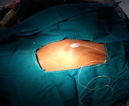
Epidural catheter being inserted in to the c7 and t1 interspace.
Surgery was started 30 minutes after Cervical Epidural Anaesthesia (CEA) administration. Monitoring was done throughout the procedure. Patients were kept in a state of conscious sedation with Midazolam 2mg IV. Vocal cord functions were monitored intermittently by verbal contact with the patient. A top up dose of 4ml, 0.5% Ropivacaine was given after 60 minutes following the initial dose. No significant decrease was observed in systolic, diastolic blood pressure or heart rate from the baseline values. Oxygen saturation remained in the range of 95-99%. The Surgery lasted for 1hour 20 minutes with an estimated blood loss of 400 ml [Table/Fig-2]. Postoperative analgesia was provided with continuous cervical epidural infusion of a mixture of 0.25% Ropivacaine and 2μg/ml Fentanyl @ 2ml/h. Vitals observed in our patient for 48 hours postoperative were stable.
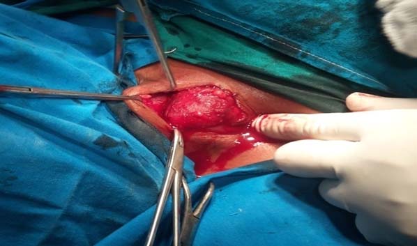
Intraoperative picture of the neck haemangioma.
Postoperative pain on rest and on movement was assessed by VAS (Visual Analogue Scale) hourly for first 24 hours and 4 hourly thereafter. Patient had good postoperative pain relief with recorded VAS scores of 4 or less with 4ml bolus of the epidural infusion solution as rescue analgesic being given 4 times during the first 48 hours postoperatively. No other complications were noted during surgery or in the immediate postoperative period.
Discussion
Dogliotti first described cervical epidural anaesthesia in 1933 for thoracic procedures using a single shot technique with local anaesthetic lignocaine [1]. Administration of local anaesthetics (with/without an opioid) into the cervical epidural space, results in anaesthesia along the cervical plexus, brachial plexus and upper thoracic dermatomes [2]. Unlike the lumbar or thoracic, the cervical epidural approach is an upcoming technique and has considerably attracted the attention of investigators to explore its viability for various surgeries. The incidence of complications in CEA is quiet low [3]. In experienced hands the use of CEA as a sole anaesthetic technique is well established [3–5]. The low cost, haemodynamic stability, reduced perioperative stress, early ambulation and excellent post-operative analgesia are the additional advantages to the patient [3]. The barorecepter reflex is partly impaired by CEA and specially benefits patients with limited cardiac reserve due to prolongation of coronary perfusion time and decreased ventricular after load [6]. Macchiarini et al., observed a reduction of heart rate, cardiac output and myocardial contractility with minimal changes in blood pressure, probably due to sympathetic blockade which reduced chances of myocardial ischemia [7]. Bacuzzi et al., observed a 10-15% reduction in the pulmonary function test after CEA compared to GA, which was clinically insignificant [8]. Stevens et al., observed a 12-16% reduction in FEV1 and FVC after CEA using lignocaine, which was clinically insignificant [9]. Capdevilla et al., reported a 21.1%, reduction in FVC in patients receiving CEA with reduction in the peak inspiratory pressures but these changes were insignificant in patients without lung pathology [10]. Our patient was haemodynamically stable intraoperatively and did not develop and pulmonary complications.
General Anaesthesia (GA) is preferred for neck surgeries but due to the GA-related cardio-respiratory and metabolic complications, the use of regional anaesthetic techniques has increased worldwide. The potential advantage of employing CEA is where GA is contraindicated. Although studies have documented the efficacy and safety of CEA for upper extremity and thoracic wall surgeries [11,12], this technique has seldom been used for neck surgeries. CEA can be used for neck (carotid artery surgery or thyroid/parathyroid surgeries) mastectomy and upper limb surgeries [5]. Singh et al., evaluated the safety and efficacy of CEA in patients undergoing modified radical mastectomy and concluded that CEA is a safer alternative to general anaesthesia providing a greater haemodynamic stability in the patient [11]. The use of cervical or high thoracic epidural anaesthesia for neck surgeries is often preferred due to decreased postoperative morbidity and superior postoperative analgesia [11]. It is now suggested that CEA is an equally safe alternative to general anaesthesia for breast, neck and upper extremity surgeries [5]. The presence of an associated laryngeal haemangioma with the potential risk of haemorrhage during airway manipulation, justify the use of CEA in our patient for the removal of neck haemangioma.
Usually the upper margin of sensory block in CEA is usually assessed at the C2 dermatome, whereas the lower margin of the sensory block is usually assessed at T3 dermatome [5]. The minimal and maximal extent of sensory block has been noted from C2-T1 and C2-T10 dermatomes [5]. Usually bupivacaine 0.25% or ropivacaine 0.5% are used as the local anaesthetic. Using 0.5% ropivacaine for CEA we were able to achieve excellent anaesthesia of the above mentioned dermatomes in our patient (VAS remained 4 or less for 48 hrs postoperatively). In our patient we noted a sensory block extending from C2 to T4 dermatomes, with a peak block onset time of 23 minutes. We preferred ropivacaine in our patient as it is less cardiotoxic.
Bonnet et al., documented dural puncture, epidural venipuncture and respiratory muscle paralysis as potential complications of CEA [4]. Hakl et al., observed bloody epidural tap, spread of local anaesthetic solution into the subarachnoid space and failed epidural puncture as the major complications in CEA [13]. Other possible complication of CEA includes bilateral phrenic nerve palsy. No procedure related complications occurred in our patient.
Conclusion
In the hands of an experienced anaesthesiologist, CEA may be a suitable choice and an alternative to general anaesthesia for neck surgeries.
[1]. Dogliotti AM, A new method of block anaesthesia: segmental peridural spinal anaesthesiaAm J Surg 1933 20:107-18. [Google Scholar]
[2]. Waldman SD, Cervical epidural nerve block. In: Waldman SD, edInterventional pain management 2001 PhiladelphiaWB Saunders:373-81. [Google Scholar]
[3]. Khanna R, Singh DK, Cervical epidural anaesthesia for thyroid surgeryKathmandu Univ Med J 2009 7:242-45. [Google Scholar]
[4]. Bonnet F, Derosier JP, Pluskwa F, Abhay K, Gaillard A, Cervical epidural anaesthesia for carotid artery surgeryCan J Anaesth 1990 37:353-58. [Google Scholar]
[5]. Michalek P, David I, Adamec M, Janousek L, Cervical epidural anaesthesia for combined neck and upper limb proceduresAnaesth Analg 2004 99:1833-36. [Google Scholar]
[6]. Dhummansure D, Kamtikar S, Haq MM, Patil SG, Efficacy and Safety of Cervical Epidural Anaesthesia for Thyroid SurgeryInt J Sci Stud 2015 3(7):245-50. [Google Scholar]
[7]. Macchiarini P, Rovira I, Ferrarello S, Awake upper airway surgeryAnn Thorac Surg 2010 89:387-90. [Google Scholar]
[8]. Stevens RA, Frey K, Sheikh T, Kao TC, Mikat SM, Morales M, Time course of the effects of cervical epidural anaesthesia on pulmonary functionReg Anaesth Pain Med 1998 23:20-24. [Google Scholar]
[9]. Bacuzzi A, Dionigi G, Del BA, Cantone G, Sansone T, Di Losa E, Anaesthesia for thyroid surgery: Perioperative managementInt J Surg 2008 6(Suppl 1):S82-85. [Google Scholar]
[10]. Capdevilla X, Biboulet P, Rubenovitch J, The effects of cervical epidural anaesthesia with bupivacaine on pulmonary function in conscious patientsAnaesth Analg 1998 86:1033-38. [Google Scholar]
[11]. Singh AP, Tewari M, Singh DK, Shukla HS, Cervical epidural anaesthesia: A safe alternative to general anaesthesia for patients undergoing cancer breast surgeryWorld J Surg 2006 30:2043-47. [Google Scholar]
[12]. Guevara-López U, Bárcenas-Olivares J, Gutiérrez-Sougarret B, Aldrete JA, Olascoaga-Ortega G, Cervical epidural anaesthesia for upper extremity surgery using three different formulations of local anaestheticsCir Cir 2005 73:273-81. [Google Scholar]
[13]. Hakl M, Michalek P, Sevcík P, Pavlíková J, Stern M, Regional anaesthesia for carotid endarterectomy: An audit over 10 yearsBr J Anaesth 2007 99:415-20. [Google Scholar]