The pancreas has both exocrine as well as endocrine functions. It arises from the endoderm as a dorsal and a ventral bud which fuse together to form the single organ. The secretions of the pancreas play an important role in the homeostasis of carbohydrate metabolism and regulating the glucose level in the blood. It extends transversely across the posterior abdominal wall from the duodenum to the spleen [1]. It has been studied in great detail in various animal species such as reptiles, mammals and amphibians. The mode of development of pancreas is similar in mammals, birds, reptiles and amphibians, while the islet cells are segregated as Brockmann bodies in some fishes [2]. Dysfunction of the pancreas results in diabetes mellitus. Diabetes itself is one of the earliest described diseases. The Eber’s Papyrus dating back to 1500 B.C, mentioned it as a disease of polyuria [3]. In 1921, the outstanding discovery of insulin molecule by Banting and Best offered an exciting prospect for the treatment of diabetes. Fetal pancreatic islet transplantation is one of the most attractive strategies for the cure of type I diabetes mellitus. This field, with its research and clinical applications, has been particularly pursued by many workers in the recent years [4]. Research into the development of the pancreas has great implications in clinical practice and treatment protocol. Knowledge of development of human pancreas is important for success of replacement therapies in treatment of diabetes mellitus [5].
The present study aimed at morphometric development of fetal pancreas at different gestational age.
Materials and Methods
This was a descriptive cross-sectional study conducted on fetal pancreas from July 2008 to January 2010 at Government Medical College, Miraj. The parameters considered were the body weight of fetus, crown rump length, pancreas weight, its length, the height of its head. Forty aborted human fetuses (25 male and 15 female) of 12-40 weeks gestational age with no obvious congenital abnormality were obtained with the permission from Department of Obstetrics and Gynaecology and prior consent of the parents. These fetuses included spontaneous abortions and stillborns. Fetuses were obtained within 4-5 hours of birth to avoid post-mortem changes. The sex, gestational age, weight and crown rump length were noted.
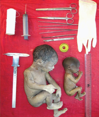
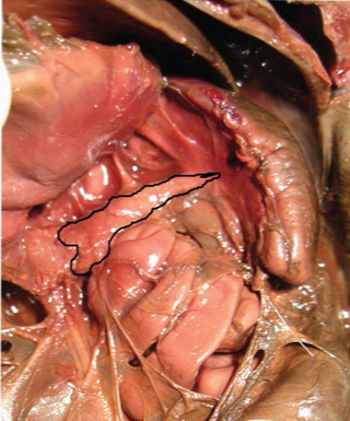
No specific reference was used to guide us for the method of dissection and measurements. A cruciate incision was taken on the anterior abdominal wall and the duodenum and pancreas were identified and removed from the abdominal cavity [Table/Fig-1,2]. The fetuses were weighed in double pan balance. The crown rump length recorded by using thread and scale. All these parameters were compared with the crown rump length and the gestational age.
Fetuses of 38 and 16 weeks with the material used for the study.
Pancreas seen in situ after reflecting the stomach in a 38th weeks fetus.
Results
The development of the pancreas was studied by considering different parameters of development. When there was more than one fetus at the given gestational age, the average of concerned parameter represented that age group. The parameters considered were the body weight of fetus, crown rump length, pancreas weight, its length, the height of its head. All these parameters were compared with the crown rump length and the gestational age.
The weight of the fetuses at different gestational age and the corresponding crown rump length were recorded in grams and centimeters respectively. It was observed that, there was increase in body weight and crown rump length with increasing gestational age. The average body weight of fetus was 125 gram at 12th weeks of gestation. It increased to 658 gram in 24th week and became 2825 gram at 40th week of gestation. The average crown rump length at 12th week was 11.50 cm. It was 19.70 cm at 24th week and 35 cm at 40th week. The weight of the pancreas at 12th week of gestation was 0.40 gram and it increased to 4.03 gram in the 40th week of gestation [Table/Fig-3]. The percentage relative weight was calculated by the following formula [6]:

Various parameters at different gestational weeks.
| Gestationalage (weeks) | Body weight(gm) | Crown rumplength (cm) | Average PancreasWeight (gm) | Relative PancreasWeight (%) | Length ofpancreas (cm) | Height ofhead (cm) |
|---|
| 12 | 125 | 11.50 | 0.40 | 0.32 | 1.80 | 0.80 |
| 14 | 150 | 12.00 | 0.50 | 0.33 | 1.80 | 0.50 |
| 16 | 200 | 12.50 | 0.60 | 0.30 | 1.90 | 0.70 |
| 18 | 276 | 14.77 | 0.67 | 0.24 | 2.60 | 0.83 |
| 20 | 445 | 16.62 | 0.81 | 0.18 | 2.28 | 0.80 |
| 22 | 535 | 18.00 | 0.74 | 0.14 | 2.75 | 1.10 |
| 24 | 658 | 19.70 | 0.92 | 0.13 | 2.23 | 0.86 |
| 26 | 800 | 23.00 | 1.15 | 0.14 | 3.15 | 0.86 |
| 28 | 1425 | 26.70 | 1.64 | 0.12 | 3.50 | 1.60 |
| 30 | 1488 | 27.80 | 1.65 | 0.11 | 2.95 | 1.15 |
| 32 | 2000 | 30.40 | 1.91 | 0.10 | 3.45 | 1.55 |
| 34 | 2200 | 29.00 | 3.40 | 0.15 | 3.90 | 2.00 |
| 36 | 2250 | 32.00 | 3.00 | 0.13 | 3.80 | 2.40 |
| 38 | 2825 | 32.00 | 2.95 | 0.10 | 4.00 | 2.50 |
| 40 | 2825 | 35.00 | 4.03 | 0.14 | 4.70 | 2.70 |
The average of percentage relative weight was calculated for particular age group wherever appropriated. The percent relative weight of pancreas was 0.32% at 12th week of gestation. It was 0.13% in 24th week and 0.14% in 40th week of gestation.
The length of pancreas and the height of its head were measured in cm using vernier calipers. It was observed that, as the gestational age increases, the length of pancreas and height of its head also increases [Table/Fig-3].
Discussion
Development includes three fundamental processes i.e., growth, differentiation and metabolism. Growth is increase in spatial dimensions and in weight. Differentiation is increase in complexity and organization [7]. Embryonically the pancreas arises between the 3rd and 4th week of gestation as a pair of evaginations of endodermal epithelium from the primitive gut. The endocrine part of the pancreas is distinguished by the 8th week of gestation. The exocrine part however is developed after 12th week. Throughout the pregnancy the pancreas shows increase in weight, length [8].
An increase in the body weight of the fetus was seen as the gestational age increased. At 12th week, the fetus weighed 125 gram. This weight increased gradually to 1425 gram at 28th week. Thereafter, the weight increased rapidly to 2825 gram at 40th week of gestation. The findings of body weight on comparison with previous studies shows, that the body weight of fetus at different gestational age reported by Greunwald [9] was more or less similar to the present study. The findings of Schultz [10] and Moore [11] showed weight less than the findings in the present study. The range of body weight given by Sadler was comparable with findings of the present study [Table/Fig-4] [12].
Showing comparison of body weight (in gram) of present study with the findings of other authors [7,9–12].
| GestationalAge (weeks) | Greunwald [9]1960 | Schulz [10]1953 | Hamilton [7]1975 | Sadler [12]2006 | Moore [11]2008 | Present Study2016 |
|---|
| 14 | - | - | - | 60-200 | 110 | 150 |
| 16 | - | - | 120 | - | 200 | 200 |
| 18 | - | - | - | 250-450 | 320 | 276 |
| 20 | - | - | 300 | - | 460 | 445 |
| 22 | - | 330 | - | 500-820 | 630 | 535 |
| 24 | 638 | - | 635 | - | 820 | 658 |
| 26 | 845 | 767 | - | 900-1300 | 1000 | 800 |
| 28 | 1020 | - | 1220 | - | 1300 | 1425 |
| 30 | 1230 | 1110 | - | 1400-2100 | 1700 | 1488 |
| 32 | 1488 | - | 1700 | - | 2100 | 2000 |
| 34 | 1838 | 1718 | - | 2200-2900 | 2900 | 2200 |
| 36 | 2165 | - | 2400 | - | 3400 | 2250 |
| 38 | 2678 | 2476 | - | 3000-3400 | - | 2825 |
| 40 | 3063 | - | 3250 | - | - | 2825 |
The crown rump length at 12th week gestation was 11.50 cm whereas, it was 35 cm at 40th week of gestation. From the [Table/Fig-5], it is seen that the values of crown rump length given by Moore and Hamilton were more or less similar to the findings of the present study. The findings of Robb about average weight of pancreas showed gradual increase in pancreas weight as age progressed but at corresponding gestational age they were less as compared to present study [Table/Fig-6] [14]. The relative percentage weight of pancreas at 12th week was 0.32% and 0.14% at 40 weeks of gestation. Detailed findings from the previous studies were not available for comparison.
Showing comparison of crown rump length (in cm) of present study with the findings of other authors [7,11,13].
| Gestationalage (weeks) | Moore[11] 2008 | Hamilton[7] 1975 | Potter & Craig[13] 1976 | Present study2016 |
|---|
| 12 | 87 | 57-84 | - | 115 |
| 14 | 120 | - | - | 120 |
| 16 | 140 | 61-100 | - | 125 |
| 18 | 160 | - | - | 150 |
| 20 | 190 | 101-200 | - | 170 |
| 22 | 210 | - | - | 185 |
| 24 | 230 | 151-200 | 209 | 197 |
| 26 | 250 | - | 234 | 230 |
| 28 | 270 | 201-260 | 254 | 267 |
| 30 | 280 | - | 271 | 277 |
| 32 | 300 | 261-320 | 284 | 304 |
| 34 | - | - | 298 | 290 |
| 36 | 340 | 321-390 | 324 | 320 |
| 38 | 360 | 391-450 | 334 | 320 |
| 40 | - | - | - | 350 |
Showing comparison of average weight of pancreas (in gm) of present study with the findings of another author [14].
| Gestational Age | Robb [14] | Present Study |
|---|
| 12 | - | 0.40 |
| 14 | - | 0.50 |
| 16 | - | 0.60 |
| 18 | - | 0.40 |
| 20 | 0.31 | 0.51 |
| 22 | - | 0.73 |
| 24 | - | 0.52 |
| 26 | 0.53 | 0.88 |
| 28 | 0.75 | 1.64 |
| 30 | - | 1.65 |
| 32 | 1.13 | 1.91 |
| 34 | 1.48 | 3.40 |
| 36 | 1.62 | 3.00 |
| 38 | 1.72 | 2.95 |
| 40 | 2.65 | 4.02 |
The present study shows that there was very less increase in the height of the head compared to increase in length of pancreas. The pancreas is situated in the concavity of the duodenum and once the duodenum becomes fixed retroperitoneally, there is very less space for the pancreas to increase vertically and so the developing pancreas grows towards the spleen. This could be attributed to faster growth of dorsal pancreatic bud than ventral bud [15].
Limitation
One of the limitations of the current study is the small sample size.
Conclusion
Fetal pancreatic islet transplantation is currently one of the most attractive strategies for the cure of Type I diabetes mellitus. The knowledge of development of pancreas helps in planning new therapeutic interventions in the treatment of various diseases related to pancreas, such as diabetes mellitus, carcinoma of pancreas, etc. The present study will help in understanding the growth and development of pancreas in fetal life. The data of the present study would also add knowledge to the existing literature.
Author’s Contribution
Abhijeet S. Dhende and Mahendra A. Kathole contributed in collection of specimen’s and dissection, analysis and interpretation of data, review of literature, writing and editing of the manuscript. Deepak S. Joshi guided us throughout this study. He mainly contributed in analysis and interpretation of data and manuscript writing.