Unusually Large Peripheral Osteoma of the Mandible – A Rare Case Report
Pulkit Khandelwal1, Vikas Dhupar2, Francis Akkara3
1 Senior Lecturer, Department of Oral and Maxillofacial Surgery, Pacific Dental College and Hospital, Udaipur, Rajasthan, India.
2 Professor and Head, Department of Oral and Maxillofacial Surgery, Goa Dental College and Hospital, Goa, India.
3 Professor, Department of Oral and Maxillofacial Surgery, Goa Dental College and Hospital, Goa, India.
NAME, ADDRESS, E-MAIL ID OF THE CORRESPONDING AUTHOR: Dr. Pulkit Khandelwal, Senior Lecturer, Department of Oral and Maxillofacial Surgery, Pacific Dental College and Hospital, Udaipur, Rajasthan, India.
E-mail: khandelwal.pulkit22@gmail.com
Osteoma is a benign tumor which is composed of mature compact or cancellous bone. Osteoma may be periosteal (arising from surface of the bone) or endosteal (develop in the medullary bone) or combination of both. Here, we present a case of unusually large osteoma present on the lingual surface of the mandible in a 40-year-old female patient. The lesion had grown slowly for 15 years and caused intra-oral swelling leading to difficulty in mastication, speech and tongue movements. Under general anesthesia, local complete surgical excision was performed.
Excision, Gardner’s syndrome, Lesion, Neoplasm
Case Report
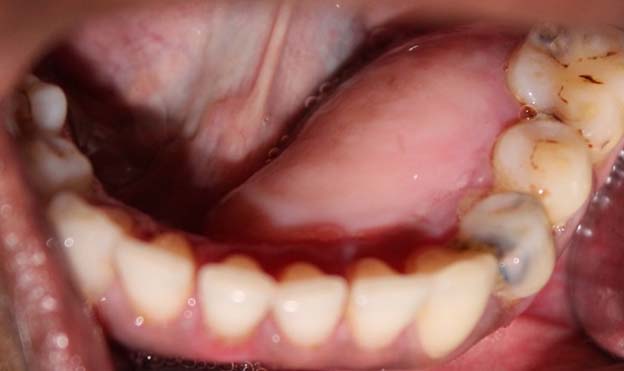
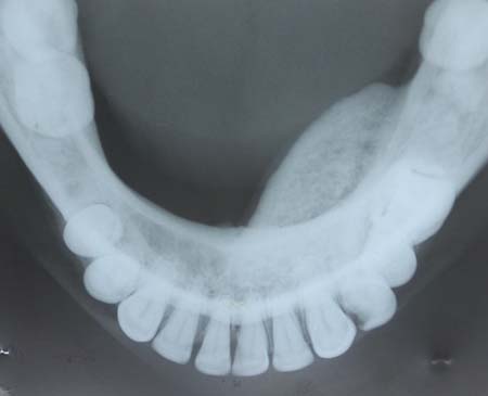
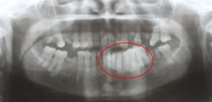
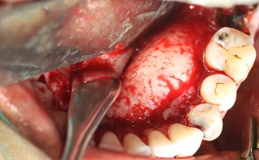
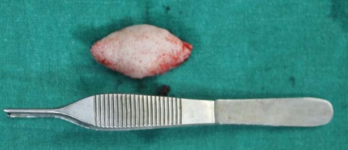
A 40-year-old female patient reported to Department of Oral and Maxillofacial Surgery with chief complaint of difficulty in eating due to growth on inner surface of the left side of lower jaw. Patient gave history of gradual growth of the swelling since 15 years before attaining the present size. There was no history of any facial trauma and medical history was non-significant. She didn’t have any other symptom like pain or bleeding from the swelling, reduced or difficulty in mouth opening. However, the patient reported difficulty in mastication and speech due to restricted lateral movements of the tongue. Extra-oral examination was non-significant with no gross asymmetry detected. Intra-orally, a well-defined oval, immobile growth was seen on the lingual aspect of mandible on leftside, extending from mid-symphysis region to second molar region. The swelling measured approximately 6x3cm and was non-tender and bony hard in consistency. The overlying mucosa was normal in colour and texture [Table/Fig-1]. There was no paresthesia of tongue or lower lip. All teeth were vital. No regional lymphadenopathy was present. Based on clinical examination, a provisional diagnosis of peripheral osteoma was made. Exostoses, osteoid osteoma, complex odontoma and ossifying fibroma were included as differential diagnoses. Mandibular occlusal radiograph and orthopantamogram revealed a well circumscribed, oval, 6x3cm radiopaque mass without any radiolucent rim, in relation to mandible. The radiopaque mass was present in relation to the lingual aspect of left parasymphysis and body of mandible extending from central incisor (41) till second molar (37) region [Table/Fig-2,3]. These clinical and radiographic features were sufficiently suggestive and supportive for the diagnosis of peripheral osteoma. There were no clinical features suggestive of Gardner’s syndrome. Complete haemogram was within normal limits. Patient was explained about the condition and need for surgery. Under general anaesthesia, the lesion was approached by making a crevicular incision and raising the mucoperiosteal flap [Table/Fig-4]. The bony mass was found to be adherent to the bone. The lesion was totally removed using rotary instruments (tapered straight fissure bur) and chisel [Table/Fig-5]. The cortical portion of the mandible was then smoothened. Excisional biopsy confirmed the diagnosis of peripheral osteoma. Following surgery, antibiotics (amoxicillin with clavulanic acid) and analgesics (diclofenac) were started. Post-operative recovery was uneventful and patient was discharged after three days of surgery. On regular follow-up, the operative site healed well without any complications or recurrences.
Peripheral osteoma – intra-oral appearance.
Mandibular occulsal radiograph.
Discussion
Osteoma is a benign osteogenic neoplasm, often asymptomatic, which consists of well-differentiated and matured bone. It is charac-terized by growth of either compact (dense lamellae) or cancellous (trabecular) bone or can be formed by a combination of both [1,2]. It is generally slow growing, painless, asymptomatic solitary mass that is palpable unless it arises within the medullary space. In 1935, Jaffe first described osteoma as a specific entity [3].
Osteoma can present as a central, peripheral or extra-skeletal form. The central type of osteoma evolves from the endosteum and the peripheral variant develops from the periosteum. The extra-skeletal soft tissue type of osteoma usually develops within the muscle mass or soft tissue [4,5]. The pathogenesis of osteomas is still not completely understood. Some authors consider it as a true neoplasm, while others consider it as a developmental disorder [6]. A few proposed it as a reactive mechanism, caused by trauma, muscle traction or infection [3,7].
Osteomas more frequently affect the cortical plate of long bones, but they can also present in the maxillofacial region. Peripheral osteoma affects paranasal sinuses most commonly. Other sites affected by this lesion include orbital wall, temporal bone, pterygoid processes and external auditory canal. Though a solitary peripheral osteoma of the jaw bones is very rare, the mandible is involved more than the maxilla. When it involves the mandible, the angle and the inferior border of the body of the mandible are more commonly involved in association with the buccal plate. The involvement of the lingual cortex of the mandible is rare. Peripheral osteomas are slow-growing lesions and they usually remain asymptomatic clinically. However, they exhibit continuous growth at adulthood and acquire a large size, producing swelling, severe dysfunction, limitation or deviation of the mandible on opening, dysphagia and facial asymmetry [8].
Osteomas can affect any age group; however, these lesions predominantly affect patients aged 40 years and above. Males are affected more often than females (2:1). It usually measures less than 2cm in size after years of slow growth and proliferation [5,8]. Literature search revealed nearly 82 well-documented cases of solitary peripheral osteoma of mandible involving various regions of the mandible [Table/Fig-6] [9].
Incidence of site involved by solitary peripheral osteoma of mandible [9].
| Region involved | Percent cases |
|---|
| Body | 41.5% |
| Condyle | 23.2% |
| Angle | 14.6% |
| Ascending ramus | 11% |
| Coronoid process | 8.5% |
| Sigmoid notch | 1.2% |
Patients with osteomas should also be evaluated for Gardner’s Syndrome. This syndrome is an autosomal dominant disorder which is characterized by colorectal polyposis, multiple maxillofacial osteomas, soft tissue tumors, cutaneous sebaceous cysts and multiple impacted supernumerary teeth [4]. If the dentists find multiple impacted supernumerary teeth associated with osteomas of the jaw, they should examine patient for the presence of the Gardner’s Syndrome [2,7].
Differential diagnoses of peripheral osteoma includes several pathological entities, such as exostoses, osteoblastoma, osteoid osteoma, complex odontoma or central ossifying fibroma. Exostoses are osseous overgrowths that usually stop growing after puberty is attained, differentiating them from osteomas. Central ossifying fibromas have well-defined borders and a thin, radiolucent line separating the lesion from the surrounding normal bone. Osteoblastomas and osteoid osteomas are most often painful and growth is rapid as compared to peripheral osteomas. A complex odontoma also presents as a well-defined radiopacity within bone, but the density is greater than that of bone and resembles that of a tooth [7].
Radiographic investigations such as occlusal radiograph, panoramic radiograph or Computed Tomography (CT) scan are used for imaging; however, the CT scan is the best imaging modality as it provides exact details about the site, dimensions and extension of the lesion. On radiograph, a peripheral osteoma appears as a classical well-circumscribed, round to oval, radiopaque mass with distinct borders. Characteristic X-ray changes seen in this lesion include focal rarefaction and reactive bone formation [7].
If peripheral osteoma is asymptomatic, excision of the lesion is not generally recommended. Surgical intervention is indicated only when osteoma becomes so large in size that it leads to facial asymmetry or any functional impairment. After surgical excision, recurrence of peripheral osteoma is extremely rare [4,7].
Our case was unique in few aspects because of its unusually large size (6x3cm) and involvement of lingual aspect of the mandible which is extremely rare. The mass was sessile and was peripheral (periosteal) in origin, made up of dense compact bone. As there was no history of trauma to mandible and the mass was present since 15 years only, so a traumatic or developmental aetiology was ruled out. Hence, the mass was supposed to be of neoplastic origin. Since patient was having difficulty while eating and speaking, local complete surgical excision was done to alleviate the symptoms. There were no other signs and symptoms suggestive of Gardner’s Syndrome. Also, there was no post-operative complication or recurrence on regular follow-up visits.
Conclusion
Osteoma is slow growing, benign tumor. Complete surgical excision is the treatment of choice. Its recurrence after surgical excision is extremely rare.
[1]. Frolich MA, Mandibular osteoma: A case of impossible rigid laryngoscopyThe Journal of the American Society of Anesthesiologists 2000 92(5):261-62. [Google Scholar]
[2]. Durao AR, Chilvarquer I, Hayek JE, Provenzano M, Kendall MR, Osteoma of the zygomatic arch and mandible: Report of two casesRev Port Estomatol Med Dent Cir Maxilofac 2012 53(2):103-07. [Google Scholar]
[3]. Chaudhary M, Kulkarni M, Osteoid osteoma of mandibleJ Oral Maxillofac Pathol 2007 11(2):52-55. [Google Scholar]
[4]. Goudar G, Kumar RR, Manjunath GA, Mahadesh J, Osteoma of mandibleJournal of Dental Sciences and Research 2011 2(1):116-22. [Google Scholar]
[5]. Roy ID, Peripheral osteoma of mandibleMedical Journal Armed Forced India 2008 64:385-86. [Google Scholar]
[6]. Sayan NB, Cook C, Karasu HA, Gunhau O, Peripheral osteoma of the maxillofacial region: A study of 35 new casesJ Oral Maxillofacial Surg 2002 60:1299-301. [Google Scholar]
[7]. Bulut E, Acikgoz A, Ozan B, Gunhan O, Large peripheral osteoma of the mandible: A case reportInt J Dent 2010 Article ID 834761 [Google Scholar]
[8]. Masuki Y, Peripheral osteoma at the mentum of mandibleRinsho Derma 2002 44:735-37. [Google Scholar]
[9]. Manjunatha BS, Das N, Sutariya R, Ahmed T, Peripheral osteoma of the body of mandibleBMJ Case Rep 2013 8:1-4. [Google Scholar]