A Rare Case of Anomalous Origin of First Lumbrical from the Tendon of Flexor Digitorum Superficialis to Index Finger
S Trivedi1, BC Satapathy2, M Rathore3, M B Sinha4
1 Assistant Professor, Department of Anatomy, AIIMS, Raipur, Chhattisgarh, India.
2 Senior Resident, Department of Anatomy, AIIMS, Raipur, Chhattisgarh, India.
3 Assistant Professor, Department of Anatomy, AIIMS, Raipur, Chhattisgarh, India.
4 Assistant Professor, Department of Anatomy, AIIMS, Raipur, Chhattisgarh, India.
NAME, ADDRESS, E-MAIL ID OF THE CORRESPONDING AUTHOR: Dr. S. Trivedi, Room No 1113, Department of Anatomy, First Floor, AIIMS Medical College Block, Tatibandh, Raipur-492099, Chhattisgarh, India.
E-mail: dr.somit@gmail.com
Human hand is involved in variety of precision work which requires a combined effort of forearm muscles as well as intrinsic muscles of hand. Lumbricals along with interossei muscles connect the tendons of flexor and extensor muscles and thus play a key role in the characteristic movements of human hands. Lumbricals originate from long flexor tendon and is inserted into dorsal digital expansion. Any variation in the attachment of these muscles can lead to deviation from the normal actions of the fingers and their proximal extension into carpal tunnel and might lead to carpal tunnel syndrome. An extremely rare case of first lumbrical taking origin solely from first tendon of Flexor Digitorum Superficialis (FDS) and having proximal attachment extending into carpal tunnel was noticed bilaterally in cadaveric dissection. These variations are always challenging for clinicians and surgeons during hand surgeries.
Carpal tunnel, Intrinsic muscles of hand, Long flexor tendons, Variations
Case Report
During routine dissection classes for the undergraduate medical students, a variable presentation of first lumbrical was noted in a male cadaver of 62 years. The variation existed bilaterally [Table/Fig-1,2]. After careful dissection of Right and Left hands using Cunningham’s manual of dissection as a guide, it was observed that the first lumbrical muscle in both right and left hand was having an unusual origin from the first tendon of Flexor Digitorum Superficialis (FDS) instead of Flexor Digitorum Profundus (FDP). The tendon, on tracing proximally, was having the fibers of origin more proximal to rest of lumbricals and was extending deep to the flexor retinaculum. The fibres of lumbrical muscle were seen taking origin from lateral and superior surface of the first tendon of FDS. A very few proximal fibres were seen gaining attachment to first tendon of FDP which got easily separated on lifting the FDS tendon along with first lumbrical suggesting that attachment to be very feeble. Although the further course of muscle and its site of insertion remained normal. The muscle received a twig from the branch of median nerve going to index finger in both hands. The course of the median nerve was normal and it was seen plastered to the undersurface of FDS lying deep to flexor retinaculum running in the carpal tunnel.
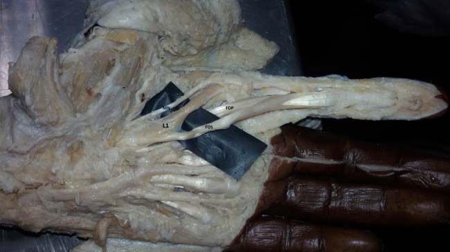
Dissected Left hand showing origin of first lumbrical (L1) from first tendon of Flexor Digitorum Superficialis (FDS) getting a twig from Median nerve (MN), FDP - first tendon of Flexor Digitorum Superficialis.
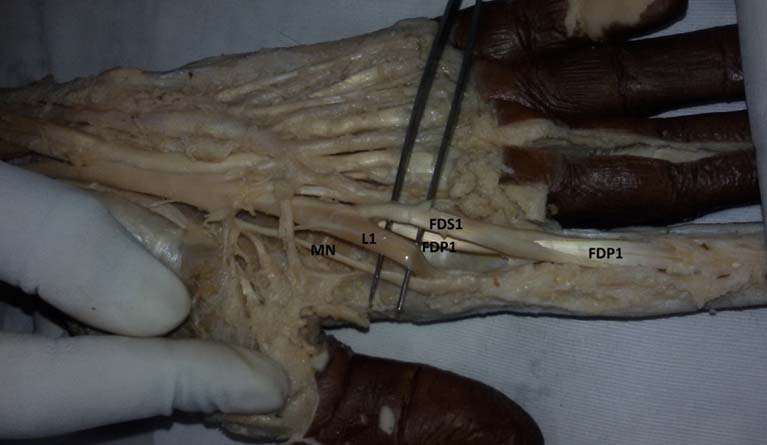
Dissected Right hand showing origin of first lumbrical (L1) from first tendon of Flexor Digitorum Superficialis (FDS1) getting a twig from Median nerve (MN), FDP1 - first tendon of Flexor Digitorum Superficialis.
Discussion
Prehensile nature of human hand as well as the precision with which a human hand can work is unique to mankind. Lumbricals are one of the intrinsic muscles of human hand that make these movements possible. Lumbricals are four in number labeled 1st, 2nd, 3rd and 4th from lateral to medial side. They take origin from the tendons of FDP and are inserted into extensor expansion of extensor digitorum. Thus, they connect the flexor muscles to extensor muscles which gives them the characteristic function of flexing metacarpophalengeal joints and extending interphalangeal joints at the same time [1,2].
The pinching action of index finger and the thumb is contributed by the 1st lumbrical which makes pulp of fingers to come in contact rather than tips of fingers as lumbrical further enhance the extension at distal Interphalangeal joint [3].
The excessive use of fingers in professional jobs can lead to hypertrophy of the lumbrical muscles and any proximal attachment of the these muscles extending into the carpal tunnel might lead to compression of median nerve thus predisposing to carpel tunnel syndrome [4,5].
Lumbrical muscles, four in number labeled numerically from lateral to medial side often present with variable picture. The first two lumbricals are unipennate taking origin from radial side of first and second tendon of FDP whereas medial two lumbricals are bipennate taking origin from adjacent sides of third and fourth tendon of FDP. In an earlier study it was reported in over 50% of the cases that first lumbrical was bulky and presented with a groove on its medial side to lodge the tendon of FDS [6]. In about 20% cases the first lumbrical had a proximal attachment extending into carpal tunnel [6]. A case of lumbrical muscles originating in the forearm from the undersurface of FDS also and passing through the carpal tunnel was reported [7]. Similarly, a case of first lumbrical arising by two bellies from tendon of both FDS as well as FDP has been reported [8]. Unusual pattern of attachment of lumbrical muscles have been seen in great numbers by earlier researchers ranging up to 86% of the cases [9]. The case presented here is extremely rare as none of the studies earlier have reported the origin of first lumbrical solely from FDS tendon.
In earlier studies, evolutionary aspect of hand muscles were studied in amphibians, reptiles and mammals to see that in lower animals, hand movements were controlled by many layers of muscles of the hand which were referred to as "brevis". Evolutionary changes with time made these brevis muscles replaced by long flexor tendons in mammals [10]. The distal muscle belly of first tendon of FDS and the first lumbrical muscle have common phylogenetic origin and as a result share intimate relationship [7]. Thus it is quite evident that the variations existing in lumbricals are of phylogenetic value [11]. In our case, the rare finding of first lumbrical taking origin from first tendon of FDS becomes of great significance as it justifies the intimate relation of FDS tendon and first lumbrical.
The variant and proximal origin of first lumbrical extending deep to the flexor retinaculum in the carpal tunnel might lead to compression of median nerve leading to carpal tunnel syndrome which becomes more pronounced at the time of flexion of wrist and fingers [4–6]. In cases of carpal tunnel decompression of median nerve beforehand knowledge of such variations should be kept in mind by clinicians and hand surgeons.
Conclusion
The variation reported in the study is extremely rare and the abnormal origin of the first lumbrical can lead to misinterpretation of facts which can be challenging for surgeons as well as clinicians. The proximal origin of first lumbrical extending into carpal tunnel may present with symptoms of carpal tunnel syndrome.
[1]. Standring S, Grey’s Anatomy: The Anatomical Basis of Clinical Practice 2013 40th EditionChurchill livingstoneElsevier:886-90. [Google Scholar]
[2]. Romanes GJ, Cunningham’s Manual of Practical Anatomy. 15th Edition. Vol 1. Upper & Lower limbs 2013 Oxford University Press:100-1. [Google Scholar]
[3]. Williams PL, Bannister LH, Berry MM, Collins P, Dyson M, Dusse KJE, Ferguson MWJ, Grey’s Anatomy 1995 38th edChurchill Livingstone, London:861-62. [Google Scholar]
[4]. Cobb TK, An KN, Cooney WP, Effect of lumbrical muscle incursion within the carpal tunnel on carpal tunnel pressure: a cadaveric studyJ Hand Surg (Am) 1995 20(2):186-92. [Google Scholar]
[5]. Siegel DB, Kuzma G, Eakins D, Anatomic investigation of the role of lumbrical muscles and Carpal tunnel SyndromeJ Hand Surg (Am) 1995 20(5):860-63. [Google Scholar]
[6]. Joshi SD, Joshi SS, Athavale SA, Lumbrical muscles and carpal tunnelJ Anat Soc India 2005 54(1):12-15. [Google Scholar]
[7]. Koizumi M, Kawai K, Honma S, Kodama K, Anomalous lumbrical muscles arising from the deep surface of flexor digitorum superficialis muscles in manAnn Anat 2002 184(4):387-92. [Google Scholar]
[8]. Potu B, Gorantla Pulakunta T, Rao M, Vollala V, Nayak S, Anomalous origin of the first lumbrical in the hand and its possible role in Carpal Tunnel SyndromeThe Internet Journal of Neurology 2006 8(1):1-3. [Google Scholar]
[9]. Mehta HJ, Gardner WU, A study of lumbrical muscle in human handAm J. Anat 1961 109:227-38. [Google Scholar]
[10]. Elliot D, Khandwala AR, Kulkarni M, Anomalies of flexor digitorum superficialis muscleJournal of Hand Surgery 1999 24(5):570-74. [Google Scholar]
[11]. Sawant SP, The cadaveric study of lumbricals of hand in 100 specimensInternational Journal of Current Sciences 2013 6E:107-10. [Google Scholar]