Determining Angle of Humeral Torsion Using Image Software Technique
Sachin Patil1, Madhu Sethi2, Neelam Vasudeva3
1 Assistant Professor, Department of Anatomy, Andaman and Nicobar Islands Institute of Medical Sciences, Port Blair, Andaman and Nicobar Islands, India.
2 Assistant Professor, Department of Anatomy, Dr Baba Sahib Ambedkar Medical College and Hospital, Delhi, India.
3 Head and Director Professor, Department of Anatomy, Maulana Azad Medical College, New Delhi, India.
NAME, ADDRESS, E-MAIL ID OF THE CORRESPONDING AUTHOR: Dr. Sachin Patil, Assistant Professor, Department of Anatomy, ANIIMS, Port Blair-744101, Andaman and Nicobar Islands, India.
E-mail: drsachin6880@gmail.com
Introduction
Several researches have been done on the measurement of angles of humeral torsion in different parts of the world. Previously described methods were more complicated, not much accurate, cumbersome or required sophisticated instruments.
Aim
The present study was conducted with the aim to determine the angles of humeral torsion with a newer simple technique using digital images and image tool software.
Materials and Methods
A total of 250 dry normal adult human humeri were obtained from the bone bank of Department of Anatomy. The length and mid-shaft circumference of each bone was measured with the help of measuring tape. The angle of humeral torsion was measured directly from the digital images by the image analysis using Image Tool 3.0 software program. The data was analysed statistically with SPSS version 17 using unpaired t-test and Spearman’s rank order correlation coefficient.
Results
The mean angle of torsion was 64.57°±7.56°. On the right side it was 66.84°±9.69°, whereas, on the left side it was found to be 63.31±9.50°. The mean humeral length was 31.6 cm on right side and 30.33 cm on left side. Mid shaft circumference was 5.79 on right side and 5.63 cm on left side. No statistical differences were seen in angles between right and left humeri (p>0.001).
Conclusion
From our study, it was concluded that circumference of shaft is inversely proportional to angle of humeral torsion. The length and side of humerus has no relation with the humeral torsion. With advancement of digital technology, it is better to use new image softwares for anatomical studies.
Digital image, Image tool, Prosthesis, Recurrent anterior dislocation of shoulder
Introduction
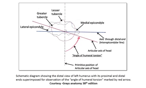
Angle of humeral torsion is defined as the angle formed between the proximal and distal articular axis of the humerus. This angle is measured at the intersection of two lines: one that evenly bisects the articular surface of the humeral head proximally and the second being the transepicondylar line distally [1,2]. But in the clinical literature, this angle is measured in the opposite direction and is referred to as humeral retroversion [3].
Since the upper extremity has developed as a prehensile appendage in humans and also has a major role in maintaining an upright posture, the humeral torsion is essential biologically [4].
Variations in the degree of torsion of the humeral head have been widely documented within anthropological literature over the course of time. The patterns of variation by age, population, side and sex have been documented. With the progress of field of sports medicine, humeral torsion has received renewed attention [5].
The variations in humeral head retroversion among individuals undergoing humeral surgery are clinically important. They may throw the light on the ways in which this parameter might be manipulated to improve surgical outcome [6].
The present study has been undertaken to study the angle of torsion by simple software technique in 250 humeri and the variation on the two sides and to determine its correlation, if any, with the length, side and circumference of the bone.
Materials and Methods
The present descriptive study was carried out on 250 dry normal adult human humeri obtained from the bone bank of Department of Anatomy, Maulana Azad Medical College, New Delhi, India. The bones showing morphological anomaly or broken ends or shaft were excluded from study. The humeri were cleaned, dried and studied in proper daylight.
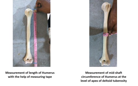
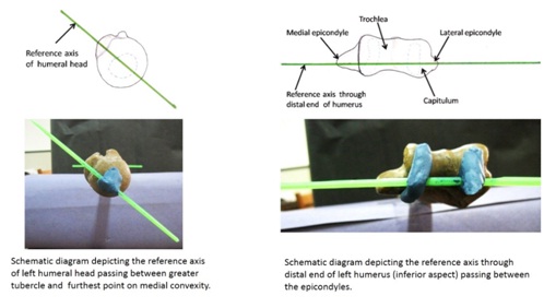
All the humeri of unknown gender were segregated as right or left side according to standard anatomical criteria. The length and mid shaft circumference of each bone was measured with the help of measuring tape [Table/Fig-1]. The upper end axis was passing through the centre of head of humerus, extending from a point where transverse diameter is maximum to the centre point on greater tuberosity of humerus [Table/Fig-2]. The lower end axis was taken as line passing between the centres of two epicondyles of humerus [Table/Fig-2].
Measurement of length and mid-shaft circumference of humerus.
Reference axes for upper and lower end of humerus.
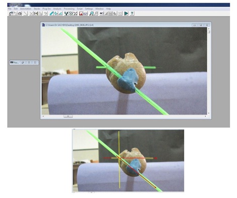
After marking the axes of upper and lower ends by fixing the plastic sticks of 0.5mm diameter with clay, the humerus was kept on flat surface and digital image was taken from the upper end for each bone as shown in [Table/Fig-3]. The camera attached with the system was kept on flat surface opposite the proximal articular of the humerus to capture the end-on image at a constant position for each sample. These images were transferred to the system and saved in JPEG format. The angle of humeral torsion was measured directly from the digital images by the image analysis using Image Tool 3.0 software program which is a multiple document interface application well supported on windows images. Each image was numbered accordingly and the measurements were taken by clicking the angle selection tool on the status bar, which facilitated marking the specified lines of intersection between the two sticks and the obtained value of the angle measured was imported and saved in the excel sheet format. [Table/Fig-4] shows schematic representation of the angle of humeral torsion adapted from Gray’s anatomy [7]. The data was analysed statistically with SPSS version 17 using unpaired t-test and Spearman’s rank order correlation coefficient.
Image analysis using Image Tool 3.0 software program.
Schematic diagram showing angle of humeral torsion.
Results
Intra- and inter-observer reliability - The principal investigator randomly selected 30 bones and erased the markings on them. All measurements were repeated on these 30 bones to assess the intra-observer variability. Another investigator also randomly selected 30 bones and repeated the whole procedure to assess inter-observer reliability.
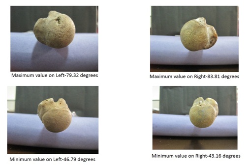
The mean angle of torsion was 64.57° ± 7.56°. On the right side it was 66.84° ± 9.69°, whereas on the left side it was found to be 63.31 ± 9.50° [Table/Fig-5]. The angle on both the sides showed great variation with maximum angle on right side was 83.81° and that on left side was 79.32° [Table/Fig-6]. Mid shaft circumference was 5.79cm on right side and 5.63cm on left side [Table/Fig-5]. On the right side the largest and lowest angles of torsion (83.81° and 43.16°) were found with bones having mid shaft circumference of 4.9cm and 6.1cm, respectively. Similarly, on left side the largest and lowest angles of torsion (79.32° and 46.79°) were found in bones with mid shaft circumference of 5.1cm and 5.4cm respectively. On comparing the mid shaft circumference with the angle of humeral torsion, a weak negative correlation on both sides was seen [Table/Fig-7]. Thus, there is a weak inverse relationship between circumference and angle of humeral torsion. The mean humeral length was 31.6cm on right side and 30.33cm on left side [Table/Fig-5]. Comparing the length with the angle of humeral torsion, the coefficient correlation showed weak negative relationship on right side and weak positive relationship on left side [Table/Fig-7]. So, we concluded that there is no relationship between length with angle of humeral torsion. No statistical difference was seen in angles between right and left humeri (p>0.001).
Mean and standard deviation of angle of torsion, mid shaft circumference and length of humerus.
| Angle of torsion | Mid shaft circumference | Length of humerus |
|---|
| Combined (n=250) | 64.57° ± 7.56°. | 5.71 ± 0.58 cm | 30.96 ± 1.98 cm |
| Right side (n=125) | 66.84° ± 9.69° | 5.79 ± 0.51 cm | 31.6 ± 2.21 cm |
| Left side (n=125) | 63.31 ± 9.50° | 5.63 ± 0.57 cm | 30.33 ± 1.87 cm |
Showing maximum and minimum value of angle of humeral torsion on both sides.
Spearman’s correlation coefficient.
| Correlation between AHT and Circumference | Correlation between AHT and Length |
|---|
| Right side | -0.19 | -0.0058 |
| Left side | -0.024 | +0.16 |
Discussion
Different aspects of the humeral torsion have been studied by several workers in different parts of the world. Studies show that the humeral torsion may be primary or secondary torsion. Primary torsion present in embryo is determined by developmental patterns and is a characteristic of various species. Secondary torsion is an outcome of the pull of muscular forces functions etc. The external and internal rotators of the shoulder and arm region exert their force on the shaft of humerus, which are responsible for addition of secondary torsion [8].
Humeral torsion is generally lower in the populations participating in more strenuous activity and increased in less active subjects [9]. [Table/Fig-8] compares the values of humeral torsion of previous studies to those of the present study [3,4,6,8–12]. The mean value humeral torsion of the present study is similar to most studies and is independent of methodology of study.
Showing mean values of angle of humeral torsion in different studies [3,4,6,8–12].
| Sr No | Author (Years) | Reference no. | Population | Mean angle of humeral torsion |
|---|
| 1 | Broca et al., 1881 | [3] | Caucasian | 74o |
| 2 | Mathews et al., 1893 | [11] | Salado-Indian | 69o |
| 3 | Martin et al., 1928 | [6] | Peruvian | 60.2o |
| 4 | Chillida et al., 1943 | [9] | Argentine Aborigine | 61o |
| 5 | Ayer and Upshon 1943 | [9] | South Indian | 62.1o |
| 6 | Krahl and Evans 1945 | [10] | Caucasian | 74.4o |
| 7 | Krahl and Evans 1945 | [10] | American | 72.6o |
| 8 | Kate et al., 1969 | [12] | Central Indian | 55o |
| 9 | Mehta et al., 1971 | [4] | Indian (Rajasthan) | 68.5o |
| 10 | Kummer et al., 1998 | [3] | American | 62.7o |
| 11 | Shah et al., 2006 | [8] | Indian (Gujarat) | 68.5o |
| 12 | Motagi et al., 2011 | [9] | Indian (Karnataka) | 59.6o |
| 13 | Present study 2016 | - | North Indian | 64.6o |
No correlation was found between length and humeral torsion in our study, similar to what have been reported by Krahl et al., and Mehta et al., [4,10]. But study by Lambert has shown that length of humerus was inversely proportional to angle of humeral torsion [13]. Present study also showed that the circumference of the shaft of humerus is inversely proportional to the angle of torsion. This finding was similar when compared to study by Shah et al., but opposite to study by Krahl et al., and Mehta et al., [4,8–10] [Table/Fig-9].
Showing correlation circumference of humerus with angle of humeral torsion in different studies [4,8–10].
| Sr No | Author (Years) | Reference no. | Population | Correlation of circumference of humerus with humeraltorsion |
|---|
| 1 | Krahl and Evans 1945 | [10] | Caucasian | Inverse proportion |
| 2 | Krahl and Evans 1945 | [10] | American | Direct proportion |
| 3 | Mehta et al., 1971 | [4] | Indian (Rajasthan) | Direct proportion |
| 4 | Shah et al., 2006 | [8] | Indian (Gujarat) | Inverse proportion |
| 5 | Motagi et al., 2011 | [9] | Indian (Karnataka) | Inverse proportion |
| 6 | Present study 2016 | - | North Indian | Inverse proportion |
The humeral torsion has been extensively studied in cases of Recurrent Anterior Dislocation of Shoulder (RADS). The increase in angle of humeral torsion is associated with increase in RADS. Even minimal force can lead to dislocation in such cases compared to normal subjects [14]. Increased frequency of RADS in such cases may be due to increased torsion predisposing to anterior dislocation by putting the head in a position of risk in the abducted and externally rotated position [15]. The rotational humeral osteotomy for RADS to decrease the angle of torsion has been found successful in many cases [16].
An increase of humeral retroversion in the throwing arm of handball players could be a protective mechanism for the anterior capsulo-labral complex by preventing anterior instability [17]. The influence of humeral torsion on posterior shoulder tightness was studied in baseball players and compared with a control group. The dominant limb of the baseball players had greater humeral torsion. The amount of humeral torsion and measures of posterior shoulder tightness showed a significant correlation [18]. An increase in thickness of the shaft of the humerus and altered humeral torsion was observed in professional baseball pitchers [19]. So the circumference of shaft is also important with measurement of angle of humeral torsion.
In living subjects Axial MRI images of the shoulder region and distal arm region can be used to get the desired value of angle of torsion. The image tool 3 software can be used to mark the separate axial angles of upper and lower end and the differences between the two will provide the angle of humeral torsion [20]. It has been found that in juvenile baseball players the repetitive load of throwing motion restricts the normal processes acting to decrease the humeral torsion angle during the growth period [21]. Hence, the humeral torsion angle can be influenced by various occupational and anthropometric factors.
Limitation
This study can be further pursued using a large collection of gender and occupation specific study sample by means of various radiographic imaging tools for a better clinically relevant comparison. For patients receiving shoulder prosthesis, pre-operative accurate and reliable estimate of the angle of humeral torsion using sophisticated instruments and complex computer-assisted three dimensional reconstructions can be very useful.
Conclusion
From our study we concluded that circumference of shaft is inversely proportional to humeral torsion. The length and side of humerus has no relation with the humeral torsion. It is very difficult to know a constant angle of humeral torsion as range of distribution is very wide. The data generated from the study of the angles of humeral torsion may give surgeons a clearer understanding of how the range of motion of shoulder may be influenced or limited by the structure of the bone, muscle and ligaments.
[1]. Krahl VE, The torsion of the humerus; its localization, cause and duration in manAm J Anat 1947 80:275-319. [Google Scholar]
[2]. Evans FG, Krahl VE, The torsion of the humerus: a phylogenetic study from fish to manAm J Anat 1945 76:303-07. [Google Scholar]
[3]. Rhodes JA, Humeral torsion and retroversion in the literature: a reply to LarsonAm J Phys Anthropol 2007 133:819-21. [Google Scholar]
[4]. Mehta L, Chaturvedi RP, Angle of humeral torsionJ Anat Soc India 1971 20:94-98. [Google Scholar]
[5]. Pieper HG, Humeral torsion in the throwing arm of handball playersAm J Sports Med 1998 26:247-53. [Google Scholar]
[6]. Dare SS, Masilili MG, Okumu G, Mohammed YG, Abba S, Okpanachi AO, Determination of angles of torsion and retroversion of the humerus of male and female skeleton specimens in ugandaAsian J Med Sci 2012 4(5):174-78. [Google Scholar]
[7]. Williams PL, Gray’s Anatomy: The Anatomical Basis of Medicine and Surgery 1995 38th edEdinburghChurchill Livingstone:1560 [Google Scholar]
[8]. Shah RK, Trivdei BD, Patel JP, Shah GV, Nirvan AB, A Study of Angle of Humeral TorsionJ Anat Soc India 2006 55(2):43-47. [Google Scholar]
[9]. Motagi M, Shankar N, Ravindranath R, Estimation of the angle of humeral torsion from digital images of dry humeri of South Indian originInternational Journal of Experimental and Clinical Anatomy 2011 6-7:34-41. [Google Scholar]
[10]. Krahl VE, Evans FG, Humeral torsion in manAm J Phys Anthropol 1945 3(3):229-53. [Google Scholar]
[11]. Mathews W, Wortrman JL, Billings JS, Human bone of the Hemenway collection in the U. S. Anny Medical MuseumMem Nat Acad Sci 1893 6:139-286. [Google Scholar]
[12]. Kate BR, Humeral torsion in IndiansJ Anat Soc India 1968 18:31 [Google Scholar]
[13]. Lambert M, Note sur la torsion de l’humerus chez l’hommeCompt Rend Soc Biol Paris 1892 T. 9(S. 4):243-244. [Google Scholar]
[14]. Symeonides PP, Hatzokos I, Humeral head torsion in recurrent anteriordislocation of the shoulderJ Bone joint Surg Am 1995 77-B(5):687-90. [Google Scholar]
[15]. Kronberg M, Brostrom LA, Humeral head retro torsion in patient with unstable humeroscapular jointsClin Orthop Relat Res 1990 260:207-11. [Google Scholar]
[16]. Weber BG, Simpson LA, Hardegger F, Rotational humeral osteotomy for recunent anterior dislocation of shoulder associated with a Large Hill-SachslesionJ Bone Joint. Surg Am 1984 66-A:l443-50. [Google Scholar]
[17]. Pieper HG, Krankenhaus AK, Retrotorsion of the humerus in the throwing arm of handball players - an adaptation to unilateral strainISBS proceedings II 1998 :321-324. [Google Scholar]
[18]. Myers JB, Oyama S, Goerger BM, Rucinski TJ, Blackburn JT, Creighton RA, Influence of Humeral Torsion on Interpretation of Posterior Shoulder Tightness Measuresin Overhead AthletesCli J Spo Med 2009 19(5):366-71. [Google Scholar]
[19]. King J, Brelsford HJ, Tullos HS, Analysis of the pitching armof the professional baseball pitcherClin Orthop Relat Res 1969 67:116-23. [Google Scholar]
[20]. Tellioglu AM, Karakas S, Taskin F, Determining torsion angle of humerus head using MRI methodTurk J Med Sci 2014 44:639-42. [Google Scholar]
[21]. Yamamato A, Kobayashi T, Tajika T, Shitara H, Kaneko T, Ichinose T, Relationship between humeral torsion angle and shoulder range of motion among juvenile baseball playersSports Orthopaedics and Traumatology 2015 31(3):182-87. [Google Scholar]