Introduction
After implant placement, a stress-free healing period of 3-6 months is a pre-requisite to achieve good osseointegration. If this duration could be reduced, the patients would feel happier. Eventhough, immediate loading of implants is a clinically feasible concept; it is not possible in certain situations. Few studies have shown that Static magnetic field is useful to promote bone formation faster after the bone is wounded.
Aim
This pilot study was intended to evaluate the tissue response after implant placement under the influence of magnetic field.
Materials and Methods
Twenty Tidal Spiral implants were used for this study. Two implants were placed in each patient in the anterior mandible corresponding to the B and D regions and the implant on the D region was exposed to magnetic field using safer magnet (Neodymium Boron Iron) and the implant on the B region served as a control. Both the implants were compared for stability using Resonance Frequency Analyzer (RFA) at Days 0, 30, 60 and 90. Mean Implant Stability Quotient (ISQ) values were compared on both sides using student’s paired t-test and repeated measures ANOVA (analysis of variance). There was a significant difference in the mean ISQ values, hence, a post-hoc test was done to evaluate whether there is any difference between the follow-ups.
Results
The average ISQ value for implants at 0 day in the B and D regions was 68.6 and 68.7 respectively. The average ISQ value at 30th day, 60th day and 90th day was 73.25, 76.05 and 78.95 respectively on the magnetic side (D region). Whereas on the non-magnetic side (B region) at 30th day, 60th day and 90th day was 68.45, 72.05 and 74.45 respectively.
Conclusion
The implant stability quotient values obtained on the magnetic side were significantly greater than on the non-magnetic side. Positive correlation exists between the magnetic field and osseointegration.
Endosseous Implants, Magnets, Osseointegration, Resonance frequency analyzer
Introduction
Osseointegrated titanium dental implants are successfully used to restore completely and partially edentulous patients [1–3]. One of the main goals in implant dentistry and a pre-requisite for clinical success is to achieve good implant stability [4,5]. Implant stability influences the healing and osseointegration process. Patients’ desire for a shorter treatment time has made clinicians to attempt loading implants early or immediately after placement [6]. The application of immediate loading protocols is not possible in all situations, in such situations, if the healing period of 3-6 months could be reduced by improving implant stability faster, the patients would feel more comfortable and satisfied. Osseointegration is a treatment concept based on stability [7]. The stability can be divided into primary and secondary phases. The primary stability plays a crucial role in implant success and is determined by various mechanical factors including density and mechanical properties of the bone, the implant design, edentulous site complications, and the surgical technique [8]. Secondary stability which results after the formation of secondary bone is a biologic phenomenon influenced by many factors like primary implant stability, surgical technique, bone quality, bone quantity, implant design, implant configuration, wound healing, implant surface coating, implant length, quality and quantity of occlusal force and prosthetic design [9]. For an osseointegrated implant, stability depends mainly on the biologic phenomenon [10].
Presently various diagnostic methods are available to assess implant stability. The limitations exhibited by traditional approaches convinced Professor Neil Meredith to introduce Resonance Frequency Analyzer (RFA) as a user friendly diagnostic technique. The results of a histomorphometric study suggested that RFA values interrelate well with the amount of implant-to-bone contact [11,12]. Many researchers have found various methods to fasten osseointegration, and the research is still going on this aspect. Few studies have shown that the static magnetic field fastens regeneration of bone after the bone is wounded [13–16]. The clinical application of magnetic fields for fracture healing began in the early 1960s [17]. From then, different technologies have been evolved and shown to promote healing of fractures using magnetic field. The mechanisms involved in this faster and improved osseointegration are yet to be confirmed at the cellular level. The evidences available from the biological safety testing suggest that the harmful effects with chronic exposure to magnets are negligible [18,19].
The aim of this study was to investigate whether Static magnetic field created by using safer magnets is useful to promote osseointegration faster after the bone is wounded during implant placement. As magnets are used commonly in prosthetic dentistry for retention purpose it is also important for us to know the tissue response under the influence of magnetic field [20–23]. The objective of this study was to comparatively evaluate the stability of the implants which were exposed to magnetic field and those not exposed to magnetic field at various time intervals.
Materials and Methods
This pilot study was conducted in the Department of Prosthodontics and Implantology at Panineeya Institute of Dental Sciences and Research Centre, Hyderabad, Telangana State, India in the year 2012. The duration of this study in each patient was three months. Before the commencement of the study, ethical clearance was obtained from the Institutional Ethical Committee Review Board.
Ten completely edentulous patients aged 50-75 years with sufficient amount of inter-arch space and enough hard tissue with D1 and D2 type of bone at the implant site to receive the implant with no jaw pathology were included in this study. Those patients who were addicted to alcohol or drugs, or have a daily smoking habit or those who have undergone or undergoing chemotherapy or radiation therapy to the head and neck region and those who have disease or condition or on any medication that might compromise healing or osseointegration process were excluded from this study.
After informing about the details of the study, written and informed consent was obtained from all the patients. Thorough history of the subjects was obtained through a medical questionnaire and oral examination was performed clinically. Routine laboratory investigations were done and noted for all the subjects.
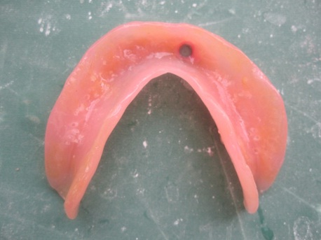
Maxillary and mandibular complete dentures were fabricated for all the patients 6 to 8 weeks before the implant placement. A surgical stent was fabricated by duplicating the mandibular denture to facilitate correct implant placement. The interforaminal region in the anterior mandible is divided into five sites equally. These five sites serve as primary implant sites, and are called as A, B, C, D and E, starting from the patient’s right side [24]. A total of 20 implants (Tidal Spiral Dental Implant Systems Huntsville, USA), two in each patient at the B and D regions were placed so that the patient always has the option to obtain additional implant support in future [Table/Fig-1] [25].
Implants placed in the B and D regions.

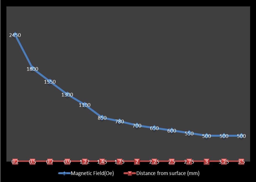
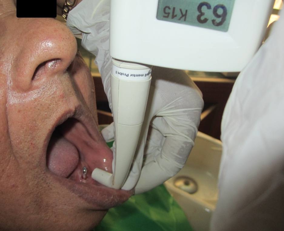
All the implants were placed at the alveolar crest level without any bone augmentation and expansion procedures by a single operator to avoid inter-operator variability. This became a split mouth study as the control and experimental group were same. The attractiveness of this split mouth design was that it removed a lot of inter-individual variability and thereby lead to less bias [26]. The primary implant stability was evaluated immediately after implant placement (at 0 day) at both the sites using RFA [27] and thereafter healing abutments were placed and soft tissues were readapted and sutured. The mandibular complete denture was relieved at the site corresponding to the D region and a circular an isotrophic Neodymium-Iron–Boron static magnet of 4.2mm diameter and 2.85mm length which maintained a flux of 500 Guass at a distance of 3.5mm from the surface was incorporated into the denture [Table/Fig-2] and the patients were asked to wear the denture for 12-15 hours daily. The measurement of the intensity of magnetic field was done using Axial Hall Probe and plotted [Table/Fig-3] as a function of distance from the surface of the magnet employed in this study. The flux decreases exponentially with distance. To avoid direct contact between the magnet and the healing abutment, soft liner material was left intact at the site of the magnet. Recall was made after one day to evaluate wound control, and after seven days for the removal of the sutures. Secondary implant stability was evaluated using RFA on both B (not exposed to magnetic field) and D (exposed to magnetic field) regions at days 30, 60 and 90 [Table/Fig-4]. After the study period of three months, an implant retained mandibular overdenture with ball attachments was delivered to all the patients thereby completing the rehabilitation procedure.
Magnet placed in the denture.
Magnetic flux (distance with significant magnetic field strength).
Checking implant stability using Resonance Frequency Analyzer.
Statistical Analysis
The basic data and mean Implant Stability Quotient (ISQ) values were tabulated. Statistical analysis was carried out using students paired t-test for comparative evaluation of implant stability values. Mean ISQ values obtained immediately following implant placement through three months follow-up in magnetic and non-magnetic sides were compared using repeated measures ANOVA [28]. The data was checked for normality before using ANOVA test. There was a significant difference in the mean ISQ values between magnetic and non-magnetic side. Hence, a post-hoc test was done to evaluate whether any differences exists between the follow-ups.
Results
The ISQ values at specific time intervals for 10 patients at magnetic and non-magnetic side are shown in [Table/Fig-5].
The ISQ values at specific time intervals for 10 patients at magnetic and non-magnetic side.
| Patient No. | 0 Day | 30 Days | 60 Days | 90 Days |
|---|
| MS | NMS | MS | NMS | MS | NMS | MS | NMS |
|---|
| 1 | 74 | 73 | 77 | 73 | 81.5 | 77.5 | 83 | 78.5 |
| 2 | 72 | 71 | 76.5 | 71.5 | 78 | 75.5 | 80 | 76 |
| 3 | 71 | 72 | 75.5 | 71 | 77 | 75.5 | 78 | 76 |
| 4 | 70 | 69 | 73.5 | 69 | 76 | 73 | 78 | 75 |
| 5 | 66 | 69 | 71 | 69 | 75 | 72 | 81 | 76 |
| 6 | 64 | 63 | 69 | 64 | 73 | 67 | 77 | 71 |
| 7 | 73 | 73 | 78 | 72 | 80 | 74 | 82 | 77 |
| 8 | 59 | 58 | 63 | 58 | 66 | 61 | 70.5 | 65 |
| 9 | 69 | 70 | 75 | 69 | 77 | 74 | 80 | 75 |
| 10 | 69 | 68 | 74 | 68 | 77 | 71 | 80 | 75 |
MS – Magnetic Side, NMS – Non-Magnetic Side
The outcomes of statistical analysis are as follows:
1. There was no significant difference in the mean ISQ values on both sides (p=0.823) on the first day (immediately after implant placement). The mean ISQ values obtained in the 1st, 2nd and 3rd month on the magnetic side were significantly higher than on magnetic side (p<0.001) [Table/Fig-6].
Comparison of ISQ values immediately following implant placement through three months follow-up in magnetic and non-magnetic side using Paired t test.
| MS | NMS | p-value |
|---|
| Mean | SD | Mean | SD |
|---|
| 0 day | 68.70 | 4.57 | 68.60 | 4.74 | 0.823 |
| 1 month | 73.25 | 4.52 | 68.45 | 4.46 | <0.001 |
| 2 month | 76.05 | 4.26 | 72.05 | 4.84 | <0.001 |
| 3 month | 78.95 | 3.50 | 74.45 | 3.83 | <0.001 |
MS – Magnetic Side NMS – Non- Magnetic Side
2. The mean ISQ values were significantly higher for the 3rd month, followed by 2nd month, 1st month and on the 0 day on the magnetic side (p < 0.001). While on the non-magnetic side, 3rd month was significantly higher than 2nd month, 1st month and after immediate implant placement. Similarly 2nd month was significantly higher than 1st month and after immediate implant placement (p<0.001). There was no significant difference between the mean ISQ values of one month and immediately after implant placement [Table/Fig-7].
Comparison of RFA values immediately following implant placement through three months follow-up in magnetic and non-magnetic side using repeated measures ANOVA with post-hoc test.
| Mean | SD | p-value | Post-hoc test |
|---|
| MS 0 day | 68.70 | 4.57 | <0.001 | 3 > 2 > 1 > 0 |
| MS 1 month | 73.25 | 4.52 |
| MS 2 month | 76.05 | 4.26 |
| MS 3 month | 78.95 | 3.50 |
| NMS 0 day | 68.60 | 4.74 | <0.001 | 2 > 1, 03 > 2, 1, 0 |
| NMS 1 month | 68.45 | 4.46 |
| NMS 2 month | 72.05 | 4.84 |
| NMS 3 Month | 74.45 | 3.83 |
Discussion
In the present study it was observed that the static magnetic field of about 500 Gauss exposed for a period of 12 weeks promoted bone healing around endosseous implants in humans. There are no such previous studies in our subcontinent; this study establishes the need for evaluation of the effect of magnetic field on bone healing around endosseous implants.
The results of this study showed significant increase in ISQ values from 0 day to 90 days on the magnetic side whereas on the non-magnetic side, the ISQ values have increased from 30 to 90 days but not to the extent as on the magnetic side and a decrease in the stability value from the 0 day to 30th day. This dip in stability occurs mostly in all implant sites due to rapid decrease in mechanical phenomenon and slow increase in biologic phenomenon during bone remodeling process at the implant bone interface. But on the magnetic side this has not happened and in turn there is increase in stability values even in the first month. This might be due to influence of magnetic field during initial bone healing process.
Magnets are of two types, they are permanent and temporary magnets. A static magnet maintains its inbuilt magnetic property for a long period of time. A temporary magnet maintains its magnetism only when it is in the magnetic field created by a permanent magnet or by an electric current so called Pulsed Electromagnetic Fields (PEMFs). Both the static magnetic field and PEMF cause similar actions within the human body but their mechanism of actions are not similar [29]. Since our body can become used to nonmoving (or static) magnetic fields, they must be applied with stronger intensity for longer intervals of time when compared to PEMF devices. Many studies have shown that PEMFs promotes bone healing both in animals and humans [30–32]. It is said that PEMFs alters the membrane permeability through the induction of electric field thereby altering the cyclic guanosine monophosphate and cyclic adenosine monophosphate activity and promote osteogenesis. On the contrary static magnetic field neither produce electric currents nor create vectorial changes, but it has potency to promote differentiation of osteoblasts and bone maturation directly [33].
Bassett et al., first used magnetic fields as a non invasive and safe method to stimulate healing of fractures [34,35]. Camilleri and McDonald evaluated the action of static magnetic field using Neodymium Iron-Boron magnet by placing over a skull suture on a rat model and showed that the mitotic activity of the cells was altered [36]. Bruce et al., showed that the stimulation with static magnetic field improved the strength of the fractured radii in rabbits [37]. The most possible mechanism behind this healing is the increased blood circulation due to dilation of blood vessels and reduction in stickiness of platelets by magnetic field [38–43]. This increased blood circulation pools oxygen and nutrients to the surgical site thereby improving the overall process of healing [44]. This also helps in bringing the natural healers to eliminate the toxic byproducts of inflammation like bradykinins and prostaglandins. Another study stated that the application of magnetic field aids in the adhesion of calcium ions to the blood clot and increase in two osteoblastic phenotype markers (alkaline phosphatase and osteocalcin) at the surgical site, which helps in the bone healing process [45,46]. From the above said evidences, it is alluring to say that magnetic fields might hasten the tissue maturation process.
Limitation
To name a few, other parameters that may influence the results include the use of various implant surface modifications, various thread designs, different edentulous regions, multiple operators etc. These were not included in this present study. Inclusion of these parameters might enhance the results obtained with this study. Many controlled clinical studies with larger number of patients and longer follow-up period are needed to make the use of magnetic field for bone healing around endosseous implants completely evidence based.
Conclusion
The results obtained from this study imply that static magnetic field may provide favorable environment for early bone healing thereby increasing the implant stability for earlier rehabilitation. The results of this study can serve as a basis for future long-term clinical studies involving the use of magnetic field not only to confirm its beneficial effects, but also to optimize the magnetic field parameters and establish a protocol for clinical use.
MS – Magnetic Side, NMS – Non-Magnetic Side
MS – Magnetic Side NMS – Non- Magnetic Side
[1]. Albrektsson T, Zarb G, Worthington P, Eriksson AR, The long-term efficacy of currently used dental implants: A review and proposed criteria of successInt J Oral Maxillofac Implants 1986 1:11-25. [Google Scholar]
[2]. Ganeles J, Wismeijer D, Early and immediately restored and loaded dental implants for single-tooth and partial arch applicationsInt J Oral Maxillofac Implants 2004 19(Suppl):92-110. [Google Scholar]
[3]. Buser D, Weber HP, Bragger U, Balsiger C, Tissue integration of one-stage ITI implants: 3-year results of a longitudinal study with hollow-cylinder and hollow-screw implantsInt J Oral Maxillofac Implants 1991 6:405-12. [Google Scholar]
[4]. LeGeros RZ, Craig RG, Strategies to affect bone remodeling: OsseointegrationJ Bone Miner Res 1993 8(Suppl.2):S583-96. [Google Scholar]
[5]. Brånemark P.-I, Osseointegration and its experimental backgroundJ Prosthet Dent 1983 50:399-410. [Google Scholar]
[6]. Degidi M, Piattelli A, Immediate functional and nonfunctional loading of dental implants: A 2 to 60-month follow-up study of 646 titanium implantsJ Periodontol 2003 74:225-41. [Google Scholar]
[7]. Atsumi M, Park SH, Wang HL, Methods used to assess implant stability: Current statusInt J Oral Maxillofac Implants 2007 22:743-54. [Google Scholar]
[8]. Molly L, Bone density and primary implant stability in implant therapyClin Oral Imp Res 2006 17(Suppl. 2):124-35. [Google Scholar]
[9]. Sim CP, Lang NP, Factors influencing resonance frequency analysis assessed by Osstell mentor during implant tissue integration: 1. Instrument positioning, bone structure, implant lengthClin Oral Implants Res 2010 21:598-604. [Google Scholar]
[10]. Simunek A, Kopecka D, Brazda T, Strnad I, Capek L, Slezak R, Development of implant stability during early healing of immediately loaded implantsInt J Oral Maxillofac Implants 2012 27(3):619-27. [Google Scholar]
[11]. Meredith N, Alleyne D, Cawley P, Quantitative determination of the stability of the implant-tissue interface using resonance frequency analysisClin Oral Implants Res 1996 7:261-67. [Google Scholar]
[12]. Scarano A, Degidi M, Vezzi G, Petrone G, Piattelli A, Correlation between implant stability quotient and bone-implant contact: A retrospective histological and histomorphometrical study of seven titanium implants retrieved from humansClin Implant Dent Relat Res 2006 8:218-22. [Google Scholar]
[13]. Steven L Henry, Matthew J Concannon, Gloria J Yee, The effect of magnetic fields on wound healing. Experimental study and review of the literatureEplasty 2008 8:e40 [Google Scholar]
[14]. Patrick D, Toto Nicholas C, Choukas Daniel D Sanders, Reaction of bone and mucosa to implanted magnetsJ Dent Res 1962 41:1438-49. [Google Scholar]
[15]. Toto PD, Choukas NC, Abati A, Reaction of bone to magnetic implantJ Dent Res 1963 42:642-52. [Google Scholar]
[16]. Costantino C, Pogliacomi F, Passera F, Concari G, Treatment of wrist and hand fractures with natural magnets: Preliminary reportActa Biomed 2007 78:198-203. [Google Scholar]
[17]. Henry SL, Concannon MJ, Yee GJ, The effect of magnetic fields on wound healing experimental study and review of the literatureEplasty 2008 8:e40 [Google Scholar]
[18]. Cerny R, The biological effects of implanted magnetic fields, part 1: Mammalian blood cellsAust Orthod J 1979 4:64-70. [Google Scholar]
[19]. Cerny R, The biological effects of implanted magnetic fields, part II: Mammalian tissuesAust Orthod J 1980 3:114-17. [Google Scholar]
[20]. Behrman SJ, Magnets implanted in the mandible: Aid to denture retentionJ Am Dent Assoc 1964 68:206-15. [Google Scholar]
[21]. Behrman SJ, The implantation of magnet in the jaw to aid denture retentionJ Prosthet Dent 1960 10:807-41. [Google Scholar]
[22]. Toto PD, Choukas NC, Abati A, Reaction of bone to magnetic implantJ Dent Res 1963 42:642-52. [Google Scholar]
[23]. Riley MA, Walmsley AD, Harris IR, Magnets in prosthetic dentistryJ Prosthet Dent 2001 86:137-42. [Google Scholar]
[24]. Misch CE, Contemporary implant dentistry 2008 3rd edSt LouisMosby [Google Scholar]
[25]. Skalak R, Biomechanical considerations in osseointegrated prosthesesJ Prosthet Dent 1983 49:843-48. [Google Scholar]
[26]. Lesaffre E, Philstrom B, Needleman I, Worthington H, The design and analysis of split-mouth studies: What statisticians and clinicians should know?Stat Med 2009 28:3470-82. [Google Scholar]
[27]. Lee HJ, Aparecida de Mattias Sartori I, Alcântara PR, Vieira RA, Suzuki D, Gasparini Kiatake Fontão F, Implant stability measurements of two immediate loading protocols for the edentulous mandible: Rigid and semi-rigid splinting of the implantsImplant Dent 2012 21:486-90. [Google Scholar]
[28]. Suzuki S, Kobayashi H, Ogawa T, Implant stability change and osseointegration speed of immediately loaded photofunctionalized implantsImplant Dent 2013 22:481-90. [Google Scholar]
[29]. Bruce GK, Howlett CR, Huckstep RL, Effect of a static magnetic field on fracture healing in a rabbit radiusClinical Orthopaedics and Related Research 1987 222:300-06. [Google Scholar]
[30]. Bassett CA, Beneficial effects of electromagnetic fieldsJ Cell Biochem 1993 51:387-93. [Google Scholar]
[31]. Bassett CA, Pawluk RJ, Becker RO, Effects of electrical current on bone in vivoNature 1964 204:652-54. [Google Scholar]
[32]. Otter MW, McLeod KJ, Rubin CT, Effects of electromagnetic fields in experimental fracture repairClin Orthop 1998 355S:90-104. [Google Scholar]
[33]. Kotani H, Kawaguchi H, Shimoaka T, Iwasaka M, Ueno S, Ozawa H, Strong static magnetic field stimulates bone formation to a definite orientation in vitro and in vivoJ Bone Miner Res 2002 17:1814-21. [Google Scholar]
[34]. Bassett CA, Effects of a static magnetic field on fracture healingClin Orthop Relat Res 1988 234:311-12. [Google Scholar]
[35]. Bassett CA, Pawluk RJ, Pilla AA, Augmentation of bone repair by inductively coupled electromagnetic fieldsScience 1974 184:575-77. [Google Scholar]
[36]. Camilleri S, McDonald F, Static magnetic field effects on the sagittal suture in rattus norvegicusAm J Orthod Dentofacial Orthop 1993 103:240-46. [Google Scholar]
[37]. Bruce GK, Howlen CA, Huckstep RL, Effect of a staticmagnetic field on fracture healing in a rabbit radiusClin Orthop 1987 222:300-06. [Google Scholar]
[38]. Boyan BD, Bonewald LF, Paschalis EP, Lohmann CH, Rosser J, Cochran DL, Osteoblast-mediated mineral deposition in culture is dependent on surface microtopographyCalcif Tissue Int 2002 71:519-29. [Google Scholar]
[39]. Greenough CG, The effects of pulsed electromagnetic fields on blood vessel growth in the rabbit ear chamberJ Orthop Res 1992 10:256-62. [Google Scholar]
[40]. Shimizu T, Zerwekh JE, Videman T, Gill K, Mooney V, Holmes RE, Bone in growth into porous calcium phosphate ceramics: Influence of pulsing electromagnetic fieldJ Orthop Res 1988 6:248-58. [Google Scholar]
[41]. Midura RJ, Ibiwoye MO, Powell KA, Sakai Y, Doehring T, Grabiner MD, Pulsed electromagnetic field treatments enhance the healing of fibular osteotomiesJ Orthop Res 2005 23:1035-46. [Google Scholar]
[42]. Ito H, Shirai Y, The efficacy of un-united tibial fracture treatment using pulsing electromagnetic fields: Relation to biological activity on nonunion bone endsJ Nippon Med Sch 2001 68:149-53. [Google Scholar]
[43]. Linovitz RJ, Pathria M, Bernhardt M, Green D, Law M, McGuire RA, Combined magnetic fields accelerate and increase spine fusion: A double-blind, randomized, placebo controlled studySpine 2002 27:1383-89. [Google Scholar]
[44]. Saifzadeh S, Hobbenaghi R, Shokouhi Sabet Jalali F, Kabiri B, Effect of a static magnetic field on bone healing in the dog: Radiologic and histopathological studiesIranian Journal of Veterinary Reseach 2007 8:8-15. [Google Scholar]
[45]. Messonnier SP, The natural health bible for dogs and cats 2001 1st. EdRosevillePrima Publishing Inc:412-15. [Google Scholar]
[46]. Yamamoto Y, Ohsaki Y, Nakasima A, Lijima T, Effects of static magnetic fields on bone formation in rat osteoblast culturesJ Dent Res 2003 89:962-66. [Google Scholar]