Recurrent inflammation or erythema and burning sensation of denture bearing tissues are relatively common in denture wearers and are described by the term denture stomatitis. It is frequently found in the maxillary ridge of elderly denture wearers. C. albicans is the predominant oral yeast associated with denture stomatitis [1]. The condition is often symptomless, but when symptoms are present they may appear as mucosal bleeding, burning or painful sensations and dryness in the mouth [2].
Resilient denture lining materials are used to limit such injury by providing cushioning effect. They have a therapeutic value in patients with thin atrophic mucosa, sharp alveolar ridge crest, deep anatomic undercuts, bony protuberances, or bruxomania, where the oral mucosa exhibits a reduced tolerance to the load applied by the denture, and in obturators for cleft palate [3]. Studies have reported that the fungi and bacterial species can enter porous spaces within the denture liner and that their colonization may reduce the intra-oral life of the material. The porosity of the denture liner also allows water absorption and the diffusion of nutrient materials that may support growth of oral yeasts [3].
Material and Methods
This in-vitro study was carried out at the Department of Prosthodontics and Crown and Bridge and Department of Microbiology of Maratha Mandal’s NGH Institute of Dental Sciences and Research Centre, Belgaum, Karnataka, India after approval by the Institutional Ethics Committee. The study consisted of three parts. I) Evaluation of retention of C. albicans to denture liners; II) Evaluation of colonization and penetration of denture liners by C. albicans; III) Evaluation of inhibition of C. albicans by denture liners. Following denture liners were used in the study [Table/Fig-1].
Test materials used in the study.
| 1 | Molloplast B (Detax, Germany) | Heat cure denture liners |
| 2 | Permaflex (Kohler, Germany) |
| 3 | GC Soft Liner (GC Corporation, Tokyo, Japan) | Self cure denture liners |
| 4 | Ufi Gel Hard C (Voco, Germany) |
I) Evaluation of Retention
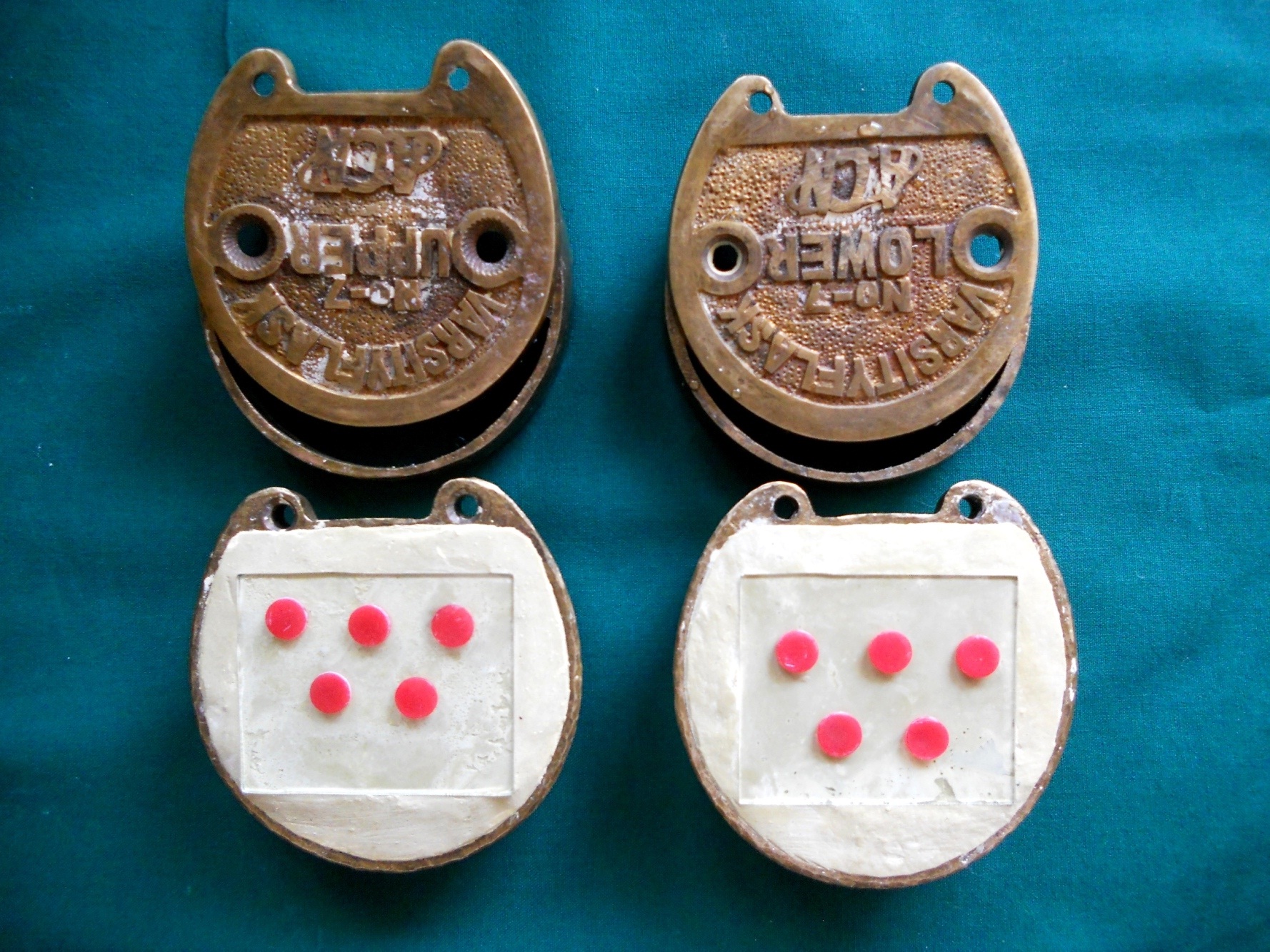
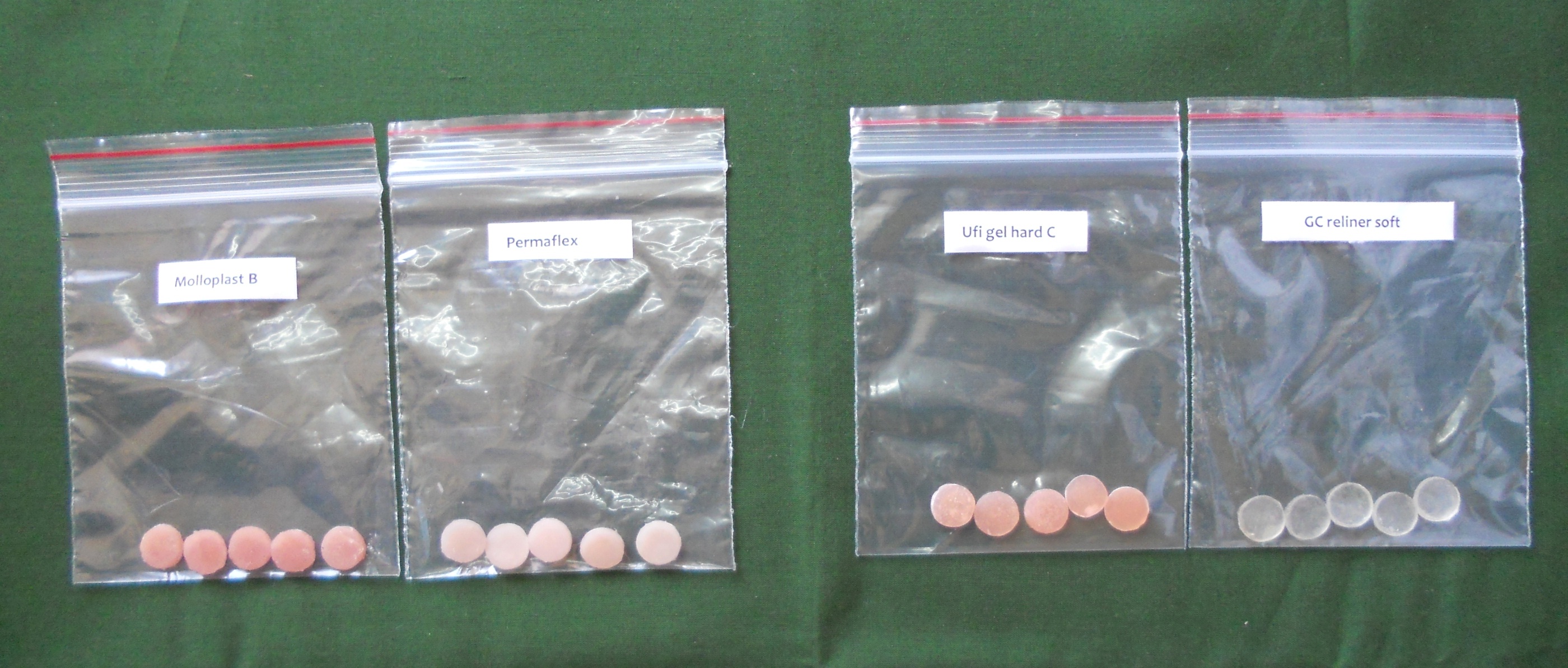
a) Preparation of test discs: Five test discs of 10mm diameter and 1.5mm thickness prepared from each of the four denture liners were used. Each test disc had smooth surface on one side and rough surface on the other. To prepare this, a glass piece was pressed onto the dental stone mixture (Asian Chemicals, Rajkot, India) in the drag of the flask. After the dental stone had set, wax discs of 10mm diameter and 1.5mm thick were punched from a sheet of modeling wax and placed on top of the glass surface [Table/Fig-2]. The cope of the flask was positioned and the dental stone poured over the wax discs. The voids produced after de-waxing were filled by denture liners. The discs thus obtained had one glass-smooth surface and one dental stone-rough surface [Table/Fig-3]. Curing of test discs of heat cure and cold cure denture liners were carried out according to standard procedures.
Wax discs positioned on glass surface.
Test discs of Permaflex, Molloplast B, Ufi gel hard and GC soft liner with smooth surface on one side and rough on the other.
b) Surface roughness measurements: Surface roughness (Ra) was measured at three areas of each surface (i.e., smooth and rough) of each specimen, yielding six measurements for each disc, using profilometer (Surtronic 3+, Taylor Hobson Ltd., UK) and the mean value was calculated. Subsequently, all discs were cleaned ultrasonically in distilled water for 15 seconds, sterilized and stored in sterile distilled water at 37°C for 24 hours prior to C. albicans contamination, and for the evaluation of retention of C. albicans to denture liners.
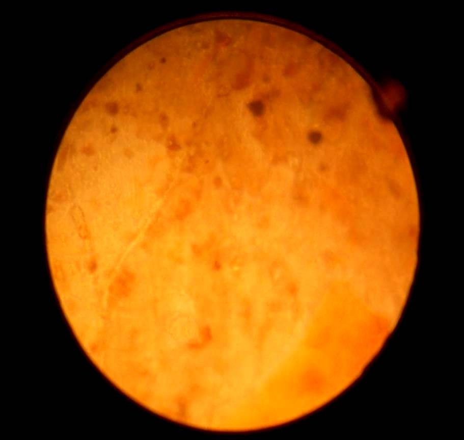
c) Microbiological procedure: About (4–6) colonies of C. albicans (ATCC 2091) from a Sabouraud’s plate were inoculated into 100ml artificial saliva (Saleva, Global Dent Aids Pvt., Ltd., India) and incubated at 37°C for 24 hours. Cells were collected by centrifugation, and re-suspended in Phosphate Buffered Saline (PBS). Each sterile test disc was placed in a sterile 5ml glass bottle in a vertical position and 2ml of the standardized cell suspension (2.8 X 10⁶ cfu/ml) were added to each bottle, the apparatus was incubated, without agitation at 37°C for one hour. Test discs were then removed from the suspension, rinsed by dipping gently three times in sterile PBS to remove loosely attached cells and left to air dry. Attached cells remaining on the discs were fixed by immersion in 100% methanol (Himedia Pvt., Ltd., India) for one minute, stained by immersing in 0.03% acridine orange (Himedia Pvt., Ltd., India) for 2 minute, followed by washing in distilled water and air drying. Test discs were viewed using inverted microscope at 40X magnification (Magnus Analytics, Italy). The number of adherent cells in 10 random fields was counted for each test disc and the mean number was obtained [Table/Fig-4].
Adherent cells as seen under inverted microscope at 40X magni-fication.
II) Evaluation of Colonization and Penetration.
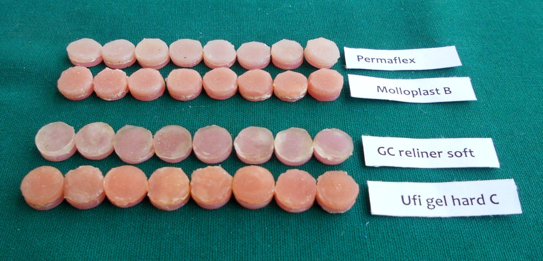
a) Preparation of test discs: Eight test discs of each of the four materials were tested. Disc shaped voids were prepared as described above. Acrylic resin powder and liquid (Trevalon, Dentsply. India) were mixed and the dough was filled into the voids in the flasks. After polymerization, acrylic discs were removed. Discs of modeling wax were anchored to each acrylic disc and flasked. This procedure produced acrylic discs surmounted by a void which was filled by the denture liners [Table/Fig-5].
Acrylic discs surmounted by liner.
b) Microbiological procedure: Test acrylic/liner discs were placed into 15 ml glass bottles containing 10 ml artificial saliva and sterilized. Each of the bottles was inoculated with 0.1 ml of an 18 hours culture of C. albicans in artificial saliva and incubated at 370C for 6 weeks.
After this, test discs were removed from the bottles and placed in 4% glutaraldehyde in PBS for at least 2 hour to fix the cells and dehydrated in alcohol. Two sections were cut across each test disc with hard tissue microtome (Leica SP 1600, Leica Microsystems Germany) to provide three replicate samples. Sections were immersed in 0.03% acridine orange for one minute, rinsed in distilled water, air dried and viewed via fluorescence microscope 25000X (Magus Analytics, Italy) [Table/Fig-6]. The penetration of C. albicans into sections of denture liners was evaluated by counting the number of blastospores/hyphae visible within each microscopic field of each section of test disc, as described below [8].
Penetration of Candida albicans into section 3 of the test discs of denture lining materials (fluorescence microscope 25000X): (a) Permaflex; (b) Molloplast; (c) GC soft liner and (d) Ufi gel hard C.
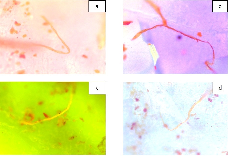
Class I: Little hyphal presence, where by mycelia forms cover up to a quarter of the microscopic field.
Class II: Moderate amount of hyphal presence, where half of the field of view was covered by mycelial forms.
Class III: Large amount of hyphal presence, whereby most of the microscopic field was covered by mycelial forms.
III) Evaluation of antifungal action.
a) Preparation of test discs: Four test discs of each of the four denture liners were tested. For this, discs of modeling wax were punched out from a sheet, dental stone was poured into the shallow part of the dental flask, and the punched wax discs were placed on the surface of the stone. After setting, separating medium was applied, the upper part of the flask was then placed into position and dental stone was poured into it. After de-waxing, denture lining materials were packed and polymerized. Subsequently, all specimens were cleaned ultrasonically in distilled water for 15 seconds, sterilized and used for the evaluation of zone of inhibition.
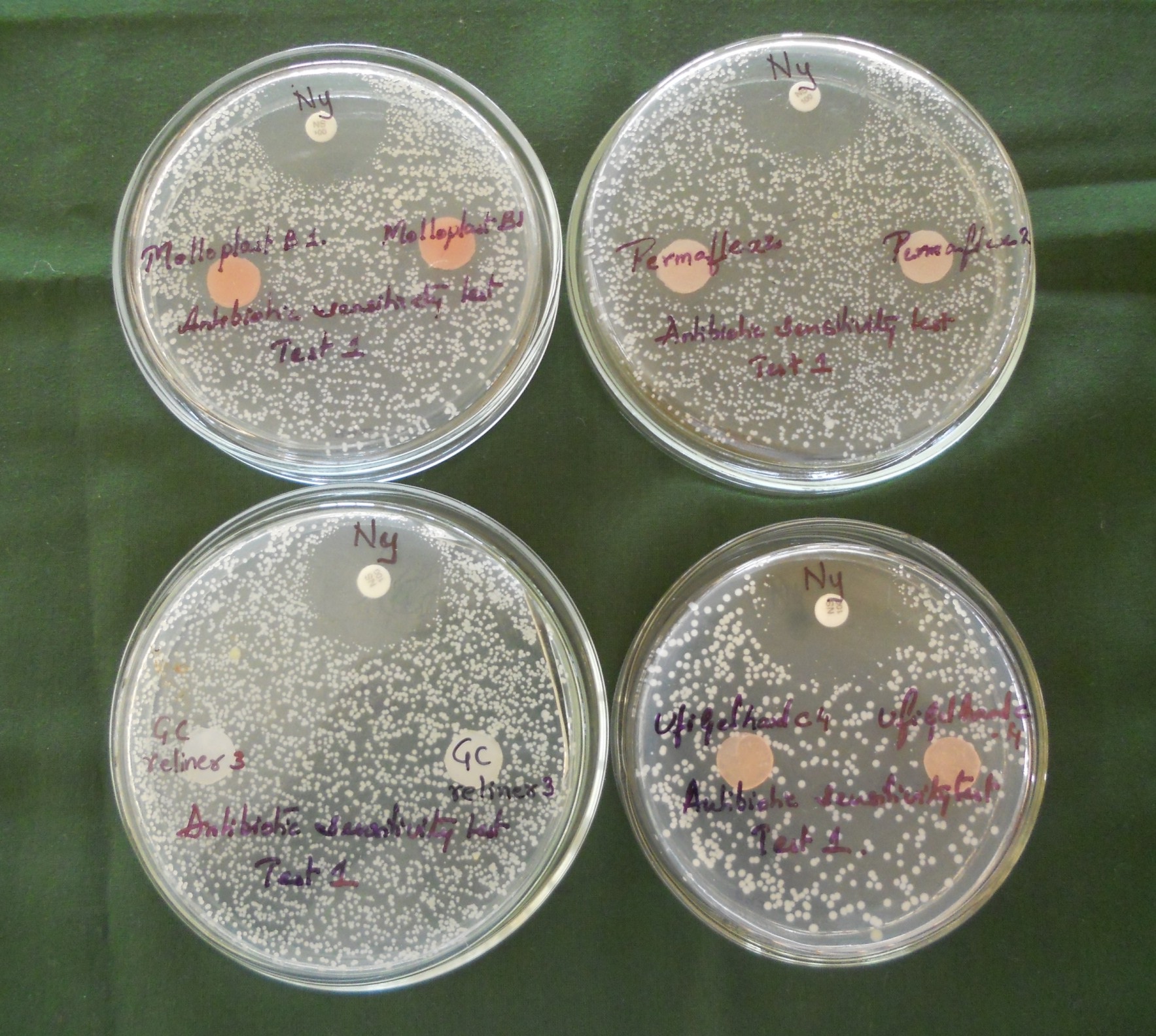
b) Microbiological procedure: An overnight culture of C. albicans diluted with 1:1000 in sterile PBS, was used to inoculate diagnostic sensitivity testing agar (Himedia laboratories Pvt., Ltd., India). The plates were dried, and two test discs of each denture liner were placed in each agar plate and the experiment was repeated twice. Nystatin susceptibility test discs (100 units/disc) (Himedia Laboratories Pvt., Ltd., India) were used as controls [Table/Fig-7]. After incubation at 37°C for 24 hours, the zone of inhibition formed around the samples were measured using a transparent ruler at four places around each test disc and the mean determined.
Agar diffusion test for Molloplast B and Permaflex, GC soft liner and Ufi gel hard C.
Statistical Analysis
The summary data was tabulated as mean values and standard deviation. One-way ANOVA and was used for comparing multiple unmatched groups for parametric data. Kruskal Wallis test was used for comparing multiple unmatched groups for non-parametric data. Pair wise comparison of groups was done by Mann-Whitney U test. Chi square test was used for analyzing categorical data. The confidence level of the study was set at 95%.
Results
As indicated in [Table/Fig-8,9], there was a significant variation in the surface roughness (Ra) values of smooth surfaces, with Permaflex and Ufi Gel Hard C showing higher values and GC Soft Liner showing the least value. The variation in the surface roughness values was not statistically significant for rough surfaces.
Comparison of smooth surface values of test discs.
| Materials | Mean± SD | Anova F* | Difference between materials† |
|---|
| F-value | p-value | Mollo-plast B | Perm-aflex | GC Soft Liner | Ufi Gel Hard C |
|---|
| Molloplast B | 0.33±0.09 | 8.9258 | 0.0010 S | 1.0000 NS | -- | -- | -- |
| Permaflex | 0.69±0.29 | 0.0279 S | 1.0000 NS | -- | -- |
| GC Soft Liner | 0.18±0.10 | 0.5638 NS | 0.0020* S | 1.0000 NS | -- |
| Ufi Gel Hard C | 0.62±0.17 | 0.0918 NS | 0.9224 NS | 0.0070 S | 1.0000 NS |
† Tukeys multiple post hoc procedure
Comparison of rough surface values of test discs.
| Materials | Mean± SD | Anova F* | Difference between materials† |
|---|
| F-value | p-value | Mollo-plast B | Perm-aflex | GC Soft Liner | Ufi Gel Hard C |
|---|
| Molloplast B | 1.47±0.18 | 2.8526 | 0.0701 | p=1.0000 | -- | -- | -- |
| Permaflex | 1.88±0.31 | p=0.3005 | p=1.0000 | | -- |
| GC Soft Liner | 1.70±0.48 | p=0.7466 | p=0.8485 | p=1.0000 | -- |
| Ufi Gel Hard C | 2.10±0.37 | p=0.0500* | p=0.7566 | p=0.3084 | p=1.0000 |
† Tukeys multiple post hoc procedure
All rough surfaces showed higher retention of C. albicans than smooth surfaces. Among the smooth surfaces, Molloplast B and GC Soft Liner showed highest and lowest retention of C. albicans respectively. Among the rough surfaces, the variation in the retention of C. albicans was not statistically significant [Table/Fig-10].
Comparison of test discs with respect to colonies of C. albicans per field on smooth and rough surfaces.
| Materials | Smooth Surface* | Rough Surface* |
|---|
| Mean± SD | Median | Mean± SD | Median |
|---|
| Molloplast B | 11.20±1.82 | 11.00 | 12.80±3.01 | 13.50 |
| Permaflex | 8.70±1.99 | 9.00 | 14.80±2.05 | 13.50 |
| GC Soft Liner | 3.50±1.22 | 3.50 | 12.30±3.23 | 11.00 |
| Ufi Gel Hard C | 9.40±3.31 | 9.00 | 14.00±3.30 | 13.50 |
| H-value | 11.9846 | 2.4384 |
| p-value | 0.0074 S | 0.4865 NS |
| Pair wise comparison of test discs† |
| Molloplast B vs. Permaflex | p=0.0758 | p=0.3472 |
| Molloplast B vs. GC Soft Liner | p=0.0090* | p=0.6761 |
| Molloplast B vs.Ufi Gel Hard C | p=0.3472 | p=0.5309 |
| Permaflex vs. GC Soft Liner | p=0.0122* | p=0.1172 |
| Permaflex vs. Ufi Gel Hard C | p=0.6761 | p=0.9168 |
| Ufi Gel Hard C vs. GC Soft Liner | p=0.0163* | p=0.4034 |
* Kruskal Wallis ANOVA, † Mann-Whitney U test, S-significant, NS-not significant
There was no statistically significant difference among the test materials with respect to penetration of C. albicans. Third (deepest) section of all the discs had candida penetration [Table/Fig-11].
Comparison of test discs with respect to penetration of C. albicans into different sections of test discs.
| Materials | Section I | Section II | | Section III |
|---|
| Class I | Class II | Class III | Class I | Class II | Class III | None | Class I | Class II | Class III |
|---|
| Molloplast B | -- | 4 | 4 | 7 | 1 | -- | 5 | 3 | -- | -- |
| Permaflex | -- | 3 | 5 | 8 | 0 | -- | 5 | 3 | -- | -- |
| GC Soft Liner | -- | 6 | 2 | 7 | 1 | -- | 4 | 4 | -- | -- |
| Ufi Gel Hard C | -- | 5 | 3 | 8 | 0 | -- | 3 | 5 | -- | -- |
| Total | -- | 18 | 14 | 30 | 2 | -- | 18 | 14 | -- | -- |
| p* | 0.4681 | 0.5452 | 0.4681 |
*Chi square test
Molloplast B and Permaflex produced a mean zone of inhibition of 16.9±4.8mm and 14.80±3.8mm respectively, GC Soft Liner and Ufi Gel Hard C did not show any zone of inhibition when compared with the positive control (Nystatin susceptibility test discs), which showed a mean zone of inhibition of 30.37±3.33mm [Table/Fig-12].
Mean values of zone of inhibition (mm) of test discs.
| Material | Mean(mm) |
|---|
| Molloplast B | 16.93±4.8 |
| Permaflex | 14.8±3.8 |
| GC Soft Liner | -- |
| Ufi Gel Hard C | -- |
| Nystatin | 30.37 ±3.33 |
Discussion
There is a rise in the number of elderly people using complete dentures which has resulted in consequent increase in the use of denture liners. The purpose of this study was to comprehensively evaluate significant aspects related to performance of commonly used denture liners such as retention, colonization and penetration and anti-fungal action.
Retention of C. albicans to denture lining materials: The adherence of C. albicans to denture liners is the first step in colonization, followed by initiation of pathogenesis and eventually causing infection [11,12]. C. albicans seen on the surfaces were mainly blastospores, as the incubation of the cells was only for one hour, though, a few hyphae were also seen. The results are in agreement with earlier studies on short-term attachment, which showed pseudo hyphae and hyphae and not yeast growth or surface colonization [8,13].
Surface roughness is a major factor in the attachment and retention of microorganisms on surfaces, with an increase in roughness causing increased retention of cells. It has been known as a factor for the entrapment of microorganisms and subsequent protection from shear forces. C. albicans were retained on rough surfaces in higher numbers than on smooth surfaces. Similar effects have been reported in the past [6–10] [Table/Fig-13]. There are clear implications for these findings in terms of surface cleanability, denture hygiene and materials fabrication. Overall, surface structure, properties and composition of biomaterials, hydrophobicity and roughness affect the adhesion of microorganisms on biomaterials [14].
Summary of studies done to evaluate adhesion and penetration of C. albicans into denture liners.
| Authors | Materials tested | Parameters evaluated | Conclusion |
|---|
| Gedik Het al., [6] | Ufi Gel P *Ufi Gel C*Mollosil*Soft-Liner*Moloplast B**Luci Soft***Room Temperature Polymerised Denture Liner**High Temperature Polymerised Denture Liner | In-vitro evaluation of surface roughness and adherence of C. albicans | High temperature polymerised denture liners showed lower surface roughness value and lower adhesion of C. albicans than room temperature polymerised denture liners |
| Nevzatoğlu EU et al., [7] | Denture Base Acrylic Resins and Silicone-Based Resilient Liners | Adherence of C. albicans to denture base acrylics and silicone-based resilient liner materials with different surface finishes | In all types of surface finishes, C. albicans adhesion on denture base acrylics was significantly less than those of silicone liners |
| Bulad K et al., [8] | Molloplast B*Flexor *Permaflex *Luci-Soft *Eversoft **Ufi Gel Hard C****Heat Curing Silicone** Chair-Side Curing Denture Liner***Cold Curing Hard Liner | • Adhesion• Colonization and penetration• Anti-fungal action | • There was more adhesion on rough surfaces. More cells were attached to Molloplast-B (silicone material) than to Ufi-Gel Hard C• Penetration was greatest into Ufi Gel Hard C and least into Eversoft.• None of the materials produced a zone of inhibition |
| Radford DR et al., [9] | Molloplast BNovus | In-vitro adherence of C. albicans to heat-cured hard and soft denture-base materials with varying surface roughness | Rough surfaces on denture-base materials promote the adhesion of C. albicans |
| Verran J,et al., [10] | Acrylic resin and silicone surfaces | Retention of C. albicans on smooth and rough surfaces | Significantly higher numbers of cells were observed on roughened surfaces (silicone > acrylic resin) than on smooth surfaces |
Colonization and penetration of denture lining materials by C. albicans: Penetration of C. albicans within denture liners was evaluated by observing all three sections of test discs of each material. Colonization was visible with the naked eye on the surface, when materials were removed from the saliva culture. Cells were observed on the outer surface of all three sections of each test disc, indicating that penetration of the lining materials had occurred. Both blastospores and hyphal forms were visible. C. albicans is a microscopic, oval shaped, thin walled fungus usually measuring 2 to 4μm; however, filamentous shapes of variable length have also been identified having 3 to 5μm rounded ends in infected tissues [14]. C. albicans colonize not only the surface of the denture liners, but they also penetrate inside the materials.
Penetration of C. albicans (blastospores and hyphae) through Molloplast B and Permaflex was least and was relatively comparable. This is probably because both are similar in composition and are heat cure denture liners. The most significant penetration of Candida blastospores was observed in Ufi Gel Hard C, a hard denture liner. This might be because of the presence of porosities inside the matrix of the cured material which facilitate the penetration of blastospores [15]. Significant hyphal formation was seen all through the other soft denture liners. The penetration of C. albicans at the acrylic-liner junction point was observed in a few samples, particularly GC Soft Liner. This can be attributed to the failure of bonding between the liner and the acrylic base material, which may contribute to deterioration of the prosthesis lining and function.
Inhibition of C. albicans by denture lining materials: In the current study, Molloplast B and Permaflex exhibited zone of inhibition against C. albicans, but the effect was small. Of the denture lining materials tested, only Molloplast B has been studied previously for any inhibitory effect on the growth of C. albicans, with differing success, one study investigated inhibitory effect of Permaflex and Ufi Gel Hard C and there are no reported studies regarding inhibitory effect of GC Soft Liner. Uncured Molloplast B material has been reported to cause a definite inhibition of Candida growth in-vitro, with the cured material showing no growth inhibition. The active constituent, methacryloloxy-propyl-trimethoxy-silane, would become inactive during the cross-linking curing process [16]. If a small excess remained, the cured material might still exhibit minimal inhibition of growth. The curing of all the test materials used in this study was done using standard procedures. One study reported that Molloplast B demonstrated such a small degree of inhibition that the zone area was not measurable [17]. Another study found that Molloplast-B had no inhibitory influence on growth of C. albicans [16]. One more study reported no inhibitory effect of Molloplast B and Permaflex. Clearly, the method of preparation of the material and its storage, and the experimental protocol appear to have some effect on findings [8].
One of the most serious disadvantages of these materials is their reported lack of antimicrobial activity. A denture liner material with anti-fungal property is yet to be developed. To overcome these problems, attempts have been made to incorporate different anti-fungal agents to the resilient liners such as Propolis, Zeolite, Chlorhexidine, Fluconazole, Punica granatum, Nystatin, Itraconazole, Miconazole, Ketoconazole, Clotrimazole [18–25] in the resilient liners with varying degree of success.
Ideal properties of soft lining material include superior biocompatibility, enduring resilience, low water sorption, high bond strength to denture base, high dimensional stability, good color stability and resistance to microbial growth. It appears that currently available denture liners are deficient in few areas. Selection of denture liner in a given situation should be based on the knowledge about how the adherence and bio-film formation takes place. This will be helpful to diminish Candida colonization in clinical practice.
Limitation
The outcome obtained and the conclusions drawn are based on in-vitro studies, correlation to clinical practice requires further in-vivo research.
Conclusion
a) All rough surfaces showed higher retention of C. albicans than smooth surfaces. Among the smooth surfaces, Molloplast B and GC Soft Liner showed highest and lowest retention of C. albicans respectively. Among the rough surfaces, the variation in the retention of C. albicans was not statistically significant.
b) All four materials showed penetration of C. albicans into the deepest section of discs.
c) Molloplast B and Permaflex exhibited zone of inhibition against C. albicans, but the effect was small.
† Tukeys multiple post hoc procedure
† Tukeys multiple post hoc procedure
* Kruskal Wallis ANOVA, † Mann-Whitney U test, S-significant, NS-not significant
*Chi square test