Bilateral Three Rooted Mandibular Premolars and Four Rooted Mandibular First and Second Molar: A Rare Anatomical Variant
Mrunal Manohar Shinde1, Sharad Basavraj Kamat2, Rutuja Vijay Chopade3
1 Postgraduate Sudent, Department of Conservative Dentistry and Endodontics, Dental College and Hospital, BVDU, Sangli, Maharashtra, India.
2 Professor and Head, Department of Conservative Dentistry and Endodontics, Dental College and Hospital, BVDU, Sangli, Maharashtra, India.
3 Assistant Professor, Department of Conservative Dentistry and Endodontics, Dental College and Hospital, BVDU, Sangli, Maharashtra, India.
NAME, ADDRESS, E-MAIL ID OF THE CORRESPONDING AUTHOR: Dr. Mrunal Manohar Shinde, Department of Conservative Dentistry and Endodontics, Dental College & Hospital, Bharti Vidyapeeth Deemed University, Wanlesswadi, Sangli-Miraj Road, Sangli-416414, Maharashtra, India.
E-mail: mrunal821@gmail.com
The mandibular premolars and molars exhibit wide variations in the form of roots and root canals. A bilateral symmetry of three rooted mandibular first and second premolar and four rooted mandibular first and second molar in a same patient is a rare entity and one such case is presented in this case report.
Anatomic variations, Cone beam computed tomography, Radiography
Case Report
A 30-year-old male patient reported to the Department of Conservative Dentistry and Endodontics with the chief complaint of pain and fractured restoration in the right lower back tooth region since one week. Pain was mild, intermittent, non-radiating in nature and aggravated on cold food and liquid intake and relieved after removal of stimulus. Intraoral examination revealed fractured restoration with right mandibular first molar on the occlusal surface. The tooth was not mobile and periodontal probing around the tooth was within physiological limits.
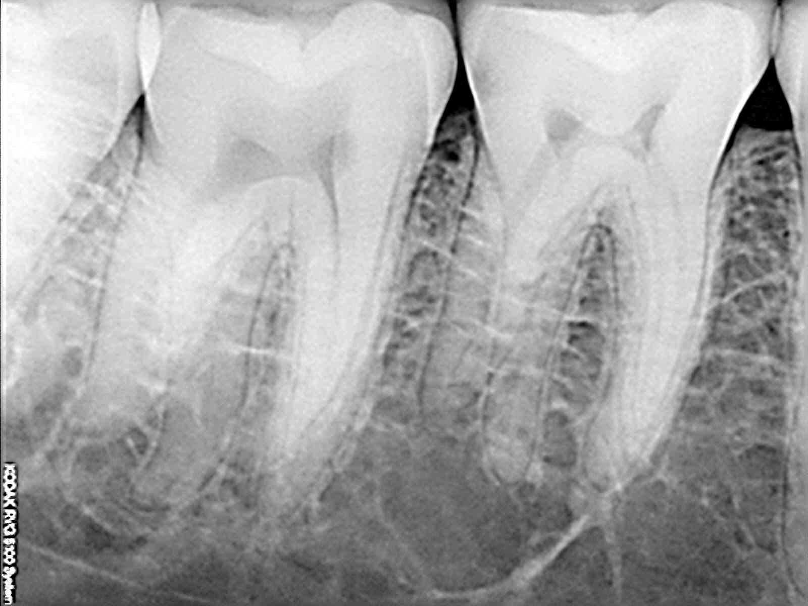
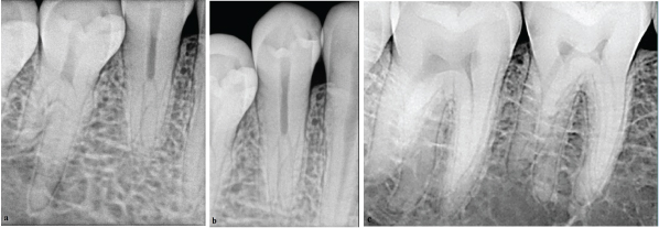
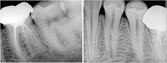
The preoperative radiograph revealed radiopacity in the enamel of mandibular first molar and also uncommon morphology with the roots of mandibular first and mandibular second molar [Table/Fig-1]. Additional 20° mesial and distal angulated Radio Visio Graphy (RVG) images revealed the presence of extra roots with right mandibular first and second premolar and first and second molar [Table/Fig-2]. The RVG images of the contralateral side were taken, which also revealed extra roots with mandibular first and second premolar as well as first and second molar [Table/Fig-3].
Four rooted mandibular first and second molar (46,47).
(a) Three rooted mandibular first premolar (44); (b) Three rooted mandibular second premolar (45); (c) Four rooted mandibular first and second molar (46,47).
(a) Four rooted mandibular first and second molar (36,37). (b) Three rooted mandibular first and second premolar (34,35).
All the images on right and left side of mandibular arch supported presence of three rooted first and second premolar and four rooted first and second molar.
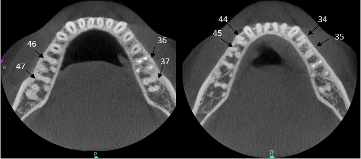
For further confirmation of this unusual morphology, the mandibular arch was subjected to Cone Beam Computed Tomography (CBCT) imaging. The mandibular arch was focused and morphology was obtained in transverse, axial, sagittal sections of 0.5mm thickness. CBCT scan confirmed the presence of three roots and three canals in relation to right and left first and second mandibular premolars and also four roots and four canals with respect to right and left first and second mandibular molars [Table/Fig-4].
CBCT images show bilateral four rooted mandibular first and second molar and bilateral three rooted mandibular first and second premolar.
Discussion
Barret has rightly stated that, “of all the phases of anatomic study in human system, one of the most complex is that of pulp canal morphology” [1].
Variation in the tooth root number and form in the dental arch are not un-common. The mandibular premolars are generally single rooted [2]. Two rooted, three rooted and four rooted mandibular first premolar are reported, but are rare [3]. The literature search on the anatomical studies has revealed 0.3% of two rooted and 0.1% of three rooted mandibular second premolar [4]. The mandibular molars are usually two rooted teeth [2]. Studies have noted 0.02%, 1% of four rooted mandibular first molar and 0.55% of four rooted mandibular second molar [5–7].
However, the incidence of three rooted lower premolars as well as four rooted lower molars is very low [4,5–7]. The bilateral occurrence is rare in nature.
This paper details an unusual occurrence of bilateral three rooted mandibular premolars and four rooted mandibular first and second molar and also highlight the use of CBCT to confirm the same.
The aetiology behind the formation of extra roots is still uncertain; however external factors during odontogenesis or to the penetration of atavic gene or polygenic system and extension of cells from the Hertwig’s epithelial root sheath towards the centre dividing the original single collar into several collars have been proposed [8,9].
The variation in the number of roots and root canals in mandibular molars makes the conventional root canal treatment challenging and mandibular premolars present the greatest difficulty of all teeth to perform successful endodontic therapy [3,5–7].
Conclusion
As there is increased awareness among the patients in retaining the natural tooth/teeth, the role of endodontist is more pivotal in treating the tooth that is pulpally involved. Therefore, a thorough understanding of the tooth morphology and potential of variation of the tooth from the norm coupled with multiple angulated radiographs help in achieving optimal success of nonsurgical endodontic treatment. This paper also highlights the role of CBCT in ascertaining the variation as an auxillary source.
[1]. Segura-Egea JJ, Jiménez-Pinzón A, Ríos-Santos JV, Endodontic therapy in a 3-rooted mandibular first molar: Importance of a thorough radiographic examinationJ Can Dent Assoc 2002 68(9):541-44. [Google Scholar]
[2]. Ash MM, Nelson SJ, Wheeler’s Dental Anatomy, Physiology and Occlusion 2003 8th editionWB SaundersPhiladelphia:239-262.Chapter 10, The permanent mandibular premolars [Google Scholar]
[3]. Cleghorn BM, Christie WH, Dong CC, The root and root canal morphology of the human mandibular first premolar: A literature reviewJ Endod 2007 33(5):509-16. [Google Scholar]
[4]. Mukhaimer RH, Bilateral mandibular second premolars with three separate rootsSaudi Endodontic Journal 2012 2(3):156-60. [Google Scholar]
[5]. Ingle JI, Bakland LK, Baumgartner JC, Ingles Endodontics 2008 Sixth editionHamiltonOntario:151-220.Chapter 6, Morphology of teeth and their root canal system [Google Scholar]
[6]. Koppolu M, Reddy K, Mathews V, Nuvvula1 S, Suneelkumar C, Anumula L, Diagnosis and management of bilateral four rooted mandibular first molarsJournal of Cranio-Maxillary Diseases 2014 3(2):172-75. [Google Scholar]
[7]. Shemesh A, Levin A, Katzenell V, Ben Itzhak J, Levinson O, Zini A, Prevalence of 3- and 4-rooted first and second mandibular molars in the Israeli populationJ Endod 2015 41(3):338-42. [Google Scholar]
[8]. Gupta A, Thakur S, Sharma V, Minocha A, Singh BP, Thakur R, Radix entomolaris: A endodotic challengeDental Journal of Advance Studies 2013 1:58-68. [Google Scholar]
[9]. Martins JNR, Ascenso J, Carames G, Endodontic treatment of a mandibular second molar with four roots - A case report and literature reviewGiornale Italiano di Endodonzia 2014 28:23-28. [Google Scholar]