Drug Induced Lupus Erythematous (DILE) is a rare adverse reaction to a large variety of drugs including Isoniazid (INH), with features resembling idiopathic Systemic Lupus Erythematosus (SLE). Diagnosis require identification of a temporal relationship between drug administered and symptom. It is an idiosyncratic reaction, with no pre-existing lupus. Our case highlights a rare presentation of isoniazid induced lupus with profound pancytopenia and mucosal ulcers, thus posing a diagnostic challenge. The patient was on multidrug treatment for pulmonary and knee joint tuberculosis. DILE was diagnosed on basis of strongly positive Anti Nuclear Antibodies (ANA), anti ds DNA and antihistone antibodies with clinical response to cessation of INH.
Case Report
A 21-year-old unmarried female was admitted with rashes over face and palms, extensive oral ulcers and low grade fever since 7 days. She also had joint pain for the last 10 days, predominantly knee and ankles and small joints of hands. Patient also complained of pain in the mouth while swallowing and loss of hair.
Treatment history was significant with a diagnosis of pulmonary and knee joint tuberculosis, for which she was on a retreatment regimen with Isoniazid, Rifampicin, Pyrazinamide, Ethambutol and Streptomycin (HRZES) under Category 2 of the Revised National Control Program (RNTCP).
On two past occasions, in 2013 and 2014 the girl had been diagnosed as having rifampicin sensitive pulmonary tuberculosis on the basis of chest X-ray findings and Gene Xpert results respectively. She was initiated on HRZE regimen under Category 1 of RNTCP. There was no history of adverse reactions noted during the course of treatment. She defaulted after 2 to 3 months of therapy each time, due to improvement in general condition and failure to follow-up after the second time.
The patient then developed left knee swelling in January 2016, which was diagnosed as tuberculous synovitis. Synovial fluid Gene Xpert and Mycobacteria Growth Indicator Tube (MGIT) culture detected presence and growth of Mycobacterium tuberculosis complex. Drug susceptibility tests did not show resistance to 1st and 2nd line drugs. High-Resolution Computed Tomography (HRCT) chest done concomitantly showed mediastinal necrotic lymph nodes and centrilobular nodules with tree in bud appearance. She was not on any treatment at this time. Hence, she was initiated on Category II regimen under RNTCP.
Thereafter, patient was on HRZES for 2 months, HRZE for 1 month, was on HRE from last 26 days. Three days prior to presentation in Pulmonary Medicine OPD, the Department of Dermatology had withheld rifampicin in view of thrombocytopenia and oral ulcers. The lesions continued to worsen despite discontinuation of the drug and she was referred to us for further management after one week.
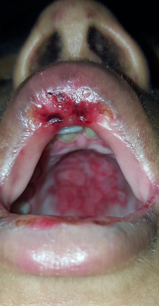
On examination, mucosal ulcers were multiple well defined erosions over lips, buccal mucosa and hard palate [Table/Fig-1]. Multiple well defined erythematous macules and papules were present over malar region of the face and both palms. Patient had tachycardia. Other vitals were within normal limits and systemic examination was unremarkable. Minimal residual swelling was present over the left knee joint with no tenderness or discharge.
Extensive oral mucosal ulcers with erosions on the lips and skin over the nose.
Laboratory Investigations: Anaemia with pancytopenia: Haemoglobin (Hb) – 8.2gm/dl, Total Leucocyte Count (TLC) – 1100cells/ml (Absolute neutrophil count- 500) with differential counts as Neutrophils-46%/Lymphocytes-45%/Eosinophils-0.4%/Monocytes-7%/Basophils-0.3%, Platelets- 57,000cells/μl.
Serum uric acid levels were raised – 9mg/dl, Serum Glutamic Oxaloacetic Transaminase (SGOT) - 133units/l, Serum Glutamic Pyruvic Transaminase (SGPT) - 57 units/l.
Iron studies showed normal iron levels, raised total and urinary iron binding capacity and raised ferritin levels (746ng/dl). Vitamin B12 level were normal.
Chest X-ray was normal. Sputum examination for Acid Fast Bacilli (AFB) for two consecutive days was negative. Ultrasound study of the abdomen was suggestive of mild hepatomegaly.
The Department of Internal Medicine was consulted, their opinion being agranulocytosis secondary to antitubercular medications. Hence, Isoniazid INH and ethambutol were also withheld and bone marrow biopsy was done to exclude alternative cause. Bone marrow biopsy showed normoblastic erythroid hyperplasia, with normal myeloid and megakaryocyte series. Dermatology reference was taken and in view of the typical malar rash possibility of Drug Induced Lupus Erythrematosus (DILE) was considered.
Autoimmune profile revealed raised ANA, Anti-ds DNA and Anti- histone antibody titres.
ANA – 120 units (normal < 20); Anti- ds DNA- > 1000 (normal < 75); Anti-histone antibody – 7.95 (normal < 1.00).
A diagnosis of INH induced lupus was arrived at.
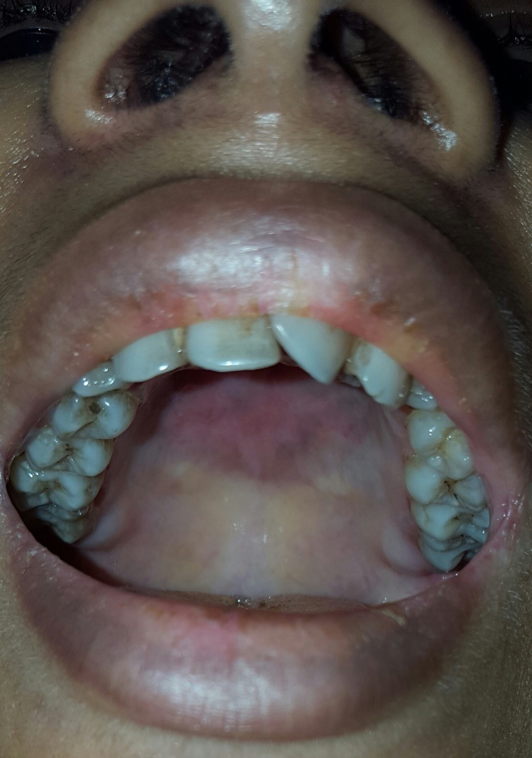
Symptomatic improvement was noted on stopping AKT. Patient improved over the next week, oral ulcers reduced significantly, with normalisation of total leucocyte count and platelets over the days. Antitubercular drugs except INH were reintroduced–Streptomycin was given for a month; rifampicin, ethambutol and levofloxacin were continued. Patient tolerated the AKT, her symptoms did not recur [Table/Fig-2].
Complete clearance of oral and skin lesions within 2 weeks of stopping INH.
Discussion
INH, one of the first-line anti-tubercular treatment drugs, is metabolized in the liver by acetylation and is excreted in urine. The lupus phenomenon, which has been reported with INH, is an idiosyncratic reaction. Up to 25% of patients taking INH have detectable ANA titres, but only 1% develop systemic DILE, which requires taking dosages of 300 to 900mg/d for a period ranging from 4 weeks to 14 months [1]. Definite mechanism by which it occurs is unknown however, theory of genetic predisposition as a slow acetylator is accepted [1]. Slow acetylators are homozygous for the recessive gene that controls expression of the liver enzyme acetyltransferase, which is involved in the metabolism of certain implicated drugs, such as procainamide, isoniazid and hydralazine [1]. Whether only slow acetylators of INH will develop the lupus syndrome remains to be determined. In comparison, acetylation rate is not a risk factor for spontaneous idiopathic lupus. Another theory for auto antibody formation is that INH may serve as substrates for myeloperoxidase in activated neutrophils which may result in formation of reactive intermediate, leading to occurrence of INH-induced lupus [2]. This patient had taken INH on two past occasions for 2-3 months within last two years. She developed symptoms of DILE only the third time after consuming INH for more than four months continuously. The time between drug exposures to onset of symptoms can vary from one month to over a decade after initiation of the drug treatment and is dose dependent and does not result in immune mediated response to the drug [3,4].
Drug induced lupus is classified into [5]:
Drug induced systemic lupus.
Drug induced subacute cutaneous lupus.
Drug induced chronic cutaneous lupus.
INH induced lupus presents with fever and pleuritis in half the number of cases and pericarditis in 30% cases [6]. Our patient presented with extensive mucosal ulcerations causing odynophagia, rash on palms and malar rash as primary complaints associated with fever and joint pains. The classic malar or discoid rash, oral ulcers and major organ involvement (renal and neurologic) seen in idiopathic SLE are notably rare in DILE [3] [Table/Fig-3]. Typical malar rash is seen in only 2% of DILE. As discussed earlier our patient had severe pancytopenia on admission, with an absolute neutrophil count of 500. Pancytopenia is extremely rare in SLE [7] and is not yet reported in case of INH induced lupus. The combination of the rash pattern, mucosal ulcers and pancytopenia is an extremely rare presentation of DILE. Pancytopenia possibly was immune complex mediated or due to Macrophage Activation Syndrome (MAS) [8]. Even though the bone marrow study was not suggestive of hemophagocytosis, a diagnosis of secondary Hemophagocytic Lymphohistiocytosis (HLH)/MAS cannot be ruled out as patient had high serum ferritin levels and mildly high SGOT and SGPT levels and mild hepatomegaly along with tricytopenia and fever. However, we lacked in having lipid profile and serum fibrinogen levels of the patient to arrive at a definite diagnosis. MAS is an acute episode of overwhelming inflammation characterized by activation and expansion of T-lymphocytes and hemophagocytic macrophages [9]. In rheumatology, it is associated with systemic lupus erythematosus and systemic juvenile idiopathic arthritis (SJIA) [9]. The main clinical manifestations include cytopenias, liver dysfunction, hypertriglyceridemia and extreme hyperferritinemia with macrophages phagocytosing hematopoietic cells in the bone marrow [9]. MAS in DILE is also yet to be reported.
Prevalence of symptoms in SLE and DILE [10,11].
| Features | SLE | DILE | Our Case |
|---|
| Fever [10] | 40 – 85% | 40-50% | Present |
| Arthralgia [10] | 70-95% | 80-95% | Present |
| Cutaneous involvement [10] | >75% | < 25% | Present |
| Rash (malar) [10] | 42% | 2% | Present |
| Rash over body [10] | 30 – 50% | 10–30% | Absent |
| Mucosal ulcers [11] | Commom | Uncommon [11] | Present |
| Hair loss [11] | Common | Uncommon [11] | Present |
| Hematological involvement [10] | Common | Unusual | Present |
| Pancytopenia [10] | Rare | Not documented | Present |
| Renal involvement [10] | 30 – 50% | 0-5% | Absent |
| ANA [10] | 90-98% | 95-100% | Present |
| Anti dsDNA [10] | 50-80% | < 5% | Present |
| Anti Histone [10] | 60-80% | 90-95% | Present |
Diagnosis of DILE is formed by detecting autoantibody levels. ANA is positive in 90% to 95 % of DILE cases [10]. ANA often persist for a greater duration than the symptoms and physical findings and in some patients autoantibodies may persist for a year or longer. Anti-histone antibodies are present in more than 95% of patients overall and is a hallmark for DILE. The anti-histone antibodies in DILE are primarily formed against a complex of the histone dimer H2A-H2B and DNA [10]. In contrast, the anti-histone antibodies in idiopathic lupus are primarily directed against the H1 and H2B histone subunits [10]. Anti-DNA antibodies are also unusual (<5 % cases) in DILE [10]. Anti-ds DNA antibodies are typically absent in DILE due to procainamide, hydralazine and isoniazid, but present in SLE [10]. Spontaneous resolution of the clinical manifestations of the disease, typically within several weeks but sometimes up to several months after the offending drug has been discontinued clinches the diagnosis of DILE and also differentiates it from SLE. Our patient had strongly positive ANA and anti-histone antibodies which are a hallmark of DILE, however she also had raised anti- dsDNA antibodies which is rare in DILE. Due to dramatic response noted after discontinuation of INH and uneventful reintroduction of other anti-tubercular drugs, a diagnosis of INH induced lupus was arrived at.
The initial step in treatment of DILE is to stop the offending drug. Systemic glucocorticoids may be required to treat cases of pericarditis and pleurisy [10]. In patients with more resistant disease, hydroxychloroquine can be used if constitutional, cutaneous and musculoskeletal symptoms persist [10]. We do not rechallenge patients with a medication that has been identified as a likely cause of DILE in that individual.
Conclusion
This case highlights the rarity and symptom severity of INH induced lupus especially pancytopenia which is yet to be reported and therefore, the importance of recognising it, as it may pose as a diagnostic challenge. Since tuberculosis is treated with a multidrug approach, it is very important to correctly identify the adverse effects of each drug and treat accordingly to keep up the patient compliance of the treatment, keeping in mind the resistant nature of the mycobacteria.
[1]. Pretel M, Marquès L, España A, Drug-induced lupus erythematosusActas Dermo-Sifiliográficas (English Edition) 2014 105(1):18-30. [Google Scholar]
[2]. Hofstra AH, Li-Muller SM, Metabolism of isoniazid by activated leukocytes. Possible role in drug-induced lupusDrug Metabolism and Disposition: The Biological Fate of Chemicals 1992 20(2):205-10. [Google Scholar]
[3]. Vasoo S, Drug-induced lupus: an updateLupus 2006 15(11):757-61. [Google Scholar]
[4]. Rubin R, Drug-induced lupusToxicology 2005 209(2):135-47. [Google Scholar]
[5]. Dyall-Smith D. Drug-induced lupus erythematosus | Derm Net New Zealand. Dermnetnz.org. 2016 [cited 12 August 2016]. Available from: http://www.dermnetnz.org/topics/drug-induced-lupus-erythematosus [Google Scholar]
[6]. Siddiqui MA, Khan IA, Isoniazid-induced lupus erythematosus presenting with cardiac tamponadeAm J Ther 2002 9(2):163-65. [Google Scholar]
[7]. Sarkar RN, Banerjee S, Dey S, Saha A, Bhattacharjee P, Banerjee TK, Haematological presentation of systemic lupus erythematosusJ Assoc Physicians India 2009 57:767-68. [Google Scholar]
[8]. Weinzierl E, Arber D, The differential diagnosis and bone marrow evaluation of new-onset pancytopeniaAmerican Journal of Clinical Pathology 2012 139(1):9-29. [Google Scholar]
[9]. Grant S. Schulert, Alexei A. Grom, Pathogenesis of Macrophage Activation Syndrome and Potential for Cytokine- Directed Therapies Annual Review of Medicine,Vol. 66: 145–159 [Google Scholar]
[10]. Merola J, Lupus-Like Syndromes Related to Drugs. In: Schur P, ed. byLupus Erythematosus Clinical Evaluation and Treatmen 2012 1st ed:211-221. [Google Scholar]
[11]. Camacho I, Kauffman C, Fredeking A, Drug-Induced Lupus Erythematosus: Background, Pathophysiology, Etiology Emedicinemedscape.com. 2016 [cited 12 August 2016]. Available from: http://emedicine.medscape.com/article/1065086-overview?pa=Ql3Sz1ZMizBayusmmHHyIrm86qyMVM8UKeI5Z9cVOVg%2F9kFaQaRlbm9VvDv8uv%2BS1xDGpL2xbRpNnHTsgL1P%2Fsfkq9g2DPn5sb65MIOIqA4%3D [Google Scholar]