Fine Needle Aspiration of a Subcutaneous Nodule Showing Eggs of Adult Filarial worm
Sandeep Ojha1, Leena Naik2, Kanchan Kothari3, Mona Agnihotri4
1 Assistant Professor, Deprtment of Pathology, Chirayu Medical College and Hospital, Bhopal, M.P, India.
2 Professor and Incharge, Department of Cytopathology, Seth G.S. Medical College and King Edward Memorial Hospital, Mumbai, India.
3 Associate Professor, Department of Cytopathology, Seth G.S. Medical College and King Edward Memorial Hospital, Mumbai, India.
4 Assistant Professor, Department of Cytopathology, Seth G.S. Medical College and King Edward Memorial Hospital, Mumbai, India.
NAME, ADDRESS, E-MAIL ID OF THE CORRESPONDING AUTHOR: Dr. Sandeep Ojha, Assistant Professor, Department of Pathology, Chirayu Medical College and Hospital Bhopal-462043, M.P, India.
E-mail: drsandy0582@gmail.com
A 22-year-old male, presented with a 2x2 cm, slightly tender nodule over medial aspect of upper arm near the elbow joint. There was no history of increase in size of the swelling, fever, cough or cold or any abscess formation.
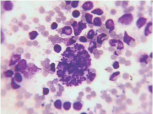
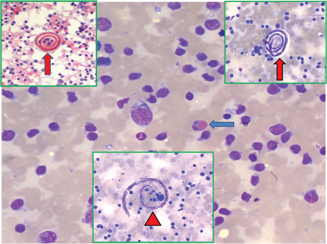
FNAC was performed using a 23G needle and smears were prepared and stained with Giemsa and Papanicolaou stain. On light microscopy, there were many non-sheathed granular ovoid structures scattered all through the smear [Table/Fig-1]. Background consisted of reactive lymphoid cells and occasional eosinophils. No granulomas were identified. On careful screening there were few coiled and uncoiled forms of microfilaria [Table/Fig-2]. These features indicated that the needle was in the uterus of a gravid female filarial worm as there were various stages of development of filarial on FNA smear. However, there were no structures resembling body wall of adult filarial worm or worm itself. We performed Peripheral Blood Examination (PBE) and wet mount of blood to look for any microfilaria, but these examinations were negative. PBE showed 3% eosinophils and few reactive lymphocytes.
Smears show non sheathed unfertilized egg of adult filarial worm. (Giemsa, X400).
Coiled forms (vertical arrow) and uncoiled form (arrow head) of microfilaria along with reactive lymphoid cells and occasional eosinophils (blue arrow). (Giemsa and Papanicolaou, X400).
Based on cytomorphology, a diagnosis of reactive lymphadenitis with adult filarial worm was offered. The patient was subsequently treated with DEC (Diethyl Carbamazine Citrate) and is doing well till date.
Discussion
Filariasis is endemic in tropical and subtropical countries including India and diagnosis is conventionally made by demonstrating the microfilaria in the PBE. The most common presentation of filariasis is fever or swelling due to lymphatic obstruction by filarial worm or lymphadenitis. Filarial worm can localize anywhere in the body but scrotal inguinal and abdominal lymphatics are common sites, from where the gravid female releases microfilaria in peripheral blood via thoracic duct leading to microfilaraemia [1]. There are various case series and reports demonstrating microfilaria on FNAC of lesions of scrotum, testes, axilla, arm, subcutaneous location and epididymis [2–5]. The incidental detection of microfilaria on a subcutaneous location of arm is rare and lodgment of filarial worms at that location is even rare. The morphology of these eggs should be well known to avoid missing the diagnosis in cases where the microfilariae are not visualized on cytology smears. It should also be differentiated from the cyst of toxoplasma where we see bradyzoites instead of nuclei.
[1]. Pant I, Singh PK, Singh SN, Agarwal A, Filariasis breast: A report of two cases, an unsual site to be involvedJ Cytol 2003 20:206-07. [Google Scholar]
[2]. Kumar B, Karki S, Yadava SK, Role of fine needle aspiration cytology in diagnosis of filarial infestationDiagn Cytopathol 2011 39(1):8-12. [Google Scholar]
[3]. Kishore B, Khare P, Gupta RJ, Bisht SP, Microfilaria of Wuchereria bancrofti in cytologic smears: a report of 5 cases with unusual presentationsActa Cytol 2008 52(6):710-12. [Google Scholar]
[4]. Pahwa R, Arora VM, Microfilaria in cytology smears from upper arm swellingJ Cytol 2010 27(4):155 [Google Scholar]
[5]. Azad K, Arora R, Gupta K, Sharma U, Lymphatic filariasis: Aspiration of adult gravid female worm from a soft tissue swellingJournal of Cytology 2010 27(4):156 [Google Scholar]