Today’s approach in dentistry is to restore any tooth even if only a small piece remains. So, in restoring endodontically treated teeth, when insufficient coronal tooth structure remains, to retain a crown the use of root canal space may be required for retention of the core and subsequent restoration. Restoration of devitalized teeth has been common since Pierre Fauchard’s first work in 1728 [1]. Prefabricated posts do not involve the laboratory phase and is time saving. Prefabricated post systems can be of Stainless steel, Titanium, Glass fiber reinforced composite posts, Carbon fiber reinforced composite posts, and Zirconia dioxide posts [2–4]. Glass fiber reinforced composite resin appears to be superior in esthetics compared to cast gold under a complete porcelain veneer crown because of its translucency, but when traumatic forces are encountered the prefabricated metal post and non metal post can differ in the pattern in which they transmit stress to the abutment tooth [5, 6]. This stress distribution pattern can be influenced by the presence (or) absence of ferrule of coronal dentin. Failure or success of a restoration may depend on how the stress is distributed to the tooth structure [7]. So it is necessary to study the stress distribution pattern around metal post and non metal post to select a suitable post system to form a foundation for a successful restoration. One of the reliable methods to study the stress distribution pattern around post is finite element analysis [8,9]. Finite element analysis is a mathematical model that allows complex structures to be drawn and divided into smaller segments with specific properties. Various loading conditions can be applied to the model and stress distribution plotted with a computer [10,11]. The advantage of finite element analysis is that it provides detailed stress information concerning a non-homogenous body such as tooth. The finite element method is a valid method for analyzing the mechanical behavior of complex structures. Three dimensional finite element models can capture the geometry of complex structures more accurately [12]. The objective of the study was to compare the stress distribution between Glass fiber and Titanium prefabricated posts using the three dimensional finite element method.
Materials and Methods
The study was conducted in the Department of Prosthodontics, Rajas Dental College, Kavalkinaru, Tamil Nadu, India. The study was ethically cleared from Institutional Human Ethics Committee (IHEC). In this study an endodontically treated maxillary central incisor with two different post materials with and without ferrule was designed three dimensionally using PRO Engineer software (Parametric Technology Corporation, USA). The models were Post-1 (Endodontically treated maxillary central incisor with a ferrule of coronal dentin and restored with parallel sided prefabricated titanium post and composite resin core), Post-2 (Endodontically treated maxillary central incisor restored with parallel sided prefabricated titanium post and composite resin core without a ferrule of coronal dentin), Post-3 (Endodontically treated maxillary central incisor with a ferrule of coronal dentin and restored with parallel sided prefabricated glass fiber reinforced composite post and composite resin core) and Post-4 (Endodontically treated maxillary central incisor restored with parallel sided prefabricated glass fiber reinforced composite post and composite resin core without a ferrule of coronal dentin).
A three dimensional finite element model of a maxillary central incisor with its internal anatomy and morphology had to be modeled first. A central incisor with its internal anatomy and morphology was modeled with geometric data. The measurements were compared with the data given in the literature. This model simulated the natural tooth with the incorporation of material properties – Young’s modulus and Poisson’s ratio [13,14]. The restored tooth was 23.5mm long with a diameter of 7mm at the level of crown margin. The cortical bone was 2mm thick. The remaining bone was modeled as spongy bone. The Post-1 was a parallel sided prefabricated post made of titanium which was 1.4mm in diameter and 12mm length with 4mm of guttapercha at the apex. The core was made of composite resin. A shoulder finish line with a 2mm ferrule of coronal dentin was modeled. Resin luting agent with thickness of 35 microns was modeled. Post-2 was a parallel sided prefabricated post made of titanium which was 1.4mm in diameter and 12mm length with 4mm of guttapercha at the apex. The core was made of composite resin. A shoulder finish line without a ferrule of coronal dentin was modeled. Resin luting agent with thickness of 35 microns was modeled. Post-3 was a parallel sided prefabricated post made of glass fiber reinforced composite. The diameter was 1.4mm and it was 12mm in length. There was 4mm of guttapercha at the apex. The core was made of composite resin. A shoulder finish line with a 2mm ferrule of coronal dentin was modeled. Resin luting agent with thickness of 35 microns was modeled. The Post-4 was a parallel sided prefabricated post made of glass fiber reinforced composite. It was 1.4mm in diameter and 12mm in length. There was 4mm of gutta-percha at the apex. The core was made of composite resin. A shoulder finish line without a ferrule of coronal dentin was modeled. Resin luting agent with thickness of 35 microns was modeled. In all the models the teeth were restored with a porcelain crown. The first step involved is modeling. The modeling was done using software called PRO Engineer. Using the software, models can be made in very short time, editing of the models can be done with a great ease, Surfaces can be created and controlled to get exact shapes at microscopic levels. Data from CT scan is required to model a tooth due to the complexity of the shape of a tooth. Tooth sections of 0.5mm interval are obtained by scanning the tooth. These scanned images were then imported into PRO/E software to various offset planes. Then manually in different sketch planes the curves were created along the tooth to get the exact shape.
From the curves, surfaces were created using a command called Boundaries. For this command the surface creation requires curves in the form of mesh i.e. curves in two directions, set of linear curves and a set of lateral curves need to be selected in order. From the surfaces solid is generated. Once the tooth is developed in similar fashion other parts are created and assembled. This assembly is then exported to an analysis package. For this study, a three dimensional model of a block section of the maxilla that included the central incisor with its supporting structures was simulated. A cylindric bone section of 20mm height and 10mm diameter was modeled. The tooth was designed to represent an endodontically treated maxillary central incisor with ferrule restored with a post and core and porcelain crown and without ferrule restored with a post and core and porcelain crown. The restored tooth was 23.5mm long with a diameter of 7mm at the level of crown margin. The cortical bone was 2mm thick. The remaining bone was modeled as spongy bone. Because their properties are similar, dentin and cementum were modeled as a single continuous material.
Finite Element Analysis: The advantage of finite element analysis compared to other stress analysis methods is that this is a non-invasive method as it is done in-vitro. When the material properties of tooth and alveolar bone are incorporated, the models simulate the natural tooth and alveolar bone. Stress value can be measured in any point of the model. Repeating the study will not alter the properties of the materials involved.
Meshing divides the entire model into smaller elements. In this work, 10-node-tetrahedral elements were used as they were better suited for the geometric structure of the tooth and more accurate results would be obtained. The model was subdivided into 18806 elements and 35575 nodes. Once meshing is done the next process is to define boundary conditions. Boundary condition means defining loads and restraints. Once, the loads were defined, then the problem was solved by incorporation of material properties and the results were reviewed [Table/Fig-1] [15].
| Material | Young’s modulus (Gpa) | Poisson’s ratio |
|---|
| Enamel | 41.0000 | 0.30 |
| Dentin | 18.6000 | 0.31 |
| Pulp | 0.00200 | 0.45 |
| Cortical bone | 13.7000 | 0.30 |
| Spongy bone | 1.37000 | 0.30 |
| Porcelain | 69.0000 | 0.28 |
| Resin composite core | 22.2000 | 0.30 |
| Guta-percha | 0.00069 | 0.45 |
| Titanium | 103.400 | 0.33 |
| Glass fiber/ Bis-GMA matrix parallel to fibers. | 50.0000 | 0.33 |
| Resin luting agent | 8.00000 | 0.30 |
Results
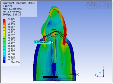
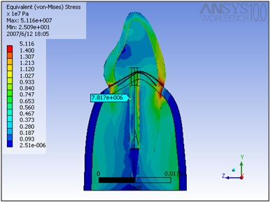
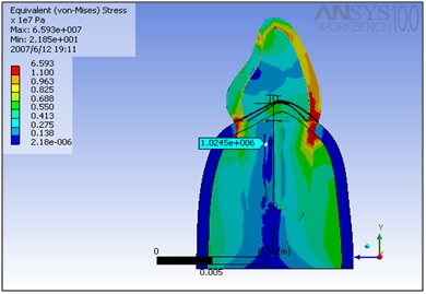
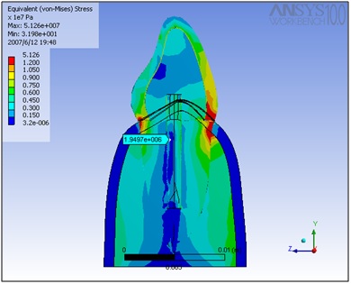
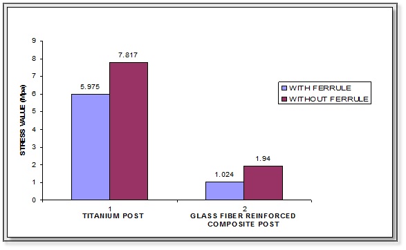
The stress distribution within a maxillary central incisor with titanium and glass fiber posts, with and without ferrule, when a load was applied was analysed using three dimensional finite element methods. The stress distribution was calculated at the post-cement-dentin interface, as this is the area where failure is more likely to initiate. Because there is increased tendency for the tooth to fracture under tensile loading, stress along the long axis of the teeth was taken for analysis in this study. Stress distribution was represented in numerical and color coding. The value for P1 model (Titanium post with ferrule) is 5.9751 Mpa [Table/Fig-2] and a value for P2 model (Titanium post without ferrule) is 7.817 Mpa [Table/Fig-3]. The values for P3 model (Glass fiber reinforced composite post with ferrule) are 1.0245 [Table/Fig-4] and values for P4 model (Glass fiber reinforced composite post without ferrule) are 1.9497 Mpa [Table/Fig-5]. The lowest stress value was obtained for Glass fiber post with a ferrule of coronal dentin. The highest stress value was obtained for Titanium post without a ferrule of coronal dentin [Table/Fig-6,7].
Titanium post with ferrule.
Titanium post without ferrule.
Glass fiber post with ferrule.
Glass fiber post without ferrule.
| Group | Mean Stress level |
|---|
| Post-1 | 5.975 |
| Post-2 | 7.817 |
| Post-3 | 1.0245 |
| Post-4 | 1.9497 |
Comparison of stress values.
Discussion
After endodontic treatment of a tooth is complete, the tooth must be protected due to risk of fracture during functional loading. The loss of tooth structure also makes retention of subsequent restorations more problematic. It is well established that after root canal treatment, dentin undergoes changes not only in its physiologic characters but also in its physical properties with a decrease in immature collagen levels. This can lead to reduced hardness and resistance to shearing [16]. Dehydration also occurs causing a decrease in Young’s modulus. Endodontically treated teeth with severe loss of coronal tooth structure are usually restored by using cast posts, prefabricated metal and fiber posts supporting composite or amalgam cores. Among metal posts prefabricated posts made of Titanium are increasingly becoming popular due to their favorable physical properties and resistance to corrosion [17]. Among non-metal posts, Glass fiber reinforced composite posts are gaining popularity because of their translucency and flexibility. In this study, the stress concentration in the post-cement-dentin boundary on the palatal side was studied. This region was considered important because according to Vasconcellos et al., post-dentin interface is the region where failure is more likely to initiate. Finite element analysis is a powerful tool in calculating stress distribution in complex structures. It provides results without variation. The validity of the study depends on the extent to which the model approaches reality. The advantage is that, it provides detailed stress information concerning a non-homogenous body such as tooth. The variables can be changed easily and simulation can be performed without the need for human material [14]. With elastic posts, the tooth, cement and post will all deform during function. So, failure will appear at the post-cement-dentin interface as this is an adhesive joint which is a weakest point. The less the remaining coronal tooth structure, the greater will be the stress on the adhesive joint [18]. So in this study it was decided to compare the stress concentration in the post-cement-dentin interface when a prefabricated post system made of titanium and another prefabricated post system made of glass fiber was used. It was also decided to study the stress concentration in the post-cement-dentin interface when these post systems were used in the presence of a ferrule of coronal dentin and without a ferrule. A ferrule prevents the fracture of the root due to the post [19].
The use of a three dimensional model was more accurate com-pared to a two- dimensional model in terms of element numbers, simulation quality and more realistic and finer representation of tooth forms. The effect of material, geometry and load variables were effectively determined in a three-dimensional model. The values of the mechanical properties were obtained from the literature and these values were widely used by researchers such as Vasconcellos et al., and Asmussen et al., [13,14,20]. A load of 100N was applied to the models at approximately 2mm from the incisal edge on the palatal surface, with an angle of 45o with respect to the longitudinal axis of the tooth to simulate the mastication force. This angle simulated the average anterior angle of function for Angle’s Class I occlusion and was used by other researchers such as Leary. The load of 100N was applied as this was the range of maximum biting force for maxillary central incisor [21].
This study was conducted by considering the three dimensional Von Mises criteria. The reason for selecting Von Mises criteria which apparently results in a tensile type normal stress, lies in the fact that the brittle materials of which, the tooth is a member, fail primarily due to tensile type normal stresses [22].
P1 model showed lesser stress levels than P2 model and P3 model showed lesser stress level than P4. This is because in P2 and P4 the absence of ferrule was found to be a determining negative factor which gave rise to considerably higher stress levels. Jefferson Ricardo Pereira et al., studied the effect of crown ferrule on the fracture resistance of endodontically treated teeth restored with prefabricated posts and found that increased amount of coronal dentin significantly increases the fracture resistance of endodontically treated teeth restored with prefabricated posts and composite resin cores, supporting the results of this study [23]. The stress levels for Titanium posts are higher than the stress levels for glass fiber posts. This is because the stiffness, elastic limit and strength of glass fiber post are significantly lower than Titanium posts [20].
Vasconcellos et al., did a study to assess how the geometry and material of the post affected maxillary central incisors [14]. Their results were similar to the results of this study as Titanium posts showed more stress than glass fiber posts. Hence, the results of this study show that it is preferable to use the glass fiber post with a 2mm ferrule of coronal dentin to avoid failure when using prefabricated posts. Irrespective of the post systems used, it is better to have a ferrule of coronal dentin to reduce the stress concentration. Titanium posts show more stress level than glass fiber posts.
Clinical Implications: Prefabricated glass fiber posts produce less stress concentration than prefabricated Titanium posts. Fracture resistance of the tooth can be improved by incorporating a ferrule. So whenever prefabricated posts are planned for restoring a tooth it is better to use glass fiber post. A ferrule effect also should be included to strengthen the remaining tooth structure.
Limitation
The limitation of this study is that it is an in-vitro study. So the oral conditions can’t be exactly simulated, but of most of the in-vitro studies the 3D finite element method gives more accurate results with no variation when the model is accurately simulated.
Conclusion
This study shows that it is preferable to use a glass fiber post when a prefabricated post is to be used. Also it is preferable to incorporate a ferrule of coronal dentin whenever posts are used. Titanium posts show more stress levels than glass fiber posts at the post-cement-dentin interface.