Mandibular Canine with Two Roots and Two Root Canals – A Rare Case
Nishita Rajeev Kulkarni1, Sharad Basavraj Kamat2, Santosh Irappa Hugar3, Girish Shankar Nanjannawar4
1 Postgraduate Student, Department of Conservative Dentistry and Endodontics, Bharati Vidyapeeth Dental College and Hospital, Sangli, Maharashtra, India.
2 Professor and Head, Department of Conservative Dentistry and Endodontics, Bharati Vidyapeeth Dental College and Hospital, Sangli, Maharashtra, India.
3 Associate Professor, Department of Conservative Dentistry and Endodontics, Bharati Vidyapeeth Dental College and Hospital, Sangli, Maharashtra, India.
4 Associate Professor, Department of Conservative Dentistry and Endodontics, Bharati Vidyapeeth Dental College and Hospital, Sangli, Maharashtra, India.
NAME, ADDRESS, E-MAIL ID OF THE CORRESPONDING AUTHOR: Dr. Nishita Rajeev Kulkarni, Department of Conservative Dentistry and Endodontics, Bharati Vidyapeeth Dental College and Hospital, Wanlesswadi, Sangli-Miraj Road, Sangli-416414, Maharashtra, India.
E-mail: nrkulkarni19@gmail.com
Anatomic variation, Periapical periodontitis, Radiography
A 54 year old female patient reported to the Department of Conservative Dentistry and Endodontics, with the chief complaint of dull aching pain in the lower right front teeth region since one month. History of present illness revealed intermittent pain with hot and cold stimuli for the past three months. Past dental history revealed asymptomatic, endodontically treated right mandibular first premolar. The patient’s medical history was noncontributory.
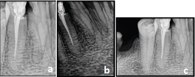
On examination, the lower right canine revealed proximal caries on disto-buccal side. The tooth was not mobile and periodontal probing around the tooth was within physiological limits. Thermal tests were positive and electric pulp testing elicited delayed response with the right mandibular canine. A diagnostic radiograph revealed a coronal disto-occlusal radiolucency involving the pulp space and widening of the periodontal ligament space. The radiograph also revealed an unusual anatomy of involved tooth. It showed presence of two roots and two root canals. For further confirmation of this unusual morphology, multiple pre-operative radiographs were taken at 10-40 degree mesial and distal angulations which confirmed the presence of two roots and two root canals [Table/Fig-1a-1c].
Multiple preoperative radiographs of the right mandibular canine.
From the clinical and radiographic findings, a diagnosis of symptomatic apical periodontitis was made, and root canal treatment was planned. Treatment was scheduled and initiated after obtaining written informed consent from the patient.
The caries on the disto-buccal surface was excavated and restored with composite resin (3M ESPE, A G Seefeld, Germany). Local anesthesia was administered and access opening was done using endo-access bur (Dentsply Tulsa, Tulsa, OK) under rubber dam isolation. The pulp chamber was opened to facilitate location of buccal and lingual canals. Working length was established using apex locator and also radiographically [Table/Fig-2a].
Working length. b) Master cone. c) Obturation.
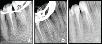
The canals were instrumented using stainless steel K- files (Dentsply Maillefer, Ballaigues, Switzerland) with master apical filing upto #35 K-file. A 5.2% solution of sodium hypochlorite and 17% EDTA were used alternatively as irrigants, at each change of file. Final irrigation was done using 2% chlorhexidine [Table/Fig-2b]. The canals were dried with absorbent paper points (Dentsply, DeTrey, Konstanz, Germany). The instrumented root canals were obturated using 2% gutta percha cones and AH Plus sealer (Dentsply, DeTrey, Konstanz, Germany) using lateral condensation technique. The final radiograph showed two well-obturated canals. After completion of root canal treatment, the tooth was restored using resin composite (3M ESPE, A G Seefeld, Germany) [Table/Fig-2c].
Discussion
Proper diagnosis and identification of the number of roots and root canals are key to success of endodontic treatment [1–3]. The studies of Greene, Hess and Vertucci revealed 13%, 15% and 18% of two canals in single root of mandibular canines respectively [1]. The occurrence of two roots and two separate root canals in mandibular canine is a rare entity and literature search has revealed 5%, 1% and 1.2% cases with two roots and two root canals respectively [4]. D’Arcangelo et al., also presented two cases of mandibular canines with two roots and two root canals [5].
The intraoral periapical radiographs plays an important role in identifying the internal anatomy of the tooth. Multiple angulated radiographs aid in locating unusual anatomical variations in the form of extra roots/canals. However, the inherent drawbacks of periapical radiography like superimposition and two dimensional image warrant their use in cases of unusual anatomy [6,7].
More recently, CBCT has become a boom in the field of Endodontics to identify the variations in tooth from norm, curvature, bifurcation and determining accurate length of the tooth in both sagittal and axial planes compared to conventional radiographs to obtain optimal success in conventional root canal therapy [7].
In the present case, standard hand files were used for instrumen-tation and root canal space was filled by lateral condensation technique. Such cases can also be managed by rotary instrumentation followed by single cone obturation technique.
This paper also presents successful management of mandibular canine with two roots and two separate canals. In view of rarity, such cases should be reported thereby enabling the clinicians to keep in mind the normal root morphology and anatomical variations.
Conclusion
Clinicians should be aware of anatomical aberrations in the teeth they are treating and should never assume that root canal morphology is simple. Although mandibular canines have a single root and single root canal, search for second canal or second root must be taken into consideration. Careful radiographic interpretation with different angulations and high end diagnostic images may also prove vital in cases of unusual root canal morphology.
[1]. Green D, Double canal in single roots. Oral SurgOral Med and Oral Pathol 1973 35:689-696. [Google Scholar]
[2]. Hess W, The anatomy of the root canals of teeth of the permanent dentition 1925 New YorkWilliams Wood Co [Google Scholar]
[3]. Vertucci FJ, Root canal anatomy of the mandibular anterior teethJ Am Dent Assoc 1974 89:369-371. [Google Scholar]
[4]. Victorino FR, Bernardes RA, Baldi JV, Moraes IG, Bernardinelli N, Garcia RB, Bilateral mandibular canines with two roots and two separate canals: case reportBrazilian Dental Journal 2009 20(1):84-86. [Google Scholar]
[5]. D’Arcangelo C, Varvara G, De Fazio P, Root canal treatment in mandibular canines with two roots: a report of two casesInt End J 2001 34:331-34. [Google Scholar]
[6]. Bharadwaj A, Bharadwaj A, Mandibular canines with two roots and two canals – A case reportInternational Journal of Dental Clinics 2011 3(3):77-78. [Google Scholar]
[7]. Betancourt P, Fuentes R, Cone beam computed tomography analysis of an unusual mandibular canine with two independent roots and two canalsBiomedical Research 2016 27(1):177-80. [Google Scholar]