Melioidosis is an emerging infection in India. It usually presents as pneumonia. Melioidosis presenting as cutaneous lesions is uncommon. We present a case of cutaneous melioidosis from Southern India. Cutaneous melioidosis can present as an ulcer, pustule or as crusted erythematous lesions. A 22-year-old gentleman known case of diabetes mellitus was admitted in our hospital with an ulcer over the left thigh. Discharge from the ulcer grew Burkholderia pseudomallei. He was successfully treated with ceftazidime. Melioidosis must be considered in the differential diagnosis of nodular or ulcerative cutaneous lesion in a diabetic patient.
B. pseudomallei, Diabetes Mellitus, Skin ulcer
Case Report
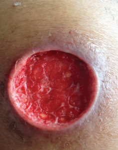
A 22-year-old gentleman was admitted in our hospital with complaints of an ulcer over the left thigh of seven days duration. History of fever was present for four days. He also complained of pain in the thigh. The patient initially noticed a nodule on the left thigh which eventually progressed to form a discharging ulcer. He was a known case of diabetes mellitus (type 1) on insulin. Clinical examination revealed a 5cm× 5cm ulcer on the left thigh with purulent discharge [Table/Fig-1].
Ulcer on the left thigh (after incision and drainage).
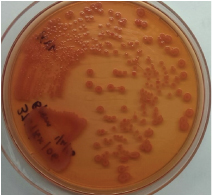
Laboratory investigations (on admission) showed: Total count: 9,500/cmm and platelets: 2, 66,000/cmm. His fasting blood sugar was 312 mg/dl. serum urea and creatinine were within normal limits. He was initially treated with injection Clindamycin. Daily dressing of the ulcer was done. Discharge from the ulcer grew a non-fermentative Gram negative Bacillus that was oxidase positive, showed pink colonies on MacConkey’s agar [Table/Fig-2] with an iridescent sheen and was resistant to Gentamicin and Polymixin B; based on which organism was presumptively identified as Burkholderia pseudomallei. Identification was confirmed by the Vitek 2 Compact system (bioMérieux). Antimicrobial drug testing showed susceptibility to Ceftazidime, Trimethoprim-sulfamethoxazole, Doxycycline, Imipenem and meropenem. Clindamycin was stopped. He was treated with injection ceftazidime for three weeks followed by oral Trimethoprim-sulfamethoxazole for six weeks. The ulcer healed completely within one month.
Colonies of B. pseudomallei on MacConkey’s agar plate.
Discussion
Melioidosis is an emerging infection in western coastal India [1]. Melioidosis is caused by the gram negative bacterium B. pseudomallei. It usually presents as pneumonia [2]. Cutaneous melioidosis is a rare entity. Cutaneous melioidosis may be primary (presenting symptom is skin infection) or secondary (melioidosis at other sites in the body with incidental skin involvement) [3]. There is limited published data from India documenting cutaneous melioidosis.
B. pseudomallei reside in soil and water [4]. Inoculation, inhalation or ingestion of infected food or water are the modes of transmission [5]. Alcohol intake, diabetes and chronic lung disease are some of the risk factors for melioidosis [1,2]. Our patient had diabetes. The disease is seasonal with majority of cases presenting during the rainy season [1]. Our patient presented during the monsoon season. Varying presentations of melioidosis is seen in clinical practice so it is called the great mimicker. The clinical manifestations range from localized infection to life threatening sepsis [3].
In a study of 486 cases of melioidosis in Australia, 58 (12%) had primary cutaneous melioidosis [3]. In the same study secondary skin melioidosis was seen in 10 patients (2%). Majority of patients with primary skin melioidosis have single lesions [3]. Cutaneous melioidosis can present as an ulcer, pustule or as crusted erythematous lesions [3]. Ulcerative form is the commonest presentation of primary skin melioidosis [3]. Our patient had a nodule that later developed into an ulcerative lesion on the thigh. The incidence of skin nodules in one of the largest case series of melioidosis from India was 3% [1]. Case reports of travel associated cutaneous melioidosis have been reported from Europe [6–8]. Cases of cutaneous melioidosis have also been reported from Taiwan and Singapore [9,10]. Cutaneous B pseudomallei infections can rarely progress to necrotizing fasciitis [11].
Diagnosis is based on isolation of B. pseudomallei from clinical specimen [12]. Ashdown’s medium is a selective medium used to isolate B. pseudomallei [1,11,13], and is often required while isolating B. pseudomallei from sputum and such other specimens which have numerous other flora as well. In our case, discharge from the ulcer grew B. pseudomallei on the primary plates and we did not use any selective media. Treatment consists of an intensive phase followed by eradication phase. In the intensive phase two weeks of Ceftazidime or Meropenem can be used followed by oral eradication therapy, usually with Trimethoprim–sulfamethoxazole for 12 weeks [3,14,15]. Our patient was treated with intravenous Ceftazidime followed by oral Trimethoprim–sulfamethoxazole. Patients with cutaneous melioidosis have been successfully treated with oral antibiotics alone (with or without incision and drainage) [3,6,7]. Prognosis is good for primary cutaneous melioidosis [3].
Conclusion
Melioidosis must be considered in the differential diagnosis of nodular or ulcerative cutaneous lesions. Empirical antibiotics that are usually used for cutaneous infections in diabetics in our country usually do not provide adequate coverage for melioidosis. Such a case report would serve to sensitize the clinicians about cutaneous melioidosis so that they can provide appropriate treatment to patients thereby decreasing morbidity.
[1]. Vidyalakshmi K, Lipika S, Vishal S, Damodar S, Chakrapani M, Emerging clinico-epidemiological trends in melioidosis: analysis of 95 cases from western coastal IndiaInt J Infect Dis 2012 16:e491-97. [Google Scholar]
[2]. Currie BJ, Ward L, Cheng AC, The epidemiology and clinical spectrum of melioidosis: 540 cases from the 20 year Darwin prospective studyPLoS Negl Trop Dis 2010 4:e900 [Google Scholar]
[3]. Gibney KB, Cheng AC, Currie BJ, Cutaneous melioidosis in the tropical top end of Australia: a prospective study and review of the literatureClin Infect Dis 2008 47:603-09. [Google Scholar]
[4]. McLeod C, Morris PS, Bauert PA, Kilburn CJ, Ward LM, Baird RW, Clinical presentation and medical management of melioidosis in children: a 24-year prospective study in the Northern Territory of Australia and review of the literatureClin Infect Dis 2015 60:21-26. [Google Scholar]
[5]. Kim SW, Kwon G-Y, Kim B, Kwon D, Shin J, Bae G-R, Imported melioidosis in south korea: a case series with a literature reviewOsong Public Health and Research Perspectives 2015 6:363-68. [Google Scholar]
[6]. Meckenstock R, Therby A, Marque-Juillet S, Monnier S, Khau D, Pangon B, Cutaneous melioidosis in adolescent returning from GuadeloupeEmerg Infect Dis 2012 18:359-60. [Google Scholar]
[7]. Bodilsen J, Langgaard H, Nielsen HL, Cutaneous melioidosis in a healthy Danish man after travelling to South-East AsiaBMJ Case Rep 2015 2015 [Google Scholar]
[8]. Ezzedine K, Heenen M, Malvy D, Imported cutaneous melioidosis in traveler, BelgiumEmerg Infect Dis 2007 13:946-47. [Google Scholar]
[9]. Tzeng WT, Hsieh CS, Recurrent cutaneous melioidosis treated with surgery and antibioticsJ Plast Reconstr Aesthet Surg 2009 62:280-81. [Google Scholar]
[10]. Yeo B, Lee J, Alagappan U, Pan JY, Cutaneous melioidosis with unusual histological featuresClin Exp Dermatol 2016 41(3):272-74. [Google Scholar]
[11]. Wang YS, Wong C-H, Kurup A, Cutaneous melioidosis and necrotizing fasciitis caused by Burkholderia pseudomalleiEmerg Infect Dis 2003 9:1484-85. [Google Scholar]
[12]. Gopalakrishnan R, Sureshkumar D, Thirunarayan MA, Ramasubramanian V, Melioidosis: an emerging infection in IndiaJ Assoc Physicians India 2013 61:612-14. [Google Scholar]
[13]. Thng TG, Seow CS, Tan HH, Yosipovitch G, A case of nonfatal cutaneous melioidosisCutis 2003 72:310-12. [Google Scholar]
[14]. Wiersinga WJ, Currie BJ, Peacock SJ, MelioidosisN Engl J Med 2012 367:1035-44. [Google Scholar]
[15]. Dance D, Treatment and prophylaxis of melioidosisInt J Antimicrob Agents 2014 43:310-18. [Google Scholar]