The primary goal of pulp therapy is to maintain the integrity and health of the teeth and their supporting tissues [1]. The objective of pulpectomy is complete removal of necrotic and irreversibly infected pulp of a tooth affected by caries, traumatic injuries or other causes, so that the tooth remains asymptomatic and functional in the oral cavity till it exfoliates normally [2,3].
Generally, all bacteria that inhabit the oral cavity have the ability to invade the pulp space during and after pulp necrosis in primary teeth. E.faecalis being a highly resilient and persistent organism plays an important role in the etiology of recurrent periradicular lesions post endodontic treatment in spite of making a small fraction of the flora in an untreated canal [4]. In spite of chemo mechanical preparation and copious irrigation of canal, there are chances of failure of pulp therapy in primary teeth due to the entrapped micro organisms in the canal space due to the tortuous and complex nature of the root canal system [2,3]. Thus, for optimal success of endodontic treatment, substances with antimicrobial properties are advocated as obturating materials in deciduous teeth. At present, the commonly used materials for obturation of deciduous teeth are Zinc Oxide Eugenol (ZOE), iodoform based pastes, triple antibiotic paste and calcium hydroxide with different vehicles.
ZOE has been traditionally used as an obturating material in the primary dentition [5] and was the first obturating material to be recommended for primary teeth. However, its rate of resorption is slower than that of deciduous teeth [6–8]. Moreover, it can irritate the periapical tissues, can cause necrosis of bone and cementum and may alter the path of eruption of succedaneous tooth [9].
Iodoform paste as an obturating material for primary teeth have received attention in the past. Commercially available iodoform based pastes are KRI paste, Maisto paste, Guedes-Pinto paste, Endoflas, Metapex and Vitapex [10,11]. Studies have shown that these iodoform combinations have better resorbability, biocompatibility and antibacterial properties [1,7,12] than ZOE but with a disadvantage that it may produce a yellowish-brown discoloration of tooth crown thereby compromising esthetics [1].
Endoflas is a resorbable paste obtained by mixing a powder containing iodoform (40.6%), zinc oxide (56.5%), calcium hydroxide (1.07%), barium sulphate (1.63%) and liquid consisting of eugenol and paramonochlorphenol.
Calcium hydroxide, which was introduced to dentistry by Hermann in 1930, has been known to promote healing in many clinical situations. It has been used as an obturating material for primary teeth alone and also in association with iodoform [13,14]. Calcium hydroxide can be used with various vehicles such as distilled water, glycerine, chlorhexidine etc.
The present study was undertaken to find out the efficacy of various obturating materials (v.i.z. ZOE, calcium hydroxide with chlorhexidine, endoflas, calcium hydroxide and iodoform in aqueous vehicle, metapex and saline) [Table/Fig-1] used in primary teeth against E.faecalis.
Materials and Methods
This in-vitro study was carried out in Department of Pedodontics and Preventive Dentistry in collaboration with Department of Pathology and Microbiology, Saraswati Dental College and Hospital, Lucknow, Uttar Pradesh, India, with an overall sample size of 60.
Preparation of broth and bacterial growth: The antimicrobial activity of obturating materials used in primary teeth against E. faecalis (ATCC 29212) was evaluated in this study by agar diffusion method. The standard bacterial strain of E. faecalis (ATCC 29212) was obtained from King George Medical University, Lucknow, Uttar Pradesh, India. The purity of test strain was confirmed using the Gram’s stain. Only 0.37 grams of Brain Heart Infusion (BHI) broth was added to 10mL of distilled water and mixed by gently shaking the container. This mixture was sterilized by autoclaving for 15 minutes at 121°C and 15lb pressure and then allowed to cool at room temperature. The bacterial strain (E. faecalis ATCC 29212) was inoculated in BHI broth and incubated at 37°C for 24 hours. Following incubation, the cultures were centrifuged at 3000 rpm for 10 minutes. The supernatant liquid was discarded and the precipitate containing microbial cells was separated from the base of test tube. The precipitate containing microbial cells was re-suspended in saline and turbidity of this culture suspension was adjusted until it was equivalent to the no.1 McFarland Standard (Approximately 3 X 10⁸ cells/mL).
Preparation of culture medium: Mueller Hinton agar (15.2gm) was added to 400mL of distilled water and mixed by gently shaking the container. This mixture was sterilized by autoclaving for 15 minutes at 121°C and 15lb pressure. After 15 minutes, the liquid was cooled to room temperature. In a laminar flow chamber, this liquid medium was poured in 20 petri dishes of size 90mm and allowed to set.
Inoculation of bacterial strain on culture media: Each agar plate with 20mL of Mueller Hinton agar was inoculated with 0.1mL microbial suspension using sterile swab. The 3 wells (4mm of depth X 6mm of diameter) were made in each of the agar plates, i.e., total 60 wells were prepared in 20 agar plates to test five different obturating materials and a control.
Placement of obturating materials and incubation: A sterile spatula and glass slab was used for mixing the obturating materials in creamy consistency. Each freshly mixed experimental obturating material was placed in 10 wells of different petri dishes. These agar plates were then incubated at 37°C under anaerobic conditions in an incubator using Microaerophilia (candle jar system). An anaerobic indicator tablet (Anaero indicator tablets) was placed in the jar to monitor oxygen contamination of the environment.
Measuring the size of zone of inhibition: A lack in bacterial colonization was observed for each obturating material. It was indicated by growth inhibitory zones (clearing of agar) around each obturating material. The most uniform diameter segment of the zone of inhibition was determined in millimeters by measuring the shortest distance between the outer margin of the well and initial microbial growth after 24 hours of incubation.
Statistical Analysis
The statistical analysis was done using SPSS Version 15.0. The Analysis of Variance (ANOVA) and Post-Hoc Tests (Tukey-HSD) were performed to know the effect of each variable and to reveal the statistical significance. The values were represented in number(%) and mean±SD.
Results
In the present study, a total of 60 samples were assessed for antimicrobial efficacy against E. faecalis [Table/Fig-2]. Antimicrobial efficacy assessment was done at 24 hours time intervals in terms of mean zones of inhibition. At 24 hours, no inhibition was seen in control group. However, in experimental groups mean zone of inhibition was maximum in Group IV (4.53±1.02 mm) followed by Group II (3.80±0.82), Group III (3.10±0.21), Group V (2.10±0.70) and Group I (1.80±0.35) respectively [Table/Fig-3].
Group wise distribution of materials.
| S No | Group | Description | No. of Samples | Percentage of groupwise distribution of material |
|---|
| 1 | I | Metapex | 10 | 16.7 |
| 2 | II | ZOE | 10 | 16.7 |
| 3 | III | Ca(OH)2 + Chlorhexidine | 10 | 16.7 |
| 4 | IV | Endoflas | 10 | 16.7 |
| 5 | V | Ca(OH)2 + Iodoform + Distilled water | 10 | 16.7 |
| 6 | VI | Control | 10 | 16.7 |
Intergroup comparison of mean zones of inhibition (in mm) in different groups at 24 hours time intervals (ANOVA).
| S No. | Group | Mean | SD |
|---|
| 1 | I | 1.80 | 0.35 |
| 2 | II | 3.80 | 0.82 |
| 3 | III | 3.10 | 0.21 |
| 4 | IV | 4.53 | 1.02 |
| 5 | V | 2.10 | 0.70 |
| 6 | VI | 0.00 | 0.00 |
| F (ANOVA) | 66.004 |
| p-value | <0.001 |
Between group comparisons (Tukey HSD test) at 24 hours [Table/Fig-4], revealed Group IV had significantly higher zone of inhibition as compared to all the other groups except Group II. Group II had significantly higher zone of inhibition as compared to Groups I, V and VI. Group III also had significantly higher mean value as compared to Groups I, V and VI. Group V had significantly higher mean value as compared to Groups I and VI. Group I had significantly higher mean value as compared to Group VI. On the basis of above evaluation, the following order of mean zone of inhibition was observed in different groups:
Between group comparisons (Tukey HSD test).
| S.No. | Group | Mean Diff. | SE | p-value |
|---|
| 1 | I vs II | -2.00 | 0.28 | <0.001 |
| 2 | I vs III | -1.30 | 0.28 | <0.001 |
| 3 | I vs IV | -2.73 | 0.28 | <0.001 |
| 4 | I vs V | -0.30 | 0.28 | 0.892 |
| 5 | I vs VI | 1.80 | 0.28 | <0.001 |
| 6 | II vs III | 0.70 | 0.28 | 0.145 |
| 7 | II vs IV | -0.73 | 0.28 | 0.120 |
| 8 | II vs V | 1.70 | 0.28 | <0.001 |
| 9 | II vs VI | 3.80 | 0.28 | <0.001 |
| 10 | III vs IV | -1.43 | 0.28 | <0.001 |
| 11 | III vs V | 1.00 | 0.28 | 0.010 |
| 12 | III vs VI | 3.10 | 0.28 | <0.001 |
| 13 | IV vs V | 2.43 | 0.28 | <0.001 |
| 14 | IV vs VI | 4.53 | 0.28 | <0.001 |
| 15 | V vs VI | 2.10 | 0.28 | <0.001 |
Endoflas ≥ ZOE ≥ Calcium hydroxide + Chlorhexidine > Calcium hydroxide + Iodoform + Glycerine ~ Metapex > Saline.
Discussion
The objective of pulp therapy in primary teeth is to preserve the tooth in functional stage until their exfoliation as the teeth act as natural space maintainers, so that the permanent teeth may have adequate space for aligning themselves [15,16]. Also, preservation of primary teeth helps in esthetics, phonetics, mastication and social acceptance. Pulpectomy is indicated when pulp has become irreversibly infected or necrotic due to caries, trauma or other causes [2]. However, tortuous and complex nature of the root canal system and change in their morphology with root resorption makes it difficult to achieve proper cleansing by mechanical instrumentation and irrigation of the canals [2]. For optimal success of pulp therapy, substances with antimicrobial properties are advocated as root filling material in deciduous teeth. Bacterial re-entry and growth of residual micro-organism may be prevented by incorporation of antimicrobial components into obturating materials [17].
In the present study, to assess the antimicrobial activity of endodontic sealers and obturating materials agar diffusion test was employed as it is one of the most commonly used methods [3,13,18]. The agar diffusion method operates on the principle that test material will diffuse from the paper disk or small well into an agar medium containing test organism. It evaluates halos of microbial growth inhibition as reference parameters. This method is advantageous since it directly compares the efficacy of different obturating materials against the micro-organisms thereby determining which material can be used for effectively disinfecting the pulp canal space [19]. However, the results can be influenced by the degree of diffusion of material across the medium [19].
Success rate of root canal treatment can be lowered significantly if there is presence of E.faecalis in pulp canal system at the time of obturation [11]. Most importantly, E.faecalis efficiently and rapidly colonizes dentinal tubules compared to other species and therefore is particularly difficult to eradicate [20]. It represents a standard against which the antimicrobial action of a medicament should be tested [20,21].
The present study was undertaken to assess the antimicrobial activity of different obturating materials used for primary teeth. For this purpose, five combinations of obturating materials and saline as control were analysed against E.faecalis by agar diffusion method. The data was collected and statistically analysed for intergroup and between group comparisons (Tukey HSD test) changes at 24 hours time intervals.
In the present study, endoflas, calcium hydroxide with chlorhexidine, fresh mix of calcium hydroxide with iodoform using distilled water as a vehicle were tested as there is very sparse literature and studies present regarding them, as an obturating material in primary teeth, whereas ZOE and metapex are regularly used in dental practice for obturation of primary teeth.
On intergroup comparison, the results revealed that all the materials tested possessed antimicrobial activity as demonstrated by the formation of growth inhibition zones. Endoflas showed maximum mean zone of inhibition against E.faecalis followed by ZOE and combinations of calcium hydroxide (calcium hydroxide + chlorhexidine, calcium hydroxide + iodoform + distilled water, metapex) [Table/Fig-5,6].
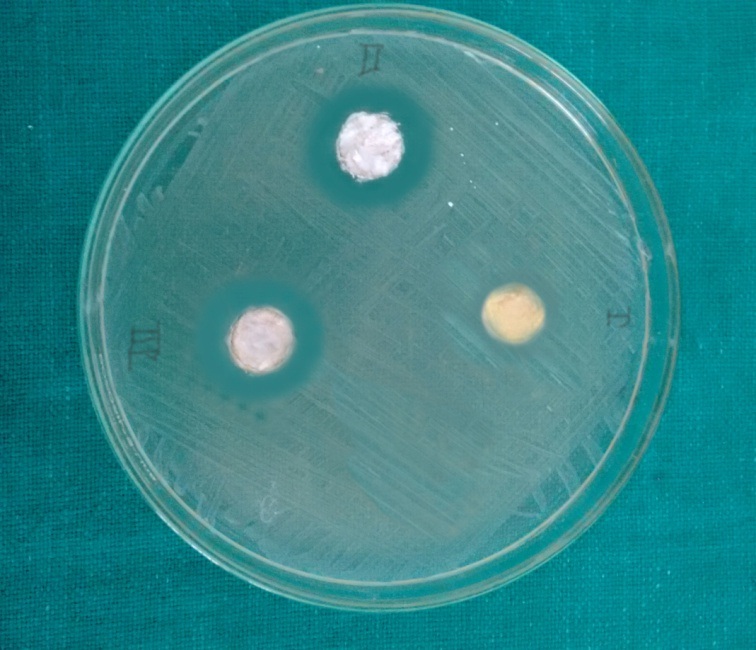
Zone of inhibition observed after 24 hours of incubation of different obturating materials (I- metapex, II-ZOE, III-calcium hydroxide+ chlorhexidine).
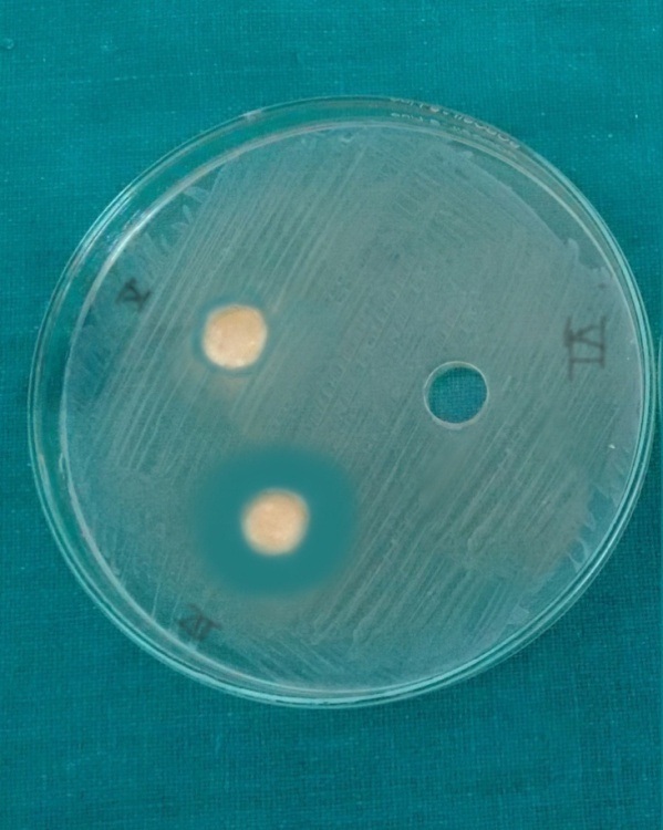
Zone of inhibition observed after 24 hours of incubation of different obturating materials (IV- endoflas, V-calcium hydroxide+ iodoform+ distilled water, VI-saline).
Eugenol based root canal obturating material (Endoflas and ZOE) showed highest antimicrobial activity as compared to the non-eugenol containing in the present study. The antimicrobial effect of eugenol based obturating material was mainly attributed to the action of eugenol. Eugenol acts on micro-organism by causing protein denaturation rendering it non-functional [22–24]. The results of the various studies performed by Gomes et al., Markowitz et al., and Saggar et al., also confirmed that eugenol containing obturating materials were more superior in inhibiting the microorganisms [22,24,25].
Endoflas FS showed marginally better, antimicrobial activity than ZOE obturating material probably due to incorporation of known bactericidal agents such as iodoform [22]. Iodoform acts by the liberation of iodine. It is believed that iodine, which is an oxidizing agent, can irreversibly oxidize and thus, inactivate essential metabolic compounds like proteins, nucleotides and fatty acid resulting in cell death, but the exact mode of action is not fully known [25]. Similar results have been reported by Gopikrishna et al., and Kayaoglu et al., [23,26].
In the present study, it was also observed that various calcium hydroxide combinations showed inhibitory action against E. faecalis. Calcium hydroxide and iodoform when mixed with aqueous vehicle performed better than metapex that contain silicone oil as a vehicle. It has been asserted that all biological actions of calcium hydroxide are due to the ionic dissociation in Ca2+ and OH- ions. The vehicle in calcium hydroxide formulations plays an important role in the overall process because it determines the velocity of ionic dissociation. Calcium hydroxide, when mixed with aqueous vehicle, allows better ionic dissociation and hence, diffusion and greater zone of inhibition in the agar than when mixed with oily vehicle [26].
Among the different preparations of calcium hydroxide, calcium hydroxide with chlorhexidine showed the best result followed by freshly prepared mix of calcium hydroxide and iodoform while metapex showed minimum inhibitory action. The possible reason could be that, chlorhexidine gluconate is a broad spectrum antimicrobial agent and is effective against bacteria and fungi [27–31]. Thus, antimicrobial activity of calcium hydroxide increases when mixed with chlorhexidine [16]. Pabla et al., in their study reported lesser antimicrobial activity of metapex against aerobic and anaerobic bacteria in comparison to ZOE, KRI paste and MAISTO paste [16]. However, Garcia-Godoy and Nurko studied the effectiveness of metapex in the root canal treatment of primary teeth and reported treatment to be deemed successful as clinically the tooth was painless, with no pathological mobility, healthy gingiva and no fistulation [1,13].
It was observed from the present study that eugenol based root canal obturating material particularly endoflas showed the highest antimicrobial activity against E.faecalis at 24 hours time intervals.
The rationale for performing this invitro study is to offer information to clinicians about antimicrobial efficacy of different root canal obturating materials used in primary tooth against E.faecalis.
Limitation
Limitation of this study could be that the antimicrobial efficacy of obturating materials has been evaluated in in-vitro conditions which may be modified in a clinical setup due to presence of dentin and serum. Hence, further in-vivo studies with larger sample size are needed to evaluate the antimicrobial efficacy of root canal obturating materials, in clinical settings.
Conclusion
Within the limitations of the study, all materials produced zones of microbial growth inhibition against the tested microorganism E. faecalis. On intergroup comparison of mean zones of inhibition in different groups at different time intervals, endoflas showed maximum mean zone of inhibition followed by ZOE, various combinations of calcium hydroxide and metapex showed least antimicrobial activity. The mean zones of inhibition can be summarised as follows:
Endoflas > ZOE > Calcium hydroxide + Chlorhexidine > Calcium hydroxide +Iodoform +Distilled water ~ Metapex > Saline.