Diagnostic aids, Middle distal canal, Middle mesial canal
A 22-year-old male patient reported to the Department of Conservative Dentistry and Endodontics, with a chief complaint of broken filling and severe spontaneous pain in the lower right back tooth region of the jaw. History revealed that the patient underwent restoration of the mandibular right first molar one year back at a local dental clinic. The patient’s medical history was non-contributory.
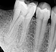
Clinical examination revealed an extensive tooth Colored resto-ration in the mandibular right first molar. Electric (Gentle Test Dental PulpTester, Pac-Dent International, Inc.,) and thermal (cold test- Endo Frost, Roeko, Langenau, Germany) pulp tests were negative; however, tenderness on vertical percussion was elicited. Periapical radiographic examination (Acteon Sopro®, Merignac, France) revealed secondary caries involving the mesial pulp horn with no signs of periapical radiolucency and aberrant anatomy [Table/Fig-1]. The clinical diagnosis of necrotic pulp with acute apical periodontitis was formulated and completion of root canal treatment was planned.
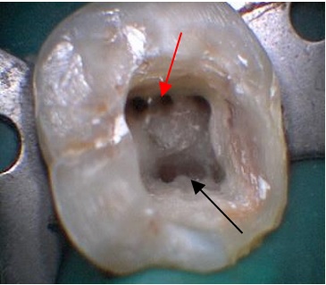
Inferior alveolar nerve block was administered using 2% Lignocaine (Lignox 2% Indoco Remedies Ltd.) with 1:80,000 epinephrine followed by isolation of the tooth using rubber dam (Hygienic Dental Dam, Coltene, Whaledent, Germany). After removal of the faulty acrylic restoration and excavation of caries, endodontic access cavity was made using an endo access bur (Dentsply, Maillefer, Ballaigues, Switzerland). The pulp chamber was repeatedly flushed with 3% sodium hypochlorite (Sigma-Aldrich Corporation, Bangalore, India) to remove necrotic tissue and microorganisms. Careful inspection of the pulp chamber revealed four canal orifices (mesiobuccal, mesiolingual, distobucal and distolingual) initially. But as the mesiobuccal and mesiolingual canals were widely separated, exploration using endodontic explorer (DG 16 Hu Friedy, Chicago, IL) at the floor of the pulp chamber revealed an additional canal orifice located between mesiobuccal and mesiolingual canals.
Further inspection using DG 16 explorer on the distal aspect of the pulp chamber revealed an additional canal orifice in close proximity to distolingual canal. Hence, a total of six canal orifices were located with three orifices in each root and the additional canals were termed middle mesial and middle distal canals in the mesial and distal roots respectively [Table/Fig-2].
Intraoral photograph revealing 6canals after biomechanical preparation.
Red arrow indicating middle distal orifice and black arrow indicating middle mesial orifice.
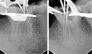
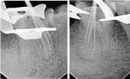
Working length radiographs were taken separately for mesial and distal root canals as six files could not be accommodated together. This also helped in preventing overlapping of files in the radiographic image. The working lengths were determined radiographically as 20mm in mesiobuccal canal and 21.5mm in mesiolingual and middle mesial canals; and 22.5mm in all the three distal canals [Table/Fig-3,4].
Working length of mesial and distal canals respectively.
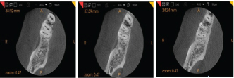
To confirm the unusual canal anatomy and to understand its configuration, Cone Beam Computed Tomography (CBCT) imaging of the tooth was performed after the tooth was temporized with temporary filling material (Orafil G, Prevest Denpro, Heidelberg, Germany). After obtaining an informed consent from the patient, CBCT of the mandible was performed with focus on the right mandibular first molar (3D Imaging Centre, Agra, India, CS 3D Imaging Software 3.3.11; 5 X 5). Axial, coronal, and sagital CBCT slices revealed six canals (three each in the distal and mesial roots) in the referred tooth. The presence of six canals was confirmed and root canal therapy was performed [Table/Fig-5-7].
CBCT images at coronal, middle and apical third of 46 respectively.
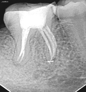
Following working length determination, biomechanical preparation was completed using Hero Shaper (MicroMega, France) rotary endodontic instrumentation in a crown down manner till their respective working length with an apical size of #25/0.04. Irrigation was performed using 5mL of 2.5% sodium hypochlorite solution and 17% EDTA (Glyde File Prep, Dentsply, Maillefer, Switzerland) alternatively. Interappointment calcium hydroxide dressing (ApexCal®, Ivoclar Vivadent, Switzerland) was placed for two weeks. In the next appointment, following a final rinse with 4mL of 2% chlorhexidine (Asep RC, Anabond Stedman Pharmaceuticals Pvt., Ltd., Chennai, India) all the canals were dried with sterile paper points (Dentsply, Maillefer, Tulsa, USA) and obturated using gutta-percha (#25/0.04) (Dentsply, Maillefer, Tulsa, USA) [Table/Fig-8,9] and AH Plus sealer (Dentsply, Maillefer, Tulsa, USA) single-cone obturation technique and subsequently the patient was referred for crown restoration after post endodontic restoration was done with light cure composite (Filtek Z 350, 3M ESPE, Minnesota, USA). [Table/Fig-10].
Master cone of mesial and distal canals respectively.
Post obturation radiograph.
Discussion
Due to the paradigm shift in the current advances in diagnostic technology in endodontics, CBCT has been a major breakthrough in dental imaging since its introduction in 1996. For the first time, the clinician is able to use a non-invasive, patient-friendly, three dimensional imaging system, with reduced radiation dose, to easily view images in three orthogonal planes namely axial, sagittal and coronal simultaneously allowing selected reconstruction of slices in all three planes, thus, allowing the area of interest to be dynamically traversed in ‘real time’ rather than being restricted to the limited two dimensional mesio-distal (proximal) plane of conventional radiography [1,2].
Most mandibular first molars have one mesial root and one distal root usually with the presence of two canals in the mesial root and one or two canals in the distal root. The incidence of three canals in the distal root is 0.2%-3% and that of the middle mesial canal is 1%-15% [Table/Fig-11,12].
Summary of incidence of three distal canals in mandibular first molar.
| Investigator | Year | Teeth | Method | Incidence (%) |
|---|
| Gulabivala et al., [3] | 2001 | 139 | Vitro | 0.7 |
| Gulabivala et al., [5] | 2002 | 118 | Vitro | 1.6 |
| Sert et al., [6] | 2004 | 20 | Vitro | 1.0 |
| Ahmed et al., [7] | 2007 | 100 | Vitro | 3.0 |
Case reports on types of middle mesial canals of first mandibular molar.
| Investigator | Side | Year | No. of canals in mesial root | Type | Imaging technique |
|---|
| Anjali et al., [8] | Right | 2013 | 4 | Independent | IOPAR |
| Anjali et al., [8] | Left | 2013 | 3 | Confluent | IOPAR |
| La et al., [9] | Right | 2010 | 3 | Independent | CBCT |
| Adrianne Freire de Paula et al., [10] | Left | 2013 | 3 | Independent | CBCT |
| Present study | Right | 2015 | 3 | Confluent | CBCT |
In the present case the axial and coronal slices of CBCT images confirmed the presence of three canals in each root which can be described as Type II (3-2) canal configuration according to Gulabivala and co-workers [3] supplemental canal configurations. Pomeranz et al., [4] morphologically classified the middle mesial canal into three categories [Table/Fig-13].
Pomeranz et al., classification of middle mesial canal [4].
| Fin | Free communication between all three canals |
|---|
| Confluent | Middle mesial canal joins one of the main canals, which most commonly joins the mesio buccal canal |
| Independent | Independent middle mesial canal from the orifice to the apex without merging with any of the main canals |
In this case the middle mesial canal is a confluent canal which originated as a separate orifice and merged with mesio buccal canal in the apical third, also the middle distal canal originated as a separate orifice and merged with the disto buccal canal in the apical third [Table/Fig-5-7].
Necessity is the mother of all inventions and CBCT has truly served the purpose of not only aiding in diagnosis but also improving management which ultimately improves treatment outcomes thereby making it a desirable addition to the endodontist’s armamentarium. When the diagnosis from clinical and conventional radiographic assessment is inconclusive, CBCT, which overcomes many of the limitations in periapical radiographic techniques, should be considered as a potentially useful imaging device.
[1]. Scarfe WC, Levin MD, Gane D, Farman AG, Use of cone beam computed tomography in endodonticsInt J Dent 2009 2009:634567 [Google Scholar]
[2]. Patel S, Dawood A, Whaites E, Pitt Ford T, New dimensions in endodontic imaging: part 1. Conventional and alternative radiographic systemsInt Endod J 2009 42:447-62. [Google Scholar]
[3]. Gulabivala K, Aung TH, Alavi A, Ng YL, Root and canal morphology of Burmese mandibular molarsInt Endod J 2001 34:359-70. [Google Scholar]
[4]. Pomeranz HH, Eidelman DL, Goldberg MG, Treatment considerations of the middle mesial canal of mandibular first and second molarsJ Endod 1981 7:565-68. [Google Scholar]
[5]. Gulabivala K, Opasanon A, Ng YL, Alavi A, Root and canal morphology of Thai mandibular molarsInt Endod J 2002 35:56-62. [Google Scholar]
[6]. Sert S, Aslanalp V, Tanalp J, Investigation of the root canal configurations of mandibular permanent teeth in the Turkish populationInt Endod J 2004 37:494-99. [Google Scholar]
[7]. Ahmed HA, Abu-bakr NH, Yahia NA, Ibrahim YE, Root and canal morphology of permanent mandibular molars in a Sudanese populationInt Endod J 2007 40:766-71. [Google Scholar]
[8]. Vats AS, Teja TS, Vats A, Grewal GS, Aggarwal S, Paul S, Middle mesial canal in mandibular molar: Two case reportDent J Adv Stud 2013 1:64-66. [Google Scholar]
[9]. La SH, Jung DH, Kim EC, Min KS, Identification of independent middle mesial canal in mandibular first molar using cone-beam computed tomography imagingJ Endod 2010 36:542-45. [Google Scholar]
[10]. de Paula AF, Brito-Júnior M, Quintino AC, Camilo CC, Cruz-Filho AM, Sousa-Neto MD, Three independent mesial canals in a mandibular molar: four-year followup of a case using cone beam computed tomographyCase Rep Dent 2013 2013:891849 [Google Scholar]