Interdisciplinary Management of Impacted Supernumerary Tooth between Roots of Permanent Teeth–A Management Dilemma?
Umapathy Thimmegowda1, Premkishore Kajapuram2, Mythri Prasanna3, Nandan Niranjanaradhya4, Ashwini Chikkanayakanahalli Prabhakar5
1 Reader, Department of Pedodontics and Preventive Dentistry, Rajarajeswari Dental College and Hospital, Bengaluru, Karnataka, India.
2 Reader, Department of Pedodontics and Preventive Dentistry, New Horizon Dental College and Research Institute, Bilaspur, Chhattisgarh, India.
3 Senior Lecturer, Department of Pedodontics and Preventive Dentistry, Bapuji Dental College and Hospital, Davangere, Karnataka, India.
4 Reader, Department of Pedodontics and Preventive Dentistry, Bangalore Institute of Dental Sciences, Bengaluru, Karnataka, India.
5 Postgraduate Student, Depatment of Oral Pathology and Microbiology, Rajarajeswari Dental College and Hospital, Bengaluru, Karnataka, India.
NAME, ADDRESS, E-MAIL ID OF THE CORRESPONDING AUTHOR: Dr. Umapathy Thimmegowda, Reader, Depatment of Pedodontics and Preventive Dentistry, Rajarajeswari Dental College and Hospital, #14 Ramohalli Cross, Kumbalgodu, Mysore Road, Bengaluru-74, Karnataka, India.
E-mail: umapathygowda@gmail.com
Anterior deep bite, Impacted teeth, Premolar
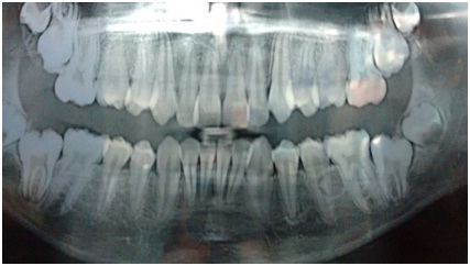
A 13-year-old boy visited a private clinic in Bangalore with a chief complaint of forwardly placed upper front teeth. His personal and past medical history was non-contributory and not associated with any syndrome. Physical and extra oral examination revealed no abnormalities. On intraoral examination, maxillary anterior crowding with labially placed teeth, increased overjet, deep bite was noted. He had Class II canine and molar relation with marked upper arch crowding. Orthopantomograph (OPG) revealed presence of all permanent teeth including third molars and developing supernumerary tooth between teeth 35 and 36 [Table/Fig-1].
OPG of the patient showing impacted supernumerary tooth between 35 and 36.

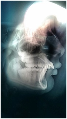
Lateral cephalogram revealed that patient has a horizontal growth pattern, increased overjet, anterior deep bite, Class II canine and molar relation [Table/Fig-2].
Lateral cephalogram of patient showing Class II Div I malocclusion.

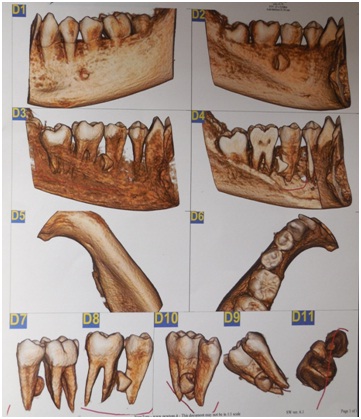
Cone Beam Computed Tomography (CBCT) examination was done and the scan report included the region of mandible in between 33 to 37. An impacted supernumerary tooth was noted in between the roots of teeth 35 and 36. The tooth measured about 8mm in length and 7mm in width with an intact follicular space and incomplete root. The crown of the tooth extended into the cortical plate to lie within the mucoperiosteum. The tooth was located lingual to and in contact with the roots of tooth 35 and mesial root of tooth 36 [Table/Fig-3].
A 3D CBCT image showing lingually impacted and incompletely formed supernumerary tooth in relation to 35 and 36.

A diagnosis of lingually impacted and incompletely formed supernumerary tooth in relation to 35 and 36 was made and confirmed with OPG and CBCT.
The most common order of frequency of supernumerary teeth are the mesiodens, maxillary fourth molars, maxillary premolars, mandibular premolars, maxillary lateral incisors, mandibular fourth molars and maxillary premolars [1].
Based on clinical, radiographic and CBCT examination impacted supernumerary teeth management is interdisciplinary, as it is planned for removal but in such cases there is always confusion whether to remove first and then followed by orthodontic management or orthodontic therapy and then extraction [2]. So here we are in dilemma, how to manage such case if encountered in clinical practice as management involves pediatric, oral and maxillofacial surgeon and orthodontist.
Pediatric Dental Surgeon is the one who expertise in recognising the complex condition, diagnose, investigate, treat them and maintain a harmonious healthy oral environment for a growing child.
Surgical management : On completion of all the above examinations, oral and maxillofacial surgeons planned for extraction of supernumerary tooth which was impacted between teeth 35 and 36 measuring 8mm in length and 7mm in width, as it was small it could be extracted easily under local anaesthesia. Since the patient requires orthodontic treatment there should be less trauma to adjacent structures like roots of premolar and molar. If patient is not willing for surgery then it is only wait and watch till tooth erupts. If impacted tooth hinders orthodontic treatment then it is to be extracted surgically, there may be more damage to adjacent structures as root formation is completed.
Orthodontic management: On clinical and radiological examination patient has horizontal growth pattern, increased overjet, anterior deep bite, Class II canine and molar relation and impacted supernumerary teeth. Diagnosis will be Angle’s Class II Division I malocclusion on skeletal Class II due to retrognathic mandible with convex profile, lip trap and impacted supernumerary tooth.
Treatment objectives are: Skeletal: To correct skeletal Class II malocclusion due to retrognathic mandible.
Dental: To correct increased over jet, correction of deep bite, Class II canine and molar relation, extraction of impacted supernumerary teeth.
Soft tissue: To correct convex profile and correction of lip trap.
Treatment involves Phase I-Therapy: Functional appliance therapy, Phase II-Therapy involves fixed appliance therapy: Preadjusted Edge wise Appliance [PEA] MBT 0.022 slot.
Phase I Therapy: Functional therapy for correction of skeletal Class II canine and molar relation, however it won’t have much effect on dental structures. So extraction of supernumerary teeth can be delayed for one year till completion of functional appliance therapy so that supernumerary teeth can still erupt further and provide better access for removal. So after functional therapy extraction of supernumerary teeth is planned followed by phase II fixed orthodontic therapy.
Any supernumerary tooth which is present clinically or impacted within bone or between teeth is usually planned for removal but in cases with impaction between roots of permanent teeth there is still a confusion whether to follow removal before orthodontic therapy or orthodontic therapy followed by removal, however, removal is final but timing of removal is crucial which decides removal, so interdisciplinary management plays a role in deciding the timing and management in such cases.
In present case orthodontist felt that early management of impacted tooth is better than delayed management as there will be lesser trauma to adjacent structures like roots of premolar and molar. Also the tooth was hindering orthodontic treatment, so, oral and maxillofacial surgeons also planned for removal of the supernumerary tooth under local anesthesia.
[1]. Khambete N, Kumar R, Genetics and presence of non syndromic supernumerary teeth- A mystery case report and review of literatureContemp Clin Dent 2012 3(4):499-502. [Google Scholar]
[2]. Masih S, Sethi HS, Singh N, Thomas AM, Differential expressions of bilaterally unerupted supernumerary teethJ Indian Soc Pedod Prev Dent 2011 29(4):320-22. [Google Scholar]