There has been a rising trend in costs for medical management of diseases. Also, there is a growing consciousness for use of environment friendly initiatives. Any change, which can bring down the costs and at the same time is more environment friendly would be a welcome step. However, any such initiative must not compromise the quality of patient care or for that matter it must not loose its intended purpose.
The trend in current day radiology practice is towards digitalization taking over the conventional film radiography. With the advent of Picture Archiving and Communication System (PACS), most of the radiologists are reporting on a console screen and no longer use films for reporting purpose [1,2]. This has led to limited utility of printing a radiographic film, which is mainly limited to supplementing the radiology report with an image for the demonstration purpose to the referring physician and for the patient’s record keeping. In developing countries like India where PACS is still not integrated in most of the departments of a hospital and also due to the presence of a large number of functioning stand-alone diagnostic centers, total avoidance of radiographic printing is not possible.
LASER films are the traditional mode of printing radiographs. However, this method involves large costs and disposal of films requires specific procedure [2,3]. Paper printers are available in most of the radiology departments, mostly used to print CT/MRI reformatted images or office work. This study was planned to assess the acceptance of paper prints to supplement the radiology reports after reporting over PACS workstations. This would serve as an environment friendly, convenient and lesser expensive form of report communication.
Few centers with integrated PACS systems abroad have reported using this technology for report communication. However, there is a significant difference in radiology practice in developing countries. The primary difference is the absence of hospital information system and PACS in all departments; and majority of radiology services being standalone. The physician acceptance level of paper printed radiographs in terms of their diagnostic accuracy is therefore more important in these set up. Considering these factors, the present study tries to address these issues to venture into the relatively unexplored territory of environment friendly, convenient and more cost effective method of radiographic report communication.
Materials and Methods
This observational analytical study was done at a tertiary care hospital of New Delhi, India during March-April 2016, after approval from Research projects approval committee and Institutional ethical committee. The primary objective of this research work was to study the image quality degradation (if any) by studying the agreement between wrist X-ray findings of traditional LASER films with paper print; screen reading on the PACS by radiologist being the gold standard. As a secondary objective, comfort level of the clinicians with paper prints in comparison with the traditional LASER films was also noted on a 1-5 point Likert scale.
Paediatric wrist radiographs for rickets were chosen as these are considered most challenging due to unfused epiphysis and changes involving the metaphyses. Many a times the metabolic bone changes are manifested as subtle radiological changes like metaphyseal fraying. A total of 58 consecutively ordered wrist radiographs of paediatric patients (6 months to 12 years of age) for ruling out rickets were retrieved from historical data using the Innowave proprietary PACS freedom IWS version 1.0, with the help of key words ‘wrist’ and ‘rickets’. All these standard antero-posterior view of both wrists were acquired by trained and certified radiographers using full room DR System (Siemens, Erlangen, Germany) with a small focal spot tube (0.6 mm), 55 kVp, and 10 mAs setting.
Out of these 58 X-rays, 21 (36.2%), had radiological evidence of rickets as noted by the primary investigator of the study from the PACS workstation with a display resolution of 2,560 × 1,536 pixels. Remaining 37 wrist X-rays were normal. Objective scoring method suggested by Thacher et al., was used to grade the severity of radiological findings of rickets [4]. Grade 1 meant widening of growth plate, irregularity of metaphyseal margin but without concave cupping and grade 2 meant metaphyseal concavity with fraying of margins. Both radius and ulna were scored separately, so a total of 4 maximum points could be given on any X-ray [Table/Fig-1,2].
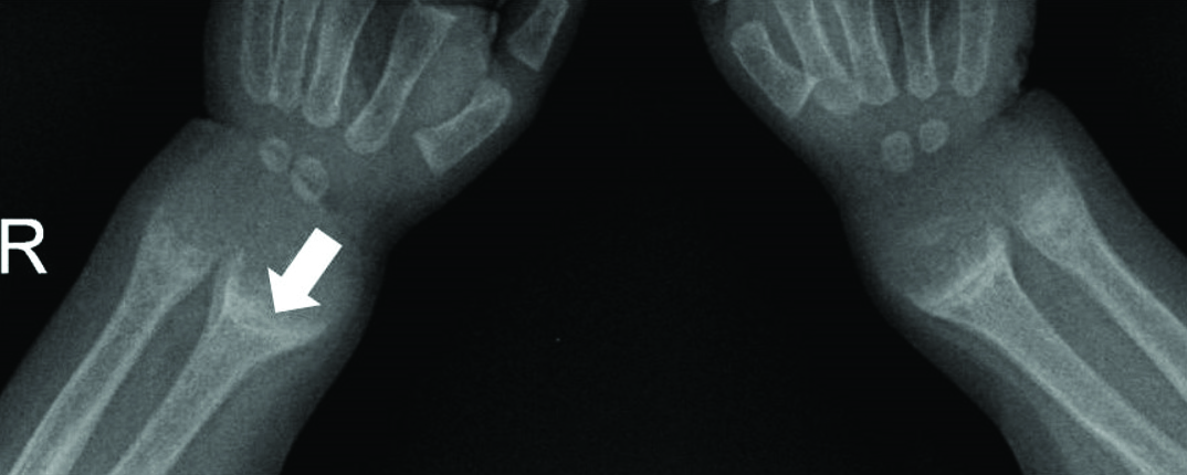
Normal metaphyseal ends of radius and ulna scored 0 for rickets (bold arrows). A note made of fracture of proximal shaft in both radius and ulna (thin arrows).

Metaphyseal concavity with fraying of margins of radius and ulna both (bold arrows), scored 4 for rickets.
The image printing on 160 GSM A4 size Glossy Photographic paper was optimized for image quality by using phantom supplied by Siemens Company. Page set up was optimized to include both wrists. The phantom included varying contrast objects producing different densities at different KVs for contrast resolution. Radiographs were obtained on two different media namely LASER film and paper using the above phantom and technique for standardization. All 58 images were printed on 8” x 10” LASER films using AGFA Drystar 2000 printer. The images were also printed on 160 GSM A4 paper using a standard digital paper printer with resolution of 5760 dpi.
All 58 images (both in LASER and paper print group) were kept in order of their printing date i.e., films with and without rickets were mixed in random and labeled as 1 to 58. These images were interpreted by total of six observers independently (one radiologist and 5 senior paediatricians), who graded the LASER films and paper prints for rickets using scoring system suggested by Thacher et al., [4]. All five paediatricians (clinicians) also mentioned their overall comfort level with the paper printed X-rays vs. LASER films on a 1-5 point Likert scale [Table/Fig-3].
Demonstration of image quality of the digital paper prints in contrast to their soft copy images; (a) and (c) are soft copy of normal wrist radiograph and one that with rickets; (b) and (d) are their respective digital paper prints.

The data was numerically coded and entered in Microsoft Excel 2007 and then transferred to STATA 14.0 (Stata Corp, College Station, TX) for analysis. Intra-observer percentage agreement i.e., the proportion of assessments in agreement to each other and Cohen’s kappa coefficient was calculated between PACS vs. LASER, PACS vs. paper print and LASER films vs. paper print for Observer 1 (primary investigator of study who is a radiologist and had access to PACS workstation). For the rest five Observers 2-6, intra-observer percentage agreement and Cohen’s kappa coefficient between LASER films and paper prints were calculated. Lastly, the intra-observer percentage agreement and Cohen’s kappa coefficient for LASER films vs. paper prints were calculated pooling the observations of all six observers.
Cohen’s kappa gives a numeric rating of the degree of agreement between the two set of observations, taking into account the degree of agreement which could be expected by chance. We used cut-offs proposed by Landis JR and Koch GG for interpretation of results [5], according to whom Cohen’s kappa coefficient ≥0.80 represent excellent agreement, between 0.61 and 0.80 represent substantial agreement, 0.41 to 0.61 moderate agreement and <0.41 fair to poor agreement.
Results
The observed intra-observer percentage agreement and value of kappa coefficient for PACS vs. LASER films and PACS vs. paper prints was equal i.e., 98.3% and 0.97, respectively. Intra-observer reliability/agreement for grading severity of rickets in LASER films vs. paper prints was excellent for all six observers; Cohen’s kappa value ranging from 0.92 to 1.00 with an observed percentage agreement ranging from 94.8% to 100%. Percentage intra-observer agreement with pooled observations of all six observers in LASER films vs. paper prints was 98%; kappa value being 0.97 (CI; 0.96-0.97) [Table/Fig-4].
Intra-observer percentage agreement and Cohen’ kappa value for radiological findings of rickets in wrist radiographs seen over PACS, laser films and paper prints.
| Observer No. | Media | N | Observed percentage agreement | Cohen’s kappa value | Confidence Interval |
|---|
| 1 | PACS vs. Laser films | 58 | 98.3% | 0.97 | 0.93-1.00 |
| PACS vs. Paper prints | 58 | 98.3% | 0.97 | 0.93-0.97 |
| Laser films vs. Paper prints | 58 | 100% | 1.00 | - |
| 2 | Laser films vs. Paper prints | 58 | 98.3% | 0.97 | 0.95-1.00 |
| 3 | Laser films vs. Paper prints | 58 | 96.5% | 0.95 | 0.94-0.98 |
| 4 | Laser films vs. Paper prints | 58 | 100% | 1.00 | - |
| 5 | Laser films vs. Paper prints | 58 | 98.3% | 0.97 | 0.96-1.00 |
| 6 | Laser films vs. Paper prints | 58 | 94.8% | 0.92 | 0.87-0.95 |
| Pooled data of all six observers | Laser films vs. Paper prints | 348 | 98.0% | 0.97 | 0.96-0.97 |
Out of 58 X-rays, 4 had associated fracture of ulna/radius. These fractures were very well demonstrated in both LASER films and paper prints and noted by all six observers. All five paediatricians rated their comfort level as 5 out of 5 for paper prints due to no requirement of any special illuminated view box and dark room.
Discussion
Over last few decades, Radiology has been one the most rapidly evolving and technologically heavy department. There is a paradigm shift from conventional film screen radiography to digital imaging and now the trend is towards a paperless department. The changes are inevitable and sticking to age-old technology is of little help [1,2]. In comparison to film interpretation, soft-copy interpretation using medical grade workstations is definitely superior in terms of sensitivity, specificity, and overall accuracy, which signifies supremacy of PACS workstation as gold standard [6].
Legal acts like Personal Privacy Act (PPA) and the Health Insurance Portability and Accountability Act (HIPAA) mandates proper disposal of LASER films only at certified disposal agencies for maintaining patient privacy and confidentiality [3]. There has been a proven advantage of doing away with LASER films in terms of cost cutting, environment friendliness, saving storage space and no need for special disposal process [2]. Due to the absence of hospital information system at most of the medical centers in India and a number of radiology centers functioning standalone, a complete paperless communication between the radiologist and clinician is not possible in India in near future. Paper printing of radiology images may serve as a convenient, less expensive and eco-friendly mode of communication between the radiologist and the treating clinician. However, only a few studies have been conducted, those too at centers outside India, which have tried to establish the utility of paper prints vs. traditional film printing in digital radiographs [7–15].
Long back in 1984, Stephenson TF et al., compared the diagnostic utility of printing CT scans on photographic paper and films against the scanner display console [7]. Pathology could be recorded adequately in all X-ray films and in 97% of photographic paper prints. Test phantom scans recorded on both media showed no observable difference in spatial or contrast resolution. Another study was published in 1998 [8] in which six CT scans were printed on paper by a high quality LASER printer and commented by seven physicians whether a printed image provide sufficient detail to assist in documenting the patient’s diagnosis and treatment and asked if the printed image was qualitatively identical to the original film image in terms of detail, contrast and noise enhancement. Out of 42 ratings of printed CT images, 40 printed images (95%) were rated as acceptable for documentation purpose. Twenty readings (48%) were rated as good as acceptable for diagnostic purpose. A suggestion was made that the LASER paper prints are as good as films for documentation purposes and in many cases for review of clinical decision making. A more recent study by Hu XY et al., in which 60 thoracic plain CT scans (28 cases with solitary pulmonary nodules) were printed by both a dry LASER printer and a high resolution paper printer, respectively, the conclusion was that the image quality obtained from a paper printer is comparable and similar to that from a dry LASER printer [9].
In older studies, there was a consistently greater success in detection of simulated caries on phantom in film images vs. thermal paper prints [10]. However, a more recent study found photographic paper comparable to the traditional radiographic films in caries diagnosis [11]. The authors also highlighted that size of the printed image also may be important in caries diagnosis. Type of printer, printer resolution, paper quality and type of ink used are other printing parameters, which must be taken care of.
Regarding the available published data about role of paper printing in full-field digital mammography, ROC analysis was significantly different between the LASER films and paper prints, finding quality of dry LASER prints being significantly superior to paper prints [12].
Digital chest radiographs of a phantom with randomly placed nodules in the mediastinum and lung which were printed on matt coated paper by a continuous inkjet printer as well as LASER films were compared with each other in a study by Lyttkens K et al., in 1994, where no significant difference was found in the performance of two printing media [13]. Maydell AT et al., compared 51 digital paediatric radiographs in paper print against reference standard of screen reading to study the diagnostic value of paper prints [14]. Specificity was excellent for all different regions (98.6-99.5%) but the sensitivity just acceptable in musculoskeletal and abdominal radiographs group (90% and 99%, respectively) and poor in chest radiographs group (66.1%).
Teixeira P et al., studied wrist trauma radiographs using LASER film, PACS workstation and paper with an optimized layout [15]. Readings were made by two independent readers who analysed 200 radiographs consecutively in one session for each type of media. The inter-technique agreement was found to be almost perfect in all cases.
In the present study, wrist radiographs for rickets were particularly chosen as they are technically challenging, require skilled reporting, an objective assessment tool is available to grade the severity of rickets on X-ray, paediatricians commonly view the printed image for their reference before starting treatment and also need the same again for post-treatment follow-up. The results of present study are motivating showing a perfect intra-observer agreement for grading the severity of rickets between conventional LASER films and paper prints. Other findings like fracture were also picked up accurately in paper prints. The comfort level of clinicians with paper prints was excellent because of no requirement of any special illuminated view box.
Limitation
The strength of present study lies in involvement of six observers independently judging the quality of paper prints vs. LASER films. This study being limited to only wrist (skeletal) radiographs, results cannot be extrapolated to chest, abdominal and other site radiography.
Conclusion
Digital paper prints of skeletal radiographs may serve as a potentially reliable, cheaper, and more convenient and environment friendly alternative to conventional LASER films for report communication between the radiologist and the clinician without significant loss of information. Further studies are warranted to demonstrate their clinical utility in chest, abdominal and other site radiography.
Conflicts of Interest: None
Source of funding: Self funded