Introduction
Smear layer removal from the root canal wall involves the use of 17% EDTA and 5.25% sodium hypochlorite, which thereby improves the adhesion of obturating materials to root dentin. But these chemical irrigants have shown to decrease micro hardness, increase roughness, cause erosion and reduce the root dentin fracture toughness. To combat these adverse effects, studies can be focussed on the remineralisation of the erosive root dentin and this novel idea has been utilized in the present study.
Aim
To evaluate the micro hardness of erosive root dentin when Casein Phosphopeptide–Amorphous Calcium Phosphate (CPP-ACP) was used as a final irrigant and its influence on resin sealer bonding tested by push-out bond strength method.
Materials and Methods
Sixty extracted maxillary incisors were divided into three groups based on the final irrigation protocol. Group 1-normal saline, Group 2-17% EDTA (Ethylene Diamine Tetraacetic Acid) + 5.25% NaOCl (Sodium Hypochlorite), Group 3 - 17% EDTA + 5.25% NaOCl + CPP-ACP; each group was divided into two subgroups. Half the specimens of each group were evaluated for Vicker’s micro hardness test after the treatment. In continuation with the above methodology the remaining specimens were tested for push-out bond strength after obturation of the specimens with self etch adhesive resin sealer and conventional 6% gutta percha cones.
Results
Micro hardness was statistically analysed using Kruskal Wallis test and push-out bond strength was evaluated using Mann Whitney test and paired t-test. CPP-ACP treated group showed increased micro hardness (p<0.05). There was no statistically significant difference between the push-out bond strength values between group EDTA + NaOCl group and EDTA + NaOCl + CPP-ACP group.
Conclusion
Within the limitations of this study it can be concluded that, CPP-ACP improved the micro hardness of erosive root dentin and is not affecting its bond strength. Therefore, CPP-ACP may be used before bonding procedures for promoting remineralization of root dentin.
Dentin, Ethylenediaminetetraacetic acid, Resin cement, Sodium hypochlorite
Introduction
Successful endodontic treatment is multifactorial. It has been emphasized that chemical and mechanical procedures of root canal instrumentation go hand in hand for obtaining the triad of complete cleaning, shaping and disinfection of root canal [1]. During these procedures, dentinal mud covering the root dentin surface known as the smear layer is formed containing organic and inorganic substances, fragments of odontoblastic processes, micro-organisms and necrotic debris [2–4]. Complete elimination of smear layer improves the adhesion of obturating materials to root dentin [2]. The maintenance of smear layer is controversial and has been addressed by many authors in various studies. The factors involved in support of its removal include: it’s thickness and volume is unpredictable as a major portion of it is made up of water, it contains necrotic tissue, bacteria and their by-products which are capable of multiplying and penetrating deeper into the dentinal tubules, it hinders the penetration of intracanal disinfectants and sealers into dentinal tubules and thereby compromising adhesion of the root canal filling [5–9]. Factor in favour of retaining the smear layer includes-it blocks dentinal tubules, preventing the exchange of bacteria and irritants altering permeability [5].
EDTA 17% along with 5.25% NaOCl is effective in complete elimination of smear layer [3,10–12]. Chelating action of EDTA causes demineralization of inorganic portion of the smear layer and root dentin surface along with exposure of collagen fibres [13–15]. NaOCl dissolves organic tissue of the smear layer and exposed collagen fibres of the root dentin [13–15]. EDTA and NaOCl when used in combination leads to inactivation of NaOCl but EDTA remain functional for several minutes [5]. EDTA causes NaOCl to lose its tissue-dissolving capacity and no free chlorine was detected in the combinations, suggesting that EDTA and NaOCl should be used separately. In an alternating irrigating regimen, copious amounts of NaOCl should be administered to wash out remnants of the EDTA. This led us to select the irrigation protocol of using EDTA prior to NaOCl [11]. According to the results of innumerable studies, authors claim that final irrigating protocols affect the adhesion of sealers to root dentin [16]. The use of these chemical irrigants show decreased microhardness, increased roughness erosion and structural changes of root dentin [10,11,17]. They can also hamper the bonding of adhesive systems and affect the dentin fracture toughness [17]. A chelating solution without erosive effect has not been introduced so far [12].
Root canal erosion and reduction in micro hardness weaken the root structure [18]. To combat the adverse effects of chelating solutions, various alternative methods have been proposed such as using an irrigant of lesser concentration for a shorter duration than the conventional, use of other irrigants such as MTAD (Mixture of Tetracycline, Acid and Detergent) and herbal irrigants etc., [18–21].
CPP-ACP has been reported to reduce the demineralization and enhance the remineralisation of human dentin [22,23]. There are no studies evaluating the role of CPP-ACP on root dentin and its properties. In the present study, evaluation of the root dentin micro hardness after its treatment with CPP-ACP as an irrigant and its effect on resin based adhesive sealers has been done.
Materials and Methods
The present study was an in-vitro study and was conducted at Drs. Sudha and Nageswara Rao Institute of Dental Sciences, Gannavaram, Andhra Pradesh, India, in the Department of Conservative Dentistry and Endodontics. The micro hardness measurement was done at Advanced Metallurgical Laboratory, Bangalore, Karnataka, India and push-out bond strength tests were carried out at Central Institute of Plastics Engineering and Technology (CIPET), Hyderabad, Andhra Pradesh, India. The entire study was completed within four months which included the collection of samples, micro hardness measurement and push-out bond strength testing. There were no ethical concerns associated with the study.
Specimen preparation and micro hardness measurement: Sixty single-rooted maxillary permanent central incisors recently extracted for periodontal reasons from patients aged between 40-55 years, were collected. The inclusion criteria were – no cracks or fractures should be present on the root surface and mature apices should be present. The teeth with structural defects, discolorations and calcifications were excluded. All the samples were stored at room temperature in buffered saline solution until use. The teeth were decoronated at the cementoenamel junction with a water-cooled high-speed diamond point. Coronal pre-flaring was done with Gates Glidden drills (Mani, Inc., Tochigi, Japan) of sizes #1, #2, and #3 in a step-back manner. The working length was established with a size 10 K-file (Mani, Inc. Tochigi, Japan) which was introduced into each canal until its tip was visible at the apex and then pulled back 1mm. The apical preparation was carried out in each root using hand files up to the size 20 K-file, followed by nickel-titanium rotary system i.e., ProTaper Universal (DENTSPLY International, Tulsa, OK), up to the size #30 - F3. All the root canals were irrigated with normal saline during cleaning and shaping. The teeth were then randomly divided into three groups based upon the final irrigation protocol as follows:
Group A – Positive control –– 10 ml of 0.9% normal saline for 5 min (n = 20).
Group B – 10 ml of 17% aqueous EDTA + 10 ml of 5.25% of NaOCl as irrigants for 5 min each (n = 20).
Group C –10 ml of 17% aqueous EDTA for 5 min + 10 ml of 5.25% NaOCl for 5 min + 10 ml of CPP-ACP as irrigants for 10 min (n = 20).
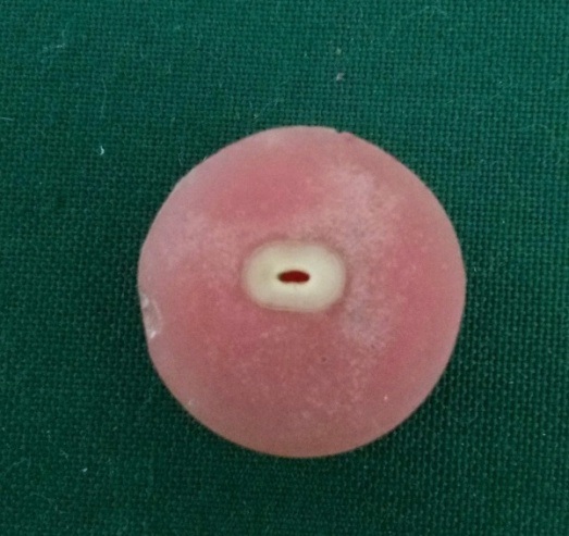
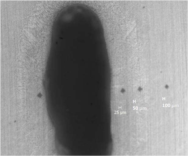
Each group was subdivided into two subgroups. Half of the samples from each group were tested for micro hardness and the remaining obturated for push-out bond strength testing. CPP-ACP (GC Tooth Mousse, GC CORPORATION, Tokyo, Japan) solution was prepared by diluting the paste to 10 times. The initial concentration of CPP-ACP was 10% w/v and after 10 times dilution its concentration was 1% w/v. Micro hardness was calculated after the final irrigation protocol. Teeth were mounted on an acrylic block and sectioned transversely at the coronal, middle and apical thirds [Table/Fig-1]. The specimens were polished using silicon carbide papers of decreasing abrasiveness and were cleaned with distilled water after polishing. Vicker’s micro hardness (High Wood Micro Vicker’s Hardness Tester, Advanced Metallurgical Laboratory, Bangalore, India) measurements were performed on each section at 25μm, 50μm and 100μm from the pulp-dentin interface [Table/Fig-2]. A 50g indentation load was applied for 15 sec. The mean value was calculated out of the three measurements for all the specimens.
Specimen prepared for micro hardness measurement.
Vicker’s micro hardness test measurements at 25, 50 and 100μm from the pulp-dentin interface.
Specimen preparation and push-out bond strength test: The methodology as described by Tuncel et al., was followed [22]. Following micro hardness measurement, the remaining half of the specimens from each group after final irrigation protocol were obturated with a #30 ProTaper master cone using a resin based sealer namely Real Seal SE (Sybron Endo, CA, USA) as per the manufacturer’s instructions. Following obturation, the root canals were sealed with composite resin. The specimens were stored at 37°C and 100% humidity for one week to allow complete setting of the test materials.
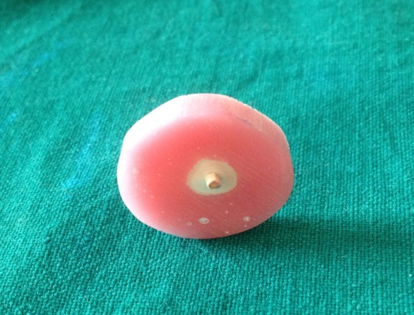
The obturated specimens were mounted in acrylic blocks and three 3mm thick horizontal sections were obtained from each specimen at coronal, middle and apical thirds using a water-cooled precision saw (Isomet, Buehler Ltd.). Following sectioning, the filling material was loaded with a stainless steel cylindrical plunger at a crosshead speed of 0.5mm/min in a Universal Testing Machine (CIPET, Hyderabad, India), which covered the filling material but not the surrounding dentin. The push-out force was applied from apical to coronal due to the convergence of the root. The force was recorded in Newtons (N) [Table/Fig-3].
Obturated specimen in acrylic blocks which has been tested for push-out bond strength causing the gutta percha to be pushed out of the canal.
Statistical Analysis
The statistical data was analysed in SPSS software version 17.0. The micro hardness data was evaluated using Kruskal Wallis with the significance level of p ≤ 0.05. The push-out bond strength data was analysed statistically using Mann Whitney U test.
Results
The Vicker’s micro hardness values have been mentioned in [Table/Fig-4]. The Push-out bond strength values have been mentioned in [Table/Fig-5].
Vicker’s micro hardness testing of three groups (VHN), p ≤ 0.05.
| Group | Mean | Standard Deviation |
|---|
| Normal Saline | 38.700 | 0.8563 |
| EDTA +NaOCl | 36.200 | 3.9595 |
| EDTA +NaOCl + CPP-ACP | 43.700 | 4.6679 |
Push-out bond strength test values of three groups (Newtons), p>0.597.
| Group | Mean | Standard Deviation |
|---|
| Normal Saline | 21.775430 | 3.2651284 |
| EDTA +NaOCl | 52.440230 | 7.8631783 |
| EDTA +NaOCl + CPP-ACP | 55.658880 | 5.5468701 |
Statistically significant difference was detected among the micro hardness of different groups by Kruskal Wallis test (p <0.05). The micro hardness of the CPP-ACP group was higher compared to the EDTA + NaOCl group.
There was no statistically significant difference detected among the push-out bond strength of CPP-ACP and EDTA + NaOCl groups by Mann Whitney test (p>0.597). Paired t-test showed no statistically significant difference between the two groups (p > 0.2662).
Discussion
Smear layer removal requires the use of chemical irrigants [1]. Complete elimination of smear layer is obtained with a combination of EDTA and NaOCl which removes inorganic as well as organic content [2,4]. The left over organic matrix of dentin mud when EDTA is used as the only irrigant hinders its penetration to the complete depth of the smear layer and results in incomplete opening of the dentinal tubules [15]. Subsequent use of NaOCl may help in the removal of the organic matrix [15]. Dentin deproteinization exposes the collagen fibres forming a porous structure with many irregularities around peri- and intertubular dentin with increased surface configuration [14]. 17% EDTA followed by 5.25% NaOCl has been used in the current study for complete removal of the smear layer which has been used by many authors [2,4,15,17,24]. But the use of these chemical irrigants leads to decreased micro hardness and weakens the root structure [18]. So to overcome these effects, root dentin remineralization can be carried out which improves its strength [18].
Decreased micro hardness of the root canal dentin was observed in the present study with the use of EDTA and NaOCl (Group 2) in compliance with many studies [15,24,25]. However, these studies did not study the importance of irrigation sequence [15]. Surface erosion of root dentin along with its dentinal tubules was seen with decreased micro hardness after final irrigation with EDTA followed by NaOCl [25]. The chelating property of EDTA shows reduced dentin micro hardness as its entire cationic receptors are saturated with Ca2+ ions of root dentin [11,13]. Many theories have been proposed to explain its chemical reaction towards the dentin. Crystalline field theory explains the electrostatic attraction force between the central metal and the ligands. The repulsive force of the ligands are suppressed by the greater attraction force of metallic ions resulting in formation of stable complex with calcium ions whereby carboxyl groups of the EDTA are ionized, hydrogen atoms are released, competing with the calcium ions [13]. Structural changes on the dentin surface along with modified properties have been noted [1,12]. Therefore, mineral loss or gain in root dentin, and its properties can be known by evaluation of micro hardness [1,2,26].
The softening of root canal dentin raises many questions regarding the prognosis of root canal treatment [1]. Root canal erosion leads to the alteration of its properties affecting its mutual interaction with filling materials and thus the coronal seal [13]. It may also cause vertical root fracture [11,15,27]. Properties like strength, hardness of dentin and adhesion of the sealer to the dentin are negatively affected and efforts should be made to minimize these aspects [12]. Studies on dentine remineralization can be focussed upon to reduce these disparities [18]. Increased numbers of studies have been focussed on the effect of CPP-ACP on enamel with limited information regarding its effect on dentin (Borges et al., 2012) [28]. But evaluating the effect of CPP-ACP on root dentin should also be studied as it has shown to affect its properties without any adverse effects. Increased potential for calcium-binding sites with CPP-ACP and thereby lesser calcium diffusion constant is observed in its mechanism of action not only towards the enamel but also dentin [28]. Prevention of dentin demineralization by CPP-ACP along with formation of protective layer for acid attacks has been studied in past [28]. In the current study, teeth treated with CPP-ACP (Group 3) as the final irrigant showed improved micro hardness of root dentin.
Bond strength of adhesives to enamel is more compared to dentin, so dentin mineralization or remineralization adds to its improved bonding [29]. CPP-ACP, treatment prior to usage of etch-and-rinse and self-etching adhesive systems can improve the bond strength of adhesive resins to enamel [29] but what about its effect on bond strength and stability to dentin? This is the first study which has evaluated the effect of CPP-ACP on root dentin. In the current study improved push-out bond strength was seen in CPP-ACP treated group but no statistically significant difference was observed between EDTA + NaOCl and EDTA + NaOCl + CPP-ACP groups. Lack of difference in push-out bond strength may be due to acid-base reaction between the hydroxyapatite deposited by the CPP-ACP paste and acidic monomers present in the self-etching monomers [29]. Elimination of washing and drying steps in self-etching adhesive systems makes the technique less sensitive which also maintains the optimum moisture of the root dentin and the residual hydroxyapatite [29]. CPP-ACP paste may be used before bonding procedures as promoting remineralization and reducing demineralization without affecting the bond strength [29].
The less sensitive vickers hardness test among the micro hardness measurement methods has been utilized in the current study [1,2]. Micro hardness has been evaluated at 25, 50 and 100μm as they are in close vicinity to the pulp up to which EDTA can penetrate. The push-out bond strength test is an efficient and reliable technique to assess bond strength of root canal filling materials to root dentin [30,31].
In contrary to the results of the present study, few authors speculate that CPP-ACP treated dentin, which is harder to condition and prime results in low bond strength [23]. According to Kamozaki et al., CPP-ACP paste has not affected the bond strength of etch and rinse and self-etch adhesives in acceptance with the current study [32,33].
Limitation
The present study has its limitations in being an in-vitro study and so it does not simulate the clinical conditions accurately in all aspects. The other limitation is that the micro hardness determination can give only an indirect evidence of mineral loss or gain from the tooth. As a future perspective of the study, the use of CPP-ACP as an irrigant, its clinical effects on all the properties of root dentin and its role in adhesion of obturating materials to CPP-ACP treated root dentin can be evaluated to get a better picture.
Conclusion
Within the limitations of the present study and literature, we can conclude use of CPP-ACP on root dentin is clinically significant because-
Improved micro hardness of erosive root dentin was observed.
No statistically significant difference between the push-out bond strength of EDTA + NaOCl and EDTA + NaOCL + CPP-ACP groups was noted.
CPP-ACP is not affecting the bond strength of resin sealer to root dentin.
[1]. Akcay I, Sen BH, The effect of surfactant addition to EDTA on microhardness of root dentinJ Endod 2012 38(5):704-07. [Google Scholar]
[2]. Mishra L, Kumar M, Rao CVS, Calcium loss from root canal dentin following EDTA and tetracycline HCL treatment with or without subsequent NaOCl irrigation and evaluation of microhardness of dentineInt J Advance Res Tech 2012 1(2):1-6. [Google Scholar]
[3]. Rossi-Fedele G, Doğramaci EJ, Guastalli AR, Steier L, Figueiredo JAPD, Antagonistic interactions between sodium hypochlorite, chlorhexidine, EDTA, and citric acidJ Endod 2012 38(4):426-31. [Google Scholar]
[4]. Görduysus M, Kücükkaya S, Bayramgil NP, Görduysus MO, Evaluation of the effects of two novel irrigants on intraradicular dentine erosion, debris and smear layer removalRestor Dent Endod 2015 40(3):216-22. [Google Scholar]
[5]. Violich DR, Chandler NP, The smear layer in endodontics – a reviewInt Endod J 2010 43(1):2-15. [Google Scholar]
[6]. Mancini M, Armellin E, Casaglia A, Cerroni L, Cianconi L, A comparative study of smear layer removal and erosion in apical intraradicular dentine with three irrigating solutions: A scanning electron microscopy evaluationJ Endod 2009 35(6):900-03. [Google Scholar]
[7]. Economides N, Liolios E, Kolokuris I, Beltes P, Long-term evaluation of the influence of smear layer removal on the sealing ability of different sealersJ Endod 1999 10(2):558-62. [Google Scholar]
[8]. Shahravan A, Haghdoost AA, Adl A, Rahimi H, Shadifar F, Effect of smear layer on sealing ability of canal obturation: a systematic review and meta-analysisJ Endod 2007 33(2):96-105. [Google Scholar]
[9]. Pèrez-Heredia M, Ferrer-Luque CM, Gonazalez-Rodriguez MP, Martin-Peinado FJ, Gonzalez-López S, Decalcifying effect of 15% EDTA, 15% citric acid, 5% phosphoric acid and 2.5% sodium hypochlorite on root canal dentineInt Endod J 2008 41(5):418-23. [Google Scholar]
[10]. Patil CR, Uppin V, Effect of endodontic irrigating solutions on the microhardness and roughness of root canal dentin: An in vitro studyIndian J Dent Res 2011 22(1):22-27. [Google Scholar]
[11]. Grande NM, Plotino G, Pomponi M, Somma F, Interaction between EDTA and sodium hypochlorite: A nuclear magnetic resonance analysisJ Endod 2006 32(5):460-64. [Google Scholar]
[12]. Turk T, Kaval ME, Şen BH, Evaluation of the smear layer removal and erosive capacity of EDTA, boric acid, citric acid and desy clean solutions: an in vitro studyBMC Oral Health 2015 3(15):105 [Google Scholar]
[13]. Cruz-Filho AM, Sousa-Neto MD, Savioli RN, Silva RG, Vansan LP, Pècora JD, Effect of chelating solutions on the microhardness of root canal lumen dentinJ Endod 2011 37(3):358-62. [Google Scholar]
[14]. Aranda-Gracia AJ, Kuga MC, Chavez-Andrade GM, Kaltzis-Sousa NG, Duarte MAH, Faria G, Effect of final irrigation protocols on microhardness and erosion of root canal dentinMicrosc Res Tech 2013 76(10):1079-83. [Google Scholar]
[15]. Qian W, Shen Y, Haapasalo M, Quantitative analysis of the effect of irrigant solution sequences on dentin erosionJ Endod 2011 37(10):1437-41. [Google Scholar]
[16]. Topçuoğlu HS, Tuncay Ö, Demirbuga S, Dinçer AN, Arslan H, The effect of different final irrigant activation techniques on the bond strength of an epoxy resin-based endodontic sealer: A preliminary studyJ Endod 2014 40(6):862-66. [Google Scholar]
[17]. Ghisi AC, Kopper PMP, Baldasso FER, Stürmer CP, Rossi-Fedele G, Steier L, Effect of superoxidized water and sodium hypochlorite, associated or not with EDTA, on organic and inorganic components of bovine root dentinJ Endod 2015 41(6):925-30. [Google Scholar]
[18]. Ulusoy ÖIA, Görgül G, Effects of different irrigation solutions on root dentine microhardness, smear layer removal and erosionAust Endod J 2013 39(2):66-72. [Google Scholar]
[19]. Torabinejad M, Cho Y, Khademi AA, Bakland LK, Shabahang S, The effect of various concentrations of sodium hypochlorite on the ability of MTAD to remove the smear layerJ Endod 2003 29(4):233-39. [Google Scholar]
[20]. Torabinejad M, Khademi AA, Babagoli J, Cho Y, Johnson WB, Bozhilov K, A new solution for the removal of the smear layerJ Endod 2003 29(3):170-75. [Google Scholar]
[21]. Machnick TK, Torabinejad M, Munoz CA, Shabahang S, Effect of MTAD on flexural strength and modulus of elasticity of dentinJ Endod 2003 29(11):747-50. [Google Scholar]
[22]. Adebayo OA, Burrow MF, Tyas MJ, Resin-dentine interfacial morphology following CPP-ACP treatmentJ Dent 2010 38(2):96-105. [Google Scholar]
[23]. Adebayo OA, Burrow MF, Tyas MJ, Dentine bonding after CPP-ACP paste treatment with and without conditioningJ Dent 2008 36(12):1013-24. [Google Scholar]
[24]. Sayin TC, Serper A, Cehreli ZC, Kalayci S, Calcium loss from root canal dentin following EDTA, EGTA, EDTAC, and tetracycline – HCl treatment with or without subsequent NaOCl irrigationJ Endod 2007 33(5):581-84. [Google Scholar]
[25]. Niu W, Yoshioka T, Kobayashi C, Suda H, A scanning electron microscopic study of dentinal erosion by final irrigation with EDTA and NaOCl solutionsInt Endod J 2002 35(11):934-39. [Google Scholar]
[26]. Aslantas EE, Buzoglu HD, Altundasar E, Serper A, Effect of EDTA, sodium hypochlorite, and chlorhexidine gluconate with or without surface modifiers on dentin microhardnessJ Endod 2014 40(6):876-79. [Google Scholar]
[27]. Sen BH, Ertürk O, Pișkin B, The effect of different concentrations of EDTA on instrumented root canal wallsOral surg Oral Med Oral Pathol Oral Radiol Endod 2009 108(4):622-27. [Google Scholar]
[28]. Poggio C, Lombardini M, Vigorelli P, Ceci M, Analysis of dentin/enamel remineralization by a CPP-ACP paste: AFM and SEM studyScanning 2013 35(6):366-74. [Google Scholar]
[29]. Barreto BDCF, Catelan A, Alexio MP, Silva GR, Xavier TA, Aguiar FHB, Effect of CPP-ACP on the bond strength of silorane and methacrylate based restorative systemsJ Res Dent (Tubarao) 2013 1(1):64-71. [Google Scholar]
[30]. Mozayeni MA, Zadeh YM, Paymanpour P, Ashraf H, Mozayani M, Evaluation of push-out bond strength of AH26 sealer using MTAD and combination of NaOCl and EDTA as final irrigationDent Res J (Isfahan) 2013 10(3):359-63. [Google Scholar]
[31]. Goracci C, Tavares AU, Fabianelli A, Monticelli F, Raffaelli O, Cardoso PC, The adhesion between fiber posts and root canal walls: comparison between microtensile and push-out bond strength measurementsEur J Oral Sci 2004 112(4):353-61. [Google Scholar]
[32]. Kamozaki MBB, Prakki A, Perote LCCC, Pagani NCGC, The effect of CPP-ACP and Nd: YAG laser on the bond strength of softened dentinBraz Oral Res 2015 29(1):1-7. [Google Scholar]
[33]. Shadman N, Ebrahimi SF, Shoul MA, Sattari H, In vitro evaluation of casein phosphopeptide-amorphous calcium phosphate effect on the shear bond strength of dental adhesives to enamelDent Res J (Isfahan) 2015 12(2):167-72. [Google Scholar]