The primary functions of the root canal obturation are sealing the in-growth of the bacteria’s from outside the canal, entombment of residual bacteria and, thorough obturation at a microscopic level to avoid stagnant fluid from collecting and aiding as nutrient for micro-organisms from any source [1]. The most common orthograde obturating method world-wide is gutta-percha. As, gutta-percha by itself cannot obturate the complete root canal system, owing to its poor sealing properties hence a sealer is used in conjunction with gutta-percha [2]. All the contemporary obturating techniques make use of the sealer to enhance the seal of the root canal filling [3]. In current method the most frequently used method in canal obturation employ a semisolid, solid or a rigid core material cemented in the canal with a root canal sealer used as a binding agent. Sealer accomplishes the objective of providing fluid tight seal. The core occupies space serving as a vehicle for sealer. Before setting, the sealer can be made to flow and fill the accessory canal and multiple apical foramina. Many root canal sealers have been developed for use and can be classified according to chemical composition that is eugenol based, calcium hydroxide based or resin based.
The aim of the present study was to evaluate the effectiveness of the apical seal obtained by using Zinc oxide eugenol sealer, Apexit, AH Plus, and RSA when used in conjugation with cold lateral condensation of obturation using gutta-percha.
Materials and Methods
The in vitro stereomicroscopic study was done to evaluate effectiveness of sealing property of different root canal sealers and conducted in the Department of Conservative Dentistry and Endodontics, Sri Aurobindo College of Dentistry (SAIMS), Indore, M.P., India for a period of one year. Prior to conducting the study the ethical clearance was obtained from the institutional review board. A total of 100 single rooted extracted human teeth with a single root canal were selected.
Inclusion Criteria - Single-rooted extracted human permanent teeth with a single root canal were included in this study.
Exclusion Criteria - Preoperative radiographs were taken and radiographs were screened and teeth were excluded if any of the following were noted:
If the curvatures was greater than 5 degrees,
If a root fracture was evident,
If the apex was incompletely formed or larger than a #25 K-type file,
If any bifurcating canals, fins, ribbon-shaped canal, or extreme calcifications could be seen.
The teeth were stored in 1% sodium hypochlorite (NaOCl) solution, for three days to remove organic debris and then they were stored in distilled water. The crowns were removed at the cement-enamel junction using a high speed fissure bur. Access preparation was done using an endo-access bur (Dentsply Maillefer, USA) and a barbed broach (Dentaire, SA) was used to remove the pulp. Then, a no.10 - K-file (Mani, Japan) was introduced into the canal and was pushed towards apical part until the tip of the instrument was just visible at the apical foramen. This length of the file was recorded and then after subtracting 1mm from the recorded length, working length of the root canal was determined.
The canal were cleaned and shaped with K-files (Mani, Japan) using a step-back technique with recapitulation of files to establish a progressively tapering root canal preparation. The apical portion of the canal was enlarged to a minimum 30 no. K-file and 50 no. K-file, depending on the size of the original canal. The coronal two thirds of each canal were prepared using number 2 and 3 Gates Glidden drills (Mani, Japan) and apical third were prepared with hand files. After each instrument was used, the canals were irrigated with 2ml of 5% NaOCl and 2ml of 15% solution of EDTA (Dental Source, North Hollywood CA, USA). The irrigating solutions were delivered through a 25-gauge needle which was placed as far as possible into the canal without allowing the needle to touch the canal walls. The total amount of irrigant used in each canal was 30 ml, on completion of the instrumentation process, a 10 no. K-file (Mani, Japan) was passed 1mm through the apical foramen to remove any dentinal plugs and to ensure that the foramen was patent for dye penetration.
After drying the canals with paper points, standardized gutta-percha cones (Dentsply, China) were selected as master points. The fit of each master point was assessed by radiographs to determine whether the point was fully seated to the working length. The teeth were randomly selected and divided into five groups of 20 teeth each (four experimental group and one control group). The sealers used were as follows-
Group I – Conventional Zinc-Oxide Eugenol.
Group II – AH Plus (Resin based)
Group III – Apexit (Calcium Hydroxide based)
Group IV – RSA (Silicon based)
Group V – Control Group- Gutta-Percha alone (no sealer).
All the teeth except the controlled teeth were filled with a root canal sealer and gutta-percha points using the cold lateral condensation technique. In the control group, sealer was not used.
The sealer were mixed according to the manufacturer’s directions and were introduced into the canal using a lentulo-spiral (Mani, Paste carriers, Japan) which was kept 3mm to 4mm short of the working length. This process was repeated twice to ensure that an adequate amount of sealer was placed in each canal. The master gutta-percha point (Dentsply, China) was coated with sealer and placed in the canal to the full working length.
Hand spreader (Dentsply, China) was then used for lateral condensation with standardized fine gutta-percha accessory points (Dentsply, China) was carried out until the entire canal was obturated. Excess gutta-percha was removed and the gutta-percha in the coronal third of the canal was vertically condensed with a plugger. Radiographs were taken to evaluate the obturation. Obturation was considered to be optimum when no voids were present in the radiograph. If the voids appeared in the radiograph, re-obturation was done.
The access cavities were sealed with Cavit G (3M ESPE, Germany) up-to 2 mm and the teeth were placed in a Humidifier (ICU Safe, Sanyo, Japan) for 1 to 3 weeks with 100% humidity at 37°C to ensure that the sealer set in an environment that simulate the clinical situation in which they are designed to be used.
The roots were coated with two layers of clear nail varnish (Lakme, New York) except for the apical 2 to 3 mm. At this stage the control group was further sub-divided into two equal groups, the positive and negative controls. Teeth in the positive control group had the roots coated with nail varnish (Lakme, New York) except for the apical 2mm to 3mm in the same manner as the experimental groups. They were used to test the sealing ability of gutta-percha when used without a sealer. Teeth in the negative control group had the entire root surface coated with nail varnish and were used to test the ability of the nail varnish to seal the root against dye penetration under the experimental conditions used in this study.
Once the nail varnish was absolutely dry, each sample was introduced in a 12-ml centrifuge tube with the apex of the root positioned in the direction of the open end of the centrifugal tube. Methylene blue dye solution 2% (pH=7) was poured into the each tube until the root was completely immersed into the solution. The samples were then centrifuged for 3 minutes at 30Xg using a centrifugal machine. The samples were taken out from the solution and were thoroughly bathed in running tap water.
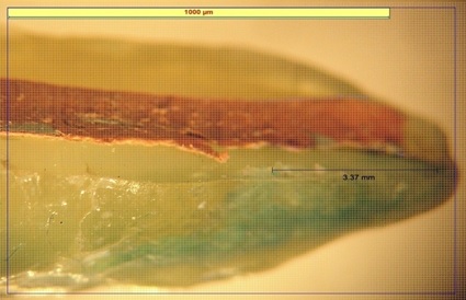
The experimental samples were sectioned longitudinally by means of a low-speed circular diamond saw (Confident, India) in a path roughly parallel to the axis of the tooth and through the apex with a coolant. After sectioning, the samples were studied under a stereomicroscope (32X Magnification, Carl Ziess). The end point of dye infilteration was calculated as the point where dye no longer penetrated the obturating material. The measurement from the apex to the end point of dye penetration was observed and documented in millimeters [Table/Fig-1].
The specimens were examined under a stereomicroscope (X32 magnification, Carl Ziess) and the end point of dye penetration was determined as the point where dye no longer penetrated the filling material. The distance from the apex to the end point of dye penetration was measured and recorded in millimeters.
Statistical Analysis
For the analysis of data Statistical Package for Social Science (SPSS) version 19.0 was used. Snedocor’s F test for the quality of variances among the experimental group and control group (One-Way ANOVA) were employed.
Results
Descriptive statistics of mean dye penetration values (in mm) for each group showed that dye penetration is maximum in Group 5 and minimum in Group 4 [Table/Fig-2]. Snedocor’s F test for the equality of variances among the experimental group and control group (One-way ANOVA) which is Significant (p<0.01) [Table/Fig-3].
Descriptive statistics of mean dye penetration values (in mm) for each group.
| Groups | Sealer | No. of specimens | Mean dye penetration (in mm) | SD | SEM |
|---|
| 1 | Zinc Oxide Eugenol | 20 | 4.49 | 0.779 | 0.174 |
| 2 | AH- Plus | 20 | 1.411 | 0.505 | 0.112 |
| 3 | Apexit | 20 | 2.47 | 0.812 | 0.181 |
| 4 | RSA | 20 | 0.543 | 0.357 | 0.070 |
| 5 | Control | | | | |
| No varnish | 10 | 0.86 | 0.943 | 0.211 |
| Varnish | 10 | 0 | - | - |
Snedocor’s F test for the quality of variances among the experimental group and control group (One-Way ANOVA).
| Groups | Sealer | No. of specimens | Mean dye penetration (in mm) | SD |
|---|
| Between groups | 129.21 | 4 | 32.30 | F=9.98 |
| Within groups | 275.06 | 85 | 3.236 | Significant |
| Total | 404.28 | 89 | | p<0.01 |
Degrees of freedom - 4,85; F tabulated value = 3.48; Level of significance α=0.01
Discussion
The entire root canal system should be filled three dimensionally following thorough cleaning and shaping of the root canal space to ensure long term clinical success. The concept of a perfect apical seal has led to search for filling and sealing materials that are stable, non-irritating and provide a flawless seal at the apical foramen. The selection of sealers is dependent on its capacity to create a comprehensive seal but it must also be well accepted by peri-radicular tissues and be comparatively easy to manipulate so that its optimum physical and biological properties can be clinically achieved. In principle the core material should push the less viscous into unreachable areas such as canal anastomosis, apical delta and into irregularities produced through canal preparation [4]. The sealing ability of various root canal filling materials and root canal sealers have been studied and it has been found that dissimilar constituents seal the canal to different extents [5]. Principle aim of canal cleaning and shaping is to eliminate all pulp material remnants and bacteria along with their substrates and optimum shaping of the root canal space [3]. The current method used in canal obturation employ a semisolid, solid or a rigid core material cemented in the canal with a root canal sealer that acts as [2]:
Apical sealing agent.
A binding agent to the well adapted master cone into a canal.
Filler for the intricacies and minor disagreements between the cones and canal space.
A lubricant to enable the placement of the gutta-percha into the canal.
Hence, this study was carried out to evaluate the sealing effectiveness of different root canal sealers by calculating the apical dye leakage.
The leakage was minimum with RSA sealer and maximum with conventional zinc oxide-eugenol sealer. RSA sealer has the better sealing ability than the other sealers used in this study when used in conjugation with cold lateral condensation using gutta-percha.
The negative control groups showed no leakage at all, indicating those two coats of nail varnish were efficient enough in preventing dye preparation. The conventional zinc oxide eugenol based specimens had the most dye penetration among all the four groups studied. The setting mechanisms of zinc oxide eugenol based cements is the outcome of equimolar mixtures of zinc oxide and eugenol, consisting of zinc oxide involved in a extended crystal matrix of zinc eugenolate chelate and is degenerated by the carbon dioxide which is present as bi-carbonate ions in the peri-radicular area and makes zinc-oxide eugenol a feeble and unbalanced cement [6]. The second possible reason would be attributed to the dimensional changes of the material upon setting. This is in concurrence with the findings of earlier study which reported a setting shrinkage of 0.3% to 1% with zinc oxide eugenol based sealers [7]. The leakage values obtained with AH-Plus group were lesser than conventional zinc-oxide eugenol and Apexit. The reason could be attributed to the slight expansion that might occur tending to wedge the sealer more tightly into the prepared root canal, thereby improving the mechanical interlocking [8]. The specimens of Apexit group had significantly lesser dye penetration. The main reason of increased leakage observed with Apexit and conventional zinc oxide eugenol would be possible due to dissolution over the time of immersion [9,10]. The reason for less amount of leakage with RSA could be attributed to the fact that it is insoluble. The second possible reason would be due to the slight expansion that might occur tending to wedge the sealer more tightly into the prepared root canal, thereby improving the mechanical interlocking. A study reported an expansion of RSA to be 0.2% [7]. In the AH Plus, and RSA groups, the sealer appeared to form a uniform structure with the gutta-percha. Under the experimental conditions RSA had a significantly better apical seal than conventional Zinc oxide eugenol, Apexit and AH plus.
Limitation
This study has the limitation that it used the classical dye-penetration method. It is recommended that future studies using dye-extraction, i.e., dissolution method and fluid filtration method on a larger sample and in vivo analysis should be performed to confirm sealing ability of newer endodontic sealers.
Conclusion
In vitro leakage studies comprise a major portion of contemporary endodontic research, yet it is difficult to draw in vivo correlation. The result of these studies should be regarded as showing a theoretical maximum amount of leakage, which may or may not occur, in-vivo, and as such, they are probably good indicators and potential for consideration of success or failure of the treatment.
Degrees of freedom - 4,85; F tabulated value = 3.48; Level of significance α=0.01