Composite resins are now-a-days one of the most commonly used materials for direct restorations and bonding brackets to the teeth [1]. With the advent of the acid-etch technique by Buonocore in 1955, its usage has increased by leaps and bounds. Over the past few years, its clinical use has expanded considerably due to the increased aesthetic demand by patients, new advances in inventions and popularization of bonding procedures [2].
The concept of direct bonding of brackets onto the teeth has revolutionized the field of orthodontics. The advantages of direct bonding include patient comfort, ease and accuracy of placement. However, composite comes with its set of disadvantages. Bonding, debonding and clean-up procedures may result in enamel alterations such as roughening due to etching, micro-cracks and enamel fractures caused by forcibly removing brackets, scratches and abrasions due to mechanical removal of the remaining composite materials [2]. The principal concern lies in re-establishing the enamel surface to as near its original state as possible following the removal of the bonded orthodontic attachments [3].
Residual composite removal on the enamel surface after debonding has been attempted in many ways. Zachrisson and Årtun used tungsten carbide burs at low speed for adhesive removal [4]. Campbell preferred to use carbide burs at high speed followed by enhance rubber points and cups, water slurry of fine pumice and finally brown and green cups at low speeds [3]. Özer and his colleagues used Sof-Lex discs and fibreglass burs for the removal of adhesive remnant [5]. There has been investigation of alternative methods such as ultrasonic applications and air abrasion techniques with aluminium oxide particles for removal of adhesive remnants [6,7].
Reflectivity of an object is a good parameter for surface finish. As the patient evaluates finishing as a function of gloss/reflectivity/shine, an attempt is made here to evaluate changes in surface finish with custom made reflectometer. The aim of the present study was to study the effect of various procedures during orthodontic treatment on the shine of enamel, using a custom made reflectometer. The instrument was designed to quantify and compare reflectivity (gloss or shine) of tooth surface after various treatment procedures.
Materials and Methods
The present study was conducted in Department of Orthodontics, ACPM Dental College and Hospital Dhule, Maharashtra, India, during period September-December 2015. In this in-vitro study freshly extracted 61 maxillary premolars for orthodontic reasons were used. Teeth were stored in distilled water and the water was changed weekly to prevent bacterial growth for a period of one month. Teeth were selected on the basis of visual observation of the solidity of labial surfaces, that is, no caries, no exposure to chemicals, no cracks and no extraction forcep marks. Sixty one teeth were embedded vertically in self-cure acrylic resin so that only crown part was exposed.
All the teeth were polished with pumice paste and water slurry to remove any residual plaque or stains by using a contra-angle micromotor handpiece and a polishing brush. One tooth was kept as standard. Standard tooth was not subjected to any procedure. Reflectivity of all the teeth before any procedure was compared with the standard tooth on the custom made reflectometer. Statistically no significant difference was found amongst all sample on analysing by single sample t-test.
The teeth were etched for 30 seconds using 37% phosphoric acid gel. Readings were taken to measure the reflectivity of enamel after acid etching on custom made reflectometer. After measuring the reflectivity of enamel after acid etching, a light cure adhesive composite (Transbond XT) was placed on the bracket bases, the brackets (3M Unitek) were bonded to the prepared enamel, excess adhesive was removed, and resin was light cured for 30 seconds. The samples were kept in a water bath for 24 hours to allow for residual polymerization of resin. All the teeth were debonded using a debonding plier and then readings were taken after this procedure to measure the reflectivity of the teeth after debonding.
Then 60 premolars were randomly divided into three groups each containing 20 teeth.
Group I: Tungsten carbide bur: Finishing and polishing was done by these burs.
Group II: Astropol: It involved a three-step system of tips of various grits ranging from finishing, polishing and high gloss polishing.
Group III: Sof-Lex discs: Finishing and polishing was done with four grits in sequential order from coarse, medium, fine and superfine.
Finishing was done using a contra-angle micro-motor handpiece according to the manufacturer’s specifications and then readings were taken after this procedure to measure the reflectivity of the teeth.
Working Principle of Reflectometer: The reflectometer was designed on principle of reflectivity of an object. The intensity of light reflected from the tooth was matched against the standard by varying former’s distance from the light source. This custom made reflectometer consisted of a single light source, partition at the centre and two scales one on each side to measure the distance of an object from light source and semi-transparent viewer for comparison of reflectivity of the object. Schematic diagram of custom made reflectometer is shown in [Table/Fig-1].
Schematic diagram of custom made reflectometer.
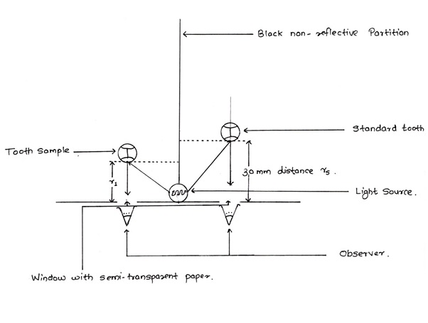
All the inner surfaces of reflectometer were black and rough i.e., non-reflective. Intensity of the light on both the sides was observed simultaneously. This gives the most accurate evaluation.
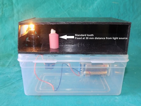
On one side the untreated natural tooth was fixed at distance of 30mm, from the light source, which was used as standard. The teeth that were evaluated were moved to and fro from the light source to match the light intensity on the observation window until the luminance i.e., intensity of light matches with the control.
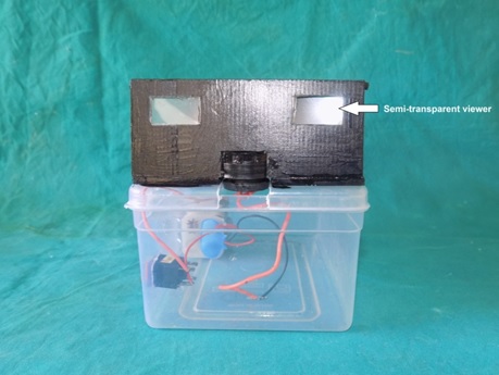
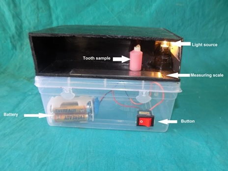
At this point, the distance of the sample tooth from light source was measured with the scale. The readings were taken in a dark room. The front view, lateral view and contra-lateral view of custom made reflectometer is shown in [Table/Fig-2,3 and 4] respectively.
Front view of custom made reflectometer.
Lateral view of custom made reflectometer.
Contra-lateral view of custom made reflectometer.
Percentage of reflectivity derived from following formula [9]:
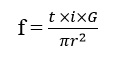
Where,
t = transparency of medium
i = intensity of light source
G = Gloss of the tooth
r = distance of tooth from light source
As t and i are constant in our study and π is a universal constant.  which is a constant
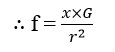
As in our study fs =f1
Gs = Gloss/Reflectivity of standard tooth which is 100% or 1 for the study
r1= distance of teeth in experiment from light source
Change in r is,
Gs-G= ∂ G (change of reflectivity).
Statistical Analysis
Data was analysed using SPSS v 16 (Statistical Package for Social Sciences Version 16). Data was tabulated and present as bar and line graphs. Descriptive statistics for percentage reflection and reflection loss of teeth was analysed and mean was compared in between the three finishing groups using ANOVA test. Inter group difference of percentage reflection and loss was analysed using Post hoc Bonferroni test. A p-value of <0.05 was considered as statistically significant.
Results
The result showed mean percentage reflection after acid etching was 31.4 % ± 2.6% standard deviation. After debonding mean reflection was 45.5% ± 3.5% [Table/Fig-5]. Due to acid etching mean reflection loss was 68.6% ± 2.6% standard deviation. Debonding caused mean reflection loss of 54.5% ± 3.5% standard deviation.
The mean percentage of reflectivity and loss after acid etching and debonding procedures.
| Sample Size (n) | Minimum | Maximum | Mean | S.D. |
|---|
| Percentage reflectivity after acid etching | 60 | 28.4% | 36.0% | 31.4% | 2.6% |
| Percentage reflectivity after debonding | 60 | 40.1% | 53.8% | 45.5% | 3.5% |
| Percentage reflectivity loss by acid etching | 60 | 64% | 71.6% | 68.6% | 2.6% |
| Percentage reflectivity loss by debonding | 60 | 46.2% | 59.9% | 54.5% | 3.5% |
[Table/Fig-6] shows percentage reflection after finishing in three groups. In Group 1, tungsten carbide finishing was able to restore the reflectivity of teeth within 53.8% to 64%, Teeth had mean reflectivity restored to 58.3% ± 3.36% standard deviation. Group 2, astropol finishing was able to restore reflectivity of teeth within 69.4% to 75.1%, Teeth had mean reflectivity restored to 72.8% ± 2.36% standard deviation. In Group 3, Sof-Lex disc finishing was able to restore reflectivity of teeth within 81% to 87.1%, teeth had mean reflectivity restored to 84.4% with 2.61% standard deviation. There was statistically highly significant difference (p<0.001) in reflectivity restored by the three finishing materials in the study.
Percentage reflection after finishing in three groups.
| Reflection | Group 1Tungsten Carbide bur (n=20) | Group 2Astropol(n=20) | Group 3Sof-Lex disc(n=20) |
|---|
| Minimum | 53.8% | 69.4% | 81.0% |
| Maximum | 64% | 75.1% | 87.1% |
| Mean | 58.3% | 72.8% | 84.4% |
| Std. Deviation | 3.36% | 2.36% | 2.61% |
| ANOVA | F value | 430.688 |
| p-value | <0.001 |
To find out the difference between two groups pair wise comparison was done by Post Hoc test as shown in [Table/Fig-7]. Mean reflectivity restored in Group 2 was 14.5% more than Group 1. Group 3 teeth had 26% more reflectivity restored after finishing than that of Group 1 teeth. Reflectivity of teeth in Group 3 was 11.5% more than that of Group 2. These difference are statistically highly significant (p<0.001). Therefore the mean reflectivity restoration Group 3 > Group 2>Group1. Thus the light reflection was better in Group 3> Group 2> Group 1.
Paired comparison of percentage reflection in groups after finishing.
| Multiple Comparisons: Bonferroni |
|---|
| Dependent Variable | (a) Group | (b) Group | Mean Difference (a-b) | p-value |
|---|
| Percent Reflection | Group 2 | Group 1 | 14.5 | <0.001 |
| Group 3 | Group 1 | 26.0 | <0.001 |
| Group 3 | Group 2 | 11.5 | <0.001 |
[Table/Fig-8] shows percentage reflectivity loss after finishing in three groups. In Group 1 of Tungsten Carbide bur finishing teeth had mean reflectivity loss of 41.7% and within the range of 36% to 46.2%. In Group 2, Astropol finishing teeth had mean reflectivity loss of 27.2% and within range of 24.9% to 30.6%. Group 3 Sof-Lex disc finishing teeth had mean reflectivity loss of 15.6% within range of 12.9% to 19%. There was statistically highly significant (p<0.001) difference of mean reflection loss in between the three finishing groups. Mean reflection loss of Group 1 > Group 2 > Group 3.
Percentage loss of reflection in three groups after finishing.
| Reflection Loss | Group 1Tungsten Carbide bur (n=20) | Group 2Astropol(n=20) | Group 3Sof-Lex disc(n=20) |
|---|
| Minimum | 36.0% | 24.9% | 12.9% |
| Maximum | 46.2% | 30.6% | 19.0% |
| Mean | 41.7% | 27.2% | 15.6% |
| Std. Deviation | 3.4% | 2.4% | 2.6% |
| ANOVA | F value | 430.688 |
| p-value | <0.001 |
[Table/Fig-9] Shows paired comparison of percentage reflection loss in groups after finishing. In Group 1, Tungsten Carbide finishing had 14.52% more reflectivity loss than Group 2 teeth having Astropol finishing. Group 1 finishing teeth had 26.04% more reflectivity loss than Group 3 finishing teeth. In Group 2 by Astropol finishing the loss was 11.52% more than in Group 3 i.e., Sof-Lex disc finishing teeth group. This difference of percent reflectivity loss of the teeth was statistically very highly significant (p<0.001). Thus reflectivity loss of Group 1 was more than Group 2 which was higher than Group 3. Group 1 had maximum and Group 3 had minimum reflectivity loss after finishing.
Paired comparison of percentage reflectivity loss in groups after finishing.
| Multiple Comparisons: Bonferroni |
|---|
| Dependent Variable | (a) Group | (b) Group | Mean Difference (a-b) | p-value |
|---|
| Reflectivity loss | Group 1 | Group 2 | 14.52% | <0.001 |
| Group 1 | Group 3 | 26.04% | <0.001 |
| Group 2 | Group 3 | 11.52% | <0.001 |
Discussion
Bonding of the brackets to enamel surface is a common practice in orthodontics. With the introduction of phosphoric acid in dentistry for etching enamel, it became possible to achieve a strong bond of composite to enamel. In 1964, Newman first used this pre-treatment technique for the bonding of orthodontic brackets [10]. As the bonding of brackets is a routine procedure in orthodontic practice, considerable researches had been devoted to bonding techniques and the removal of adhesive remnants from enamel surface. For an optimal orthodontic bonding system, the bond strength must be high enough to prevent bond failure. In addition, it should be possible to remove the bracket in such a way that it should cause minimal damage to the teeth. The influence on the enamel surface after orthodontic treatment is inevitable. Regardless of the method used, some scarring of the enamel occurs after bracket debonding and removal of resin remnants [11].
After debonding of brackets, the primary concern is to return the enamel surface as closely as possible to its original state, numerous techniques are offered to remove the adhesive remaining on the enamel surface after debonding [3]. These include most common methods like ultrasonic scalers, a low-speed handpiece with a tungsten carbide bur, a high- speed handpiece with a diamond bur, etc [3,6,11]. The quest for the best method has resulted in the introduction of lasers such as, carbon-dioxide and Nd: YAG laser radiation application for selective removal of residues of bonding resins [12,13]. There has been a surge in the development of new instruments, such as specially designed burs, discs and diamond or silicone coated polishers which are thought to be less aggressive [14].
Various methods have been used to relate enamel surfaces after remnant removal: visual inspection by photography, scanning electron microscopy and adhesive remnant index, etc [15,16]. The other methods that can be used for assessment of the surface roughness and enamel loss include 3-dimensional laser scanning [17,18], 3-dimensional surface profilometry [7] and atomic force microscopy etc., [19,20]. In clinical practice, the surface luster is usually judged without magnification. Though most of the time smoothness is correlated with the luster, but in cases such as resin based composite restorations, the smoothest surface does not necessarily provide the most lustrous surface.
For industrial applications, reflectometers are used to measure the luster. However, it is difficult to use them successfully for dental applications because of the irregular contour and small size of dental restorations [21]. Thus, a custom made reflectometer was designed to be used on tooth surface.
Reflectivity of an object is a good parameter for surface finish. In the literature search, the reflectivity parameter for the teeth was never studied before. Reflectivity is demarcated as the capability of a surface to reflect incident light [9].
There are two types of reflection:
A. Specular reflection (mirror like, regular reflection).
B. Diffused reflection (irregular reflection).
If the reflecting surface is very smooth and flat, the reflection of light that occurs is called specular or regular reflection.
When light raids the surface of a material it rebounds off in all directions due to reflections in multiple directions by the microscopic irregularities on the surface of the material (e.g., the grain boundaries of a polycrystalline material, or the cell or fiber boundaries of an organic material) and by its surface itself, if it is rough it is called as diffused or irregular reflection.
As the patient evaluates finishing as a function of gloss/reflectivity/shine, an attempt was made to evaluate changes in surface finish with custom made reflectometer. Hence, this study was formulated with the objective to compare the reflectivity of the teeth before and after finishing with various methods, taking untreated tooth as the standard for reflectivity.
In the present study, widely accepted finishing methods were used. In the earlier work, tungsten carbide bur clean-up was considered the gold standard for residual resin removal against which other methods could be compared [4,8]. In our study in Group 1 tungsten carbide bur was used at low speed because studies by Zachrisson and Arthun, [4] Hannah and Smith [22] found that tungsten carbide bur at low speed was most effective in removal of residual resin.
In Group 2 Astropol was used. It is a three-step polishing system for finishing and polishing namely finishing (F-Grey, 40 μm), polishing (P-Green, 20-40 μm) and high gloss (HP-pink, 10 μm) polishing [23,24]. It is available in the following four shapes: small flame, large flame, cup and disk. Astropol F and Astropol P consist of silicone rubber, silicon dioxide particles and colour pigments. Astropol HP contains silicon rubber, diamond particles, aluminium oxide, titanium oxide and iron oxide. The shanks are made of stainless steel.
In Group 3 Sof-Lex disc was used. The Sof-Lex finishing and polishing discs are made from a urethane coated paper that gives the discs their flexibility. This multi-step system is comprised of four individual aluminium oxide grits ranging from coarse, medium, fine and superfine. This sequence finishes and then polishes the surface. The coarse abrasive disc is coated with 100μm aluminium oxide particles, the medium grit disc with 40μm aluminium oxide particles, the fine grit with 24μm, and extra–fine grit with approximately 8μm [25]. The discs have a square brass eyelet to which the mandrel attaches. It also facilitates easy removal of the discs from the mandrel.
The present study was conducted to quantify and compare reflectivity (gloss or shine) of tooth surfaces treated with different cleanup protocols in which tungsten carbide bur, Astropol, and Sof-Lex disc using custom made reflectometer. The mean percentage of reflectivity after acid etching was 31.4%, debonding 45.5%, Tungsten carbide bur finishing (Group 1) was 58.8%, Astropol (Group 2) 72.8%, and SofLex disc (Group 3) 84.4% as that to the standard. There was statistically very highly significant (p<0.001) difference in reflectivity restored by the three finishing materials in the study. Thus, the light reflection was better in Group 3> Group 2> Group 1.
In this study the mean reflectivity loss in Tungsten Carbide bur (Group 1) finishing was 41.7%, Astropol (Group 2) was 27.2% and Sof-Lex (Group 3) was 15.6%. There was statistically very highly significant (p<0.001) difference of mean reflection loss in between the three finishing groups. Mean reflection loss of Group 1 > Group 2 > Group 3. The findings of the present study suggest that reflectivity of teeth do not reach to the pre-treatment level. Therefore, the techniques used in this study left enamel relatively dull as compared to its original state. This was consistent finding of previous studies [3,5,8,26,27].
According to Özer T et al., no cleanup procedure restored the enamel to its original smoothness [5]. The most successful was Sof-Lex disks, which restored the enamel closer to its original smoothness. In present study we took reflectivity as parameter and we observed the similar results. Sof-Lex disc finish was closest to the enamel original reflectivity.
Karan S et al., assessed difference between the effects of two burs on the surface roughness of enamel after orthodontic debonding [19]. In half of samples, adhesive remnants were removed with a tungsten carbide bur, whereas a fiber-reinforced composite bur stainbuster was used in the other half. The atomic force microscopy measurements were made after removal of resin. The results indicated that the composite bur used for resin removal produces smoother surfaces after orthodontic bonding as related to the carbide bur which increased enamel roughness. In present study tungsten carbide burs provided the least reflectivity amongst all group.
Mahdavie NN et al., evaluated the enamel scarring by debonding burs using an SEM and profilometer and concluded that the use of a less expensive, more durable bur such as white stone might seem economically advantageous, it could come at the price of inflicting more damage to the enamel [28]. That none of the enamel surfaces in study were restored to their original condition after removal of residual adhesive suggests the need for safer debonding burs.
Thus, none of the finishing and polishing systems restore the natural enamel reflectivity completely. Sof-Lex produced the best finish with Astropol coming a close second. Tungsten carbide burs produced the least reflectivity. This was in accordance with previous studies [5,14,24,29–33].
Smaller particle offer smoother and shinier surfaces. The polishing speed attains a luster, which however depends on the hardness and size of the abrasive particles and the method of abrasion. At the end of this process, there should be no visible scratches. The enamel is composed of enamel rods or prisms, rod sheaths, and in some regions a cementing interprismatic substance. The diameter of the enamel rods averages 4μm [34]. The particle thickness of aluminium oxide grits of extra fine grit with approximately 8μm. which is much bigger than the size of enamel rod. As Al2O3 (aluminium oxide) is very hard substance, scratching of enamel occurs. Finishing with larger diameter, hard particles leave a rougher surface. More is the surface rough lesser will be the reflectivity.
The removal of residual resin with rotary instruments at low speed produces more vibration and generates discomfort for patients [35]. Another disadvantage of removal of residual resin with rotary instruments is the generation of aerosols and heat. It is stated that the potentially hazardous action of adhesive particulate aerosol formed by grinding, composite resin particulates may turn as endocrinological disruptors [36].
Thus according to this study, despite of our longstanding efforts, we are unable to restore natural reflectivity of tooth enamel completely. This develops challenge to emphasize on creating newer biomaterial, techniques and their application to overcome this orthodontic scar to maintain health and integrity of normal tooth. Advantages and disadvantages of custom made reflectometer are mentioned in [Table/Fig-10].
Advantages and disadvantages of custom made reflectometer.
| S.No | Advantages | Disadvantages |
|---|
| 1 | It is very economical and very efficient tool. | Method is very subjective may differ from person to person. |
| 2 | Reflectometer is very easy for fabrication and less armamentarium required. | Dark room is needed. |
| 3 | | Time consuming method. |
Limitation
The bracket debonding technique which was employed for this study was manual. Therefore, there might be scope for error with respect to the amount remnants remaining on the enamel surface. This might be a reason for getting varied results for the composite removing. Finishing and polishing system increase the intra-pulpal temperature. However, in the current study, the temperature changes during the remnant resin removal were not recorded. Therefore, the damage caused to the pulpal tissues was not evaluated. The custom made reflectometer is technique sensitive, but with proper care and attention can yield reproducible and highly useful information about the performance of the finishing and polishing system.
Conclusion
The primary goal was to restore the enamel to its original state after orthodontic treatment. The methods tested in this study could not restore the original enamel reflectivity. The closest reproduction of the enamel reflectivity was done by Sof-Lex. Thus, the Sof-Lex disk performance was superior to that of all the other methods tested. Astropol came second to Sof-Lex. Thus, finishing and polishing done by Astropol showed that it was better than Tungsten carbide burs but not as good as Sof-Lex. Tungsten carbide burs provided the least reflectivity of all the finishing and polishing systems evaluated.