Odontogenic cysts have been traditionally classified into developmental including Odontogenic Keratocysts (OKC), Dentigerous Cysts (DC) and inflammatory including Radicular Cysts (RC) [1,2]. The recent WHO classification has classified OKCs as Keratocystic Odontogenic Tumour (KCOT). However, till date there is no international consensus on its neoplastic nature or its renomenclature [3,4].
Odontogenic cysts are routinely diagnosed through clinical, radiological and histopathological examination. The histomorphology provides valuable information but at times these cysts may produce overlapping features making it difficult to arrive at a diagnosis. In such cases immunohistochemical expression of cytokeratins can aid in diagnosis of these cysts [5].
Cytokeratins (CK) or the intermediate filaments are considered to be the fundamental markers of differentiation of epithelial cells. Changes in their expression not only vary from region to region, but may also be modified by pathologic processes during histogenesis and tissue maturation [6]. Cytokeratin 18 (CK 18) is the lower molecular weight acidic cytokeratin which shows expression in histogenetic structures such as dental lamina and enamel epithelium along with expression in respiratory and oesophageal squamous epithelium [7]. Cytokeratin 19 (CK 19) is the smallest known acidic type of cytokeratin and is discernible in simple epithelia and basal cells of non-keratinized stratified squamous epithelia. It also shows an almost obligatory expression in all normal and pathological odontogenic epithelia like cell rests of Malassez, cell rests of Serre and junctional epithelium [8,9].
There are many studies which were done to determine the cytokeratin expression patterns in odontogenic cysts to establish their reliability as diagnostic markers. Researchers have however provided consistent results with regards to certain CK and contradictory results for other cytokeratins. Similarly highly variable results for CK18 and 19 expressions in OKC, DC and RC have been obtained from existing literature search [10]. Hence the present study was planned to confirm the earlier research findings and a research hypothesis stating: CK 18 and 19 could prove valuable aid in diagnosis of OKC, DC and RC.
Materials and Methods
Samples and Procedures: The study was conducted in the Department of Oral Pathology, K. M. Shah Dental College and Hospital, Sumandeep Vidyapeeth, in the month of January 2013. A total of 60 cases, 20 each of OKC, DC and RC with data including clinical characteristics, radiographic interpretations and confirmed histopathological diagnosis were retrieved from archives of the department. Evaluation was done for immunohistochemical expression of CK 18 (DAKO, Monoclonal mouse anti-human, RTU, clone DC 10) and CK 19 (DAKO, Monoclonal mouse anti-human, RTU, clone RCK 108). Both the CK products were RTU (Ready to Use), so they did not require dilution which assisted to maintain the uniformity.
Immunohistochemistry: Formalin-fixed, paraffin-embedded tissues sectioned at 4 microns thickness were obtained from each block and subjected to immunohistochemical staining by using Polymer Horseradish Peroxidise (poly-HRP) detection system. This system offers the great advantages such as ‘minimal background noise’ and ‘minimal incubation time’. Antigen retrieval was carried out by ‘heat induced antigen retrieval method’ in which tissue sections were placed in pressure cooker along with 10 mM aqueous citrate buffer (pH 6.0) and pressure cooker operated at 120°C with full pressure. Tissue sections were then immersed in 3% hydrogen peroxidase for 10 min to block endogenous peroxidise and subsequently incubated with antibody to CK18 and CK19 overnight at 4°C. HRP-labelled rabbit anti-mouse antibody was added to the tissue sections at room temperature for 1 hour. Reaction product was developed by adding 3, 3’ Diaminobenzidine Tetrahydrochloride (DAB) to the tissue sections. Tissue sections were then counterstained with Haematoxylin and Eosin stain and evaluated under light microscope (LABOMED, CXR5) at a 100- and 250-fold magnification. Presence of brown end product at the site of target antigen indicated positive and absence of staining indicated negative immunoreactivity. Tissue sections of breast carcinoma were taken as positive control. The CK expressions were graded as negative, mild, moderate and intense as given in [Table/Fig-1]:
CK 18 and CK 19 intensity grading.
| ‘+’ | Negative | No staining |
| ‘+’ | Mild | Staining restricted to single epithelial layer. |
| ‘++’ | Moderate | More than one layer of epithelium stained but not its entire thickness. |
| ‘+++’ | Intense | staining in the entire thickness of epithelium. |
The expression patterns were further assessed as “ALL” or “FOCAL” as per specified below:
“ALL” Expression pattern - staining confined in entire layer of the epithelium. (Either basal, middle, upper or all the layers).
“FOCAL” expression pattern - staining confined in scatter areas of the epithelium. (Either basal, middle, upper or all the layers).
Each slide was scored independently by three different observers and inter-observer variability was calculated by using Kappa statistics test which was not found significant.
Statistical Analysis
Statistical analysis was done using SPSS software (version 17, IBM Corporation, US). Z-test was done to compare the CK expressions of both CK among the three cysts and Pearson’s chi-square test was done to evaluate the expression of each CK.
Results
CK 18 expression was found positive in few of the 60 cases and mostly in OKCs compared to DC and RC. A “FOCAL” expression pattern with mild intensity was observed predominantly among these positive cases [Table/Fig-2,3,4 and 5]. CK 19 expression was positive in 100% of DC and RC and about 75% in OKC. An intense and “ALL” expression was predominant in DC and RC [Table/Fig-6,7,8 and 9] while moderate and “ALL” expression was found more in OKC [Table/Fig-10]. Some of the cases of DC and RC were recorded with moderate intensity and very few cases were reported with mild intensity [Table/Fig-11,12].
Cytokeratin 18 expression in Odontogenic keratocyst, Dentigerous cyst and Radicular cyst.
| Cysts | Positive expression | Negative expre-ssion | Chi-square value | p-value |
|---|
| + | ++ | +++ | Total |
|---|
| Odontogenic keratocyst 20 (100%) | 4(80%) | 1(20%) | 0 (0%) | 5(25%) | 15 (75%) | 2.947 | 0.567 |
| Dentigerous cyst 20 (100%) | 3(100%) | 0(0%) | 0 (0%) | 3 (15%) | 17 (85%) |
| Radicular cyst 20 (100%) | 2(100%) | 0 (0%) | 0 (0%) | 2 (10%) | 18 (90%) |
Cytokeratin 18 positive expression patterns within the epithelial layers.
| Cysts | Epithelial layers(ALL Expression) | Chi-square value | p-value | Epithelial layers(FOCAL Expression) | Chi-square value | p-value |
|---|
| Total | Basal | Middle | Upper | Total | Basal | Middle | Upper |
|---|
| Odontogenic keratocyst 5 (25%) | 2(40%) | 0(0%) | 2(100%) | 1(50%) | NA* | NA* | 3(60%) | 0(0%) | 0 (0%) | 3(100%) | 0.288 | 0.866 |
| Dentigerous cyst 3 (15%) | 0(0%) | 0(0%) | 0(0%) | 0 (0%) | 3(100%) | 2 (67%) | 0 (0%) | 1 (33%) |
| Radicular cyst 2 (10%) | 0(0%) | 0(0%) | 0(0%) | 0 (0%) | 2(100%) | 0 (0%) | 1(50%) | 1 (50%) |
*NA-Not available
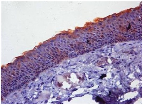
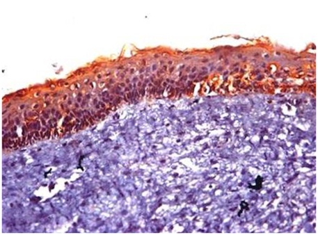
Photomicrograph of OKC showing cytokeratin 18 expression with mild (+) intensity and “FOCAL” distribution. (Upper cell layer) (40X magnification).
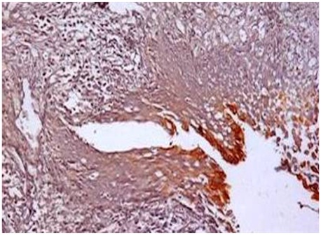
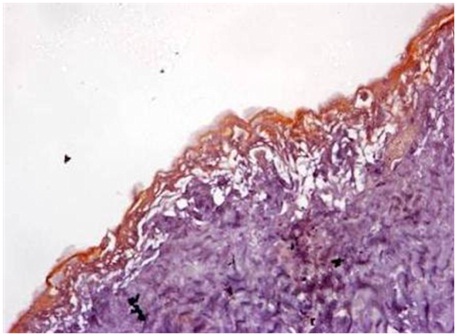
Photomicrograph of RC showing cytokeratin 18 expression with mild (+) Intensity and “FOCAL” Distribution (Upper cell layer) (20X magnification).
Cytokeratin 19 expression in Odontogenic keratocyst, Dentigerous cyst and Radicular cyst.
| Cysts | Positive expression | Negative expre-ssion | Chi-square value | p-value |
|---|
| + | ++ | +++ | Total |
|---|
| Odontogenic keratocyst 20(100%) | 4 (27%) | 11(73%) | 0 (0%) | 15 (75%) | 5 (25%) | 26.128 | 0.000 |
| Dentigerous cyst 20(100%) | 2 (10%) | 5 (25%) | 13 (65%) | 20 (100%) | 0 (0%) |
| Radicular cyst 20 (100%) | 4 (20%) | 7 (35%) | 9 (45%) | 20 (100%) | 0 (0%) | | |
Cytokeratin 19 positive expression patterns within the epithelial layers.
| Cysts | Epithelial layers(ALL Expression) | Chi-square value | p-value | Epithelial layers(FOCAL Expression) | Chi-square value | p-value |
|---|
| Total | Basal | Middle | Upper | Total | Basal | Middle | Upper |
|---|
| Odontogenic keratocyst 15 (75%) | 10(67%) | 9(90%) | 7(70%) | 3 (30%) | 10.133 | 0.006 | 5(33%) | 3(60%) | 3(60%) | 2(40%) | 0.784 | 0.676 |
| Dentigerous cyst 20 (100%) | 18(90%) | 17(94%) | 14(78%) | 16(89%) | 2(10%) | 2(100%) | 2(100%) | 0 (0%) |
| Radicular cyst 20 (100%) | 17(85%) | 12(71%) | 15(88%) | 16(94%) | 3(15%) | 2(67%) | 1(33%) | 1(33%) |
Photomicrograph of DC showing cytokeratin 19 expression with intense (+++) intensity and “ALL” distribution (basal, middle and upper cell layer) (20Xmagnification).
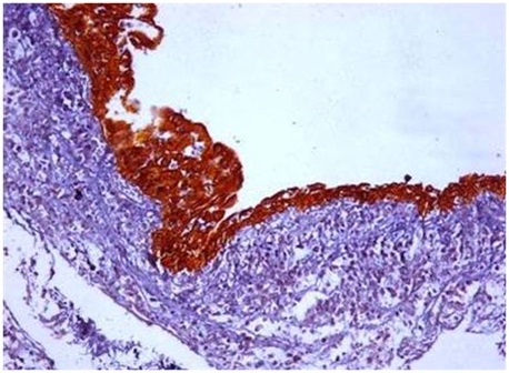
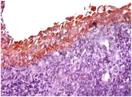
Photomicrograph of RC showing cytokeratin 19 expression with intense (+++) intensity and “ALL” distribution (basal, middle and upper cell layer) (40X magnification).
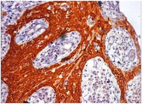
Photomicrograph of OKC showing cytokeratin 19 expression with moderate (++) intensity and “ALL” distribution (basal and upper cell layer) (40X magnification).
Photomicrograph of DC showing cytokeratin 19 expression with moderate (++) intensity and “ALL” distribution. (basal and upper cell layer) (40X magnification).
Photomicrograph of DC showing cytokeratin 19 expression with mild (+) intensity and “ALL” distribution. (upper cell layer) (40X magnification).
Comparison of CK18 and CK 19 expression in odontogenic keratocysts was statistical significant (p-value-0.001) and in dentigerous cysts and radicular cysts it was highly significant. (DC, p-value-0.001), (RC, p-value-0.001) [Table/Fig-13].
Comparison of Cytokeratin 18 and Cytokeratin 19 in Odontogenic keratocyst, Dentigerous cyst and Radicular cyst.
| Cysts | Cytokeratin 18 | Cytokeratin 19 | Z-test | p-value |
|---|
| Odontogenic keratocyst | 5(25%) | 15 (75%) | 3.65 | 0.001 |
| Dentigerous cyst | 3 (15%) | 20 (100%) | 10.65 | 0.001 |
| Radicular cyst | 2 (10%) | 20 (100%) | 13.42 | 0.001 |
Discussion
CK are constituents of complex network extending from the surface of the nucleus to the peripheral cell sector where they get inserted into different cell junctions like desmosomes and hemidesmosomes. The presence of these CKs in tooth development has lent credence to suggestions that they have an active role in the embryonic development of the dental organ and hence, their expression in odontogenic cysts and tumours has been a subject of study for various authors. Identity of a cell as an epithelial cell and also its different stages during differentiation can be studied through the patterns of expression of keratins. The utility of CK 18 and 19 to identify odontogenic epithelium have been demonstrated by numerous biomedical studies and therefore in cases where an odontogenic origin of neoplasms or cysts is suspected, they have been proven to be a useful tool in diagnosis [11–13].
In the present study, CK 18 expression was found positive in 25% of OKCs. These findings are in contrast to Meara et al., who found CK 18 positive in 17% of OKC associated with Nevoid Basal Cell Carcinoma (NBCC) syndrome, and 57% in non syndromic OKCs [14]. Nevoid basal cell carcinoma is the syndrome which not only includes OKC but also other defects within multiple body systems such as the skin, nervous system, eyes, endocrine system and bones. Because keratin expression is generally regulated by cell activation and differentiation, it can be inferred that its expression may shed light on the differentiation trends of skin neoplasms. That’s why in such cases variations are found within KC expression pattern. Apart from that, technique sensitivity also plays an important role in variation regarding the expression pattern, hence the variation in results.
MacDonald and Fletcher reported 100% positivity with monoclonal antibody LP 34 in OKC and DC but Brown et al. suggested that LP 34 stains CK other than CK 18 too. Furthermore LP 34 reactivity in paraffin sections of OKCs is particularly laboratory and technique dependent [15,16]. Santos et al., noted CK 18 expression within basal cell layer but in NBCC syndromic cases, while Hormia et al., did not find any positive case in their study [17,18].
CK 18 positivity in the present study was found in 15% of DC and 10% in RC. Meara et al., reported 75% and Hormia et al., found 50% positive cases in their study, while Mathews et al., Gao et al., and Shruthi et al., reported all the cases negative [14,18–21].
A “FOCAL” expression within the basal and upper cell layers was observed in DCs and in the middle and upper cell layers in RCs similar to Meara et al., who reported a predominant “FOCAL” expression pattern in upper cell layers [14]. These noticeable differences observed in the expression pattern of CK 18 in DC could not only be attributed to the specific histogenic origin and distinct functional characteristics of cells but alternately could also be a sign of oncofetal transformations [18]. In RC, metaplastic epithelial changes within the epithelium could be a plausible explanation for their expression [22].
CK 19 expression was positive in 75% of OKCs, finding in accordance with those of Hormia et al., Gao et al., Morgan et al., Matthews et al., Li et al., Hayakawa et al., Tsuji et al., Dos santos et al., Aragaki et al., Kamath et al., and Yarlagadda et al., who reported nearly all the cases positive with CK 19 [7,13,17–20,23–27]. However in contrast Stoll et al., Wagner et al., and de Berardinis et al., found negative expressions in their studies [5,28,29].
About 67% positive cases showed an “ALL” expression pattern predominantly within basal and middle cell layers and 33% cases depicted “FOCAL” expression pattern mainly within superficial cell layers. Similarly CK 19 expression was noted in all layers of epithelia but predominantly in basal and few suprabasal cells in the study of Hormia et al., Gao et al., and Aragaki et al., in superficial and spinous cell layer as reported by Hayakawa et al., Tsuji et al., and Kitano et al., in superficial and basal cells by Yarlagadda et al., and in superficial cells by Kamath et al., [13,18,20,23–27]. The fact that CK 19 is a minor component of basal cells of stratified squamous epithelia could be one of the possible explanations for the predominant expression in basal cell layer [7].
CK 19 expression was 100% positive in both DCs and RCs, similar to the findings of Hormia et al., Gao et al., Morgan et al., and Tsuji et al., who also reported nearly all the cases positive with CK 19 [7,18,20,25]. On the other hand Stoll et al., found 50% positive cases in DC and 47% cases in RC, while Wagner et al., recorded 48% positive cases in both the cysts [5,28].
An “ALL” expression pattern in the entire epithelial layer was seen in 90% of DCs and 85% of RCs, similar to the findings of Hormia et al., Morgan et al., Mathhews et al., and Gao et al., who also found the expression of CK 19 in all the layers of DC [7,18–20]. Tsuji et al., Stoll et al., and Kamath et al., noted predominant expression in the superficial cell layer and Katuri et al., noted in basal cells in DCs [5,25,27,30]. Also, Gao et al., Katuri et al., and Tsuji et al., reported higher expression in superficial cell layers, Stoll et al., in suprabasal cells and De berardinis et al., reported strong positivity in the basal and parabasal layers in RC [5,20,25,29,30].
However, certain minor differences observed in expression within cell layers of these cysts could be explained by proliferating odontogenic epithelium which exhibits certain phenotypical differences when compared to normal epithelia [31]. Gradual maturation of the epithelial cells as they migrate to upper layers (basal to apical differentiation) could result in expression of CK19 in OKC, a fact which is unobserved in dentigerous and radicular cysts [18].
Since there is always a possibility that masking of some or all epitopes on a particular keratin in certain cells can occur, Smith et al., and Mathhews et al., emphasized the need for care in the interpretation of negative results in immunocytochemistry, particularly in studies of keratins. Further, differences in reactivity between different monoclonal antibodies for the same keratin were well documented and this was certainly the case with CK 19 and CK 18 [32]. A comparative table of various studies has been presented in [Table/Fig-14,15].
Expression of CK 18 in OKC, DC and RC in previous studies.
| Authors (Years) | Odontogenic Keratocyst | Dentigerous cyst | Radicular cyst |
|---|
| Hormia et al., [18] (1987) | No reactivity noted with antibody PKK3 and Ks18.18 for cytokeratin 18 in simple epithelia | A distinct layer of positive cells were noted with antibody PKK3 and Ks18.18 for cytokeratin 18 in simple epithelia | No reactivity noted with antibody PKK3 and Ks18.18 for cytokeratin 18 in simple epithelia |
| Matthews et al., [19] (1988) | No Expression was observed | No expression was observed | Rare expression was observed in the superficial cells in areas of mucous metaplasia |
| Gao et al., [20] (1989) | No staining reaction was noted with mAb LE61 for K 18 in simple epithelia | The mAbs against K 18 either failed to react or only reacted weakly with the epithelium | Not Described |
| Meara et al., [14] (2000) | Trace staining with Focal expression pattern mainly in the upper cell layer in OKC without NBCC syndrome cases, while No staining was noted in OKC with NBCC syndrome | Positive expression was noted, albeit weakly and inconsistently. The pattern and intensity was too inconsistent to draw any diagnostic conclusion. | Not studied |
| Shruthi et al., [21] (2014) | Found negative in all the cases |
Expression of CK 19 in OKC, DC and RC in previous studies.
| Authors (Years) | Odontogenic Keratocyst | Dentigerous cyst | Radicular cyst |
|---|
| Hormia et al., [18] (1987) | PKK2 for CK 7, 17, 19 gave bright staining throughout all the epithelial layers | PKK2 for CK 7, 17, 19 reacted with all the cell layers of epithelium | PKK2 for CK 7, 17, 19 reacted with all the cell layers of epithelium |
| Morgan et al., [7] (1987) | LP2K for CK 19 expressed at all levels of the epithelium | LP2K for CK 19 stained most of the cells at all levels of the epithelium | |
| Matthews et al., (1988) [19] | CK 19 (‘simple’) strong reactivity within epithelial cell layer | CK 19 (‘simple’) strong reactivity within epithelial cell layer | CK 19 (‘simple’) strong reactivity within epithelial cell layer |
| Mcdonald & Fletcher [15] (1989) | LP 34 antibody reactivity found negative for basal cells while moderately to strongly positive for other cell layers | LP 34 antibody reactivity found positive for all the cell layers | |
| Matthews and Browne [16] (1989) | Did not notice the reactivity of LP 34, so raised uncertainty against the result of Mcdonald and Fletcher | | |
| Mcdonald & Fletcher [15] (1989) | Response to Matthews and Browne Highlight on the need for standardised technique | | |
| Gao et al., [20] (1989) | Patchy staining reaction predominantly with suprabasal cells and some basal cell | Strongly positive staining reaction either with full thickness of epithelium or with superficial cells | |
| Kitano et al., [23] (1998) | Reactivity to AE1 antibody (Acidic, Type 1, Cytokeratin) was limited to the upper suprabasal and surface parakeratinized cell layers. | | |
| Wagner et al., [28] (1999) | No expression was noted | Found positive expression but not specified layers | Found positive expression but not specified layers |
| Stoll et al., [5] (2005) | Completely Negative | Positive staining observed in all the cell layers but predominantly within superficial cell layer | Positive staining observed in all the cell layers but predominantly within suprabasal cell layer |
| Okada et al., [24] (2006) | Almost entirely positively expressed in the superficial and spinous cell layer in KCOT | | |
| Santos [17] (2009) | Strong expression was noted affecting mainly suprabasal and intermediate layer cells. | | |
| Aragaki et al., [26] (2010) | Strong positive expression predominantly observed in basal and suprabasal cells. | | |
| Tsuji [25] (2014) | Expression was noted predominantly within superficial cell layer followed by spinous cell layer followed by basal cell layer | Expression was observed predominantly within superficial cell layer followed by spinous cell layer followed by basal cell layer | Expression was observed predominantly within superficial cell layer followed by spinous cell layer followed by basal cell layer |
| Katuri et al., [30] (2015) | Slightly positivity noted in all the cell layers | Positive expression was noted in all the cell layersMean number of positive basal cells were predominant | Positive expression was noted in all the cell layersMean number of positive suprabasal cells and superficial cells were predominant |
| Kamath et al., [27] (2015) | Predominant expression staining grade was negative and ‘+’ with less extent of ‘++’. Positive specimen showed staining mainly within superficial cells of epithelium | Predominant expression staining grade was ‘++’ and ‘+++’ with less extent of ‘+’. Positive specimen showed staining mainly within superficial and suprabasal cells of epithelium | |
| Yarlagadda et al., (2015) [13] | Consistently positive throughout the epithelium lining and diffuse staining pattern in superficial cell layer and basal cell layer | Homogenous expression within all the cell layer | Diffuse staining reaction in all the cell layers of epithelium |
Conclusion
CK 18 and 19 expressions in OKC, DC and RC thus could prove valuable aid in diagnosis of these wherever overlapping features pose a problem. Furthermore, the differences in reports of expressions of CK 18 & CK 19 in OKC, DC and RC by different researchers may be due to different antigen retrieval methods used, different monoclonal antibodies used, or the smaller sample size. Also standardization of laboratory techniques could help in gaining a deeper insight into behaviour as well as pathogenesis of these lesions.
*NA-Not available