The goal of root canal obturation is to obtain a three dimensional seal of the root canal system. An inadequate filling during obturation can results in reentry and re-growth of microorganisms in the root canal system which irritates the periapical tissue and compromises the treatment success [1].
To accomplish this many endodontic obturation materials and sealers are being used [2]. Guttapercha is a commonly used root canal filling material which is used with different types of endodontic sealers. Sealers aim to prevent ingress of bacteria in the root canal space [3]. There are different types of endodontic sealers available that have been introduced in the market with varying physical properties [4]. According to their bases, root canal sealers are calcium hydroxide based sealers i.e., Sealapex, Resin based sealers i.e., AH26 and Adseal, Solvent based sealers i.e., Chloropercha, Glass inomer based sealers i.e., Ketac Endo, Silicone based sealers i.e., Lee Endo Fill and MTA based sealers i.e., Pro Root MTA, MTA Fillapex.
One of the recent epoxy resin based sealer is Adseal, with excellent chemical, physical properties and sealing ability. These characteristics are responsible for the superiority of this sealer over the other epoxy resin based sealers [5].
MTA is being used for pulp capping, apexification, perforation repair, root-end filling material and for pulpotomy. In 1993, US Federal Drug Administration gave acceptance to MTA and it became commercially available as ProRoot MTA. Pro Root MTA is calcium silicate-containing MTA that is used as an endodontic sealer [6].
Recently, several new products of MTA have been introduced such as MTA Fillapex, Micro Mega MTA and Bioaggregate [7]. The use of these MTA based sealers is being considered as a revolution in Paediatric and Preventive Dentistry [4].
The present study was conducted to evaluate and compare the apical microleakage of a resin based sealer; Adseal with MTA based sealers, Pro Root MTA and MTA Fillapex using dye penetration technique under Stereomicroscope at 40X magnification.
Materials and Methods
The present study was conducted in the Department of Paediartic and Preventive Dentistry, Surendera Dental College and Research Institute, Sri Ganganagar, Rajasthan, India from April 2015 to November 2015 time period. The present study was an in-vitro cross-sectional study. A total of 75 freshly extracted human single rooted teeth were used as study samples. Teeth with root fracture, root caries, open apices, developmental anomaly and external and internal root resorption were excluded from the study. These teeth were cleaned with hand scalers and soaked in 5.25% sodium hypochlorite for two hours and then stored in a solution containing thymol crystals. The teeth were decoronated using diamond disk at the cement-enamel junction uniformly and were then mounted in freshly mixed alginate in uniformly sized plastic containers. The root canal access was prepared using endo access bur and working length was determined using appropriate K-file. Standardized wooden block was constructed for taking radiovisiograph, so as to maintain the standardization for all the study samples. Then the biomechanical preparation was done with the use of NiTi rotary protaper files till size F2.
The irrigation protocol followed was use of 5.0% sodium hypochlorite in between each instrumentation and 17% Ethylenediaminetetracetic Acid (EDTA) was left in the root canals for four minutes, followed by final rinsing with normal saline. The root canals were then dried with paper points. The teeth were randomly divided into five groups of 15 specimens each [Table/Fig-1] and obturation was done as follows:
Division of study samples (n=15) in five study groups.
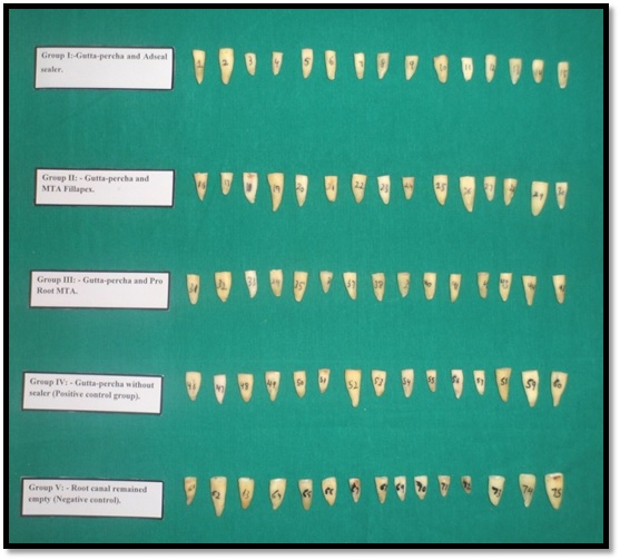
Group I: - Gutta-percha and Adseal sealer.
Group II: - Gutta-percha and MTA Fillapex.
Group III: - Gutta-percha and Pro Root MTA.
Group IV: - Gutta-percha without sealer.
Group V: - Root canal remained empty.
The tested materials were handled according to manufacturer’s instructions. Adseal sealer, MTA Fillapex and Pro root MTA was applied over the entire working length of the canal using lentulo spirals. The selected gutta-percha cone was lightly coated with sealer and placed slowly in the canal to full working length. Excess gutta-percha cone was seared off from the canal orifice using a heated instrument. After canal obturation, the teeth were radiographed to make sure the canals were fully obturated. The teeth were then stored at 37°C with 100% humidity for one week to allow the sealers to get fully set.
All the root surfaces, except the apical 2mm were covered with two layers of nail varnish. In negative control, the root surfaces including the apical foramen were completely coated with two layers of nail varnish, to test the impermeability of nail varnish to methylene blue. The sticky wax was then applied on the varnish area; teeth were immersed in 2% methelene blue dye and then were stored in an incubator for 72 h at 37°C.
The roots were rinsed in running water and dried with paper towels. The varnish and sticky wax coating was removed with a scalpel blade and a guide groove was prepared with a diamond disc in a crown-apex direction in middle of tooth till the depth of the canal. The roots were split longitudinally using a large spoon excavator.
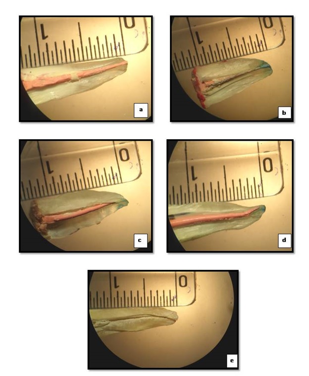
The linear dye penetration was measured from root apex to the most coronal extent under Stereomicroscope (Labo Med CMZ4, India) at 40X magnification. The depth of dye penetration was evaluated [Table/Fig-2] using criteria given by W.P. Saunders et al., [8].
Depth of dye penetration, according to degree of microleakage.
| Degree of Leakage | Depth of Dye Penetration |
|---|
| 0 | No leakage detected |
| 1 | Less than 0.5 mm |
| 2 | 0.5-1 mm |
| 3 | Greater than 1 mm |
The whole study was repeated three times and the readings were calculated. The data obtained was then subjected to statistical analysis using SPSS software (version 20.0). The tests used for statistical analysis used were ANOVA, paired t-test and interclass correlation.
Results
The apical microleakage for study groups was evaluated and results obtained were subjected to statistical analysis using SPSS software (version 20.0). Besides the study groups, two control groups (positive and negative controls) were evaluated with dye penetration test. The positive control demonstrated the penetration capacity of the dye, whereas negative control showed no dye penetration by confirming the insulating capacity of the nail varnish and yellow sticky wax.
According to the dye penetration scores, the numbers of study samples was distributed for all the five study groups [Table/Fig-3]. The apical dye penetration was observed minimum in Group V followed by Group I, then Group III, IV and II (V< I< III< IV< II).
Distribution of number of samples in all the five study groups using dye penetration scoring criteria.
| Score | Group I | Group II | Group III | Group IV | Group V |
|---|
| 0 | 13 | 1 | 6 | 1 | 15 |
| 1 | 1 | 0 | 1 | 2 | 0 |
| 2 | 1 | 4 | 8 | 12 | 0 |
| 3 | 0 | 10 | 0 | 0 | 0 |
The mean values for apical microleakage, according to dye penetration test for all the five study groups was observed. The maximum mean was found in Group II followed by Group IV, Group III, Group I and least in Group V. The analysis of variance for microleakage values used in the study exhibited highly significant correlation (p-value <0.01) between all the study groups [Table/Fig-4].
Mean values of five study groups and their intergroup comparison using ANOVA statistical analysis according to dye penetration test
| Score | Group I | Group II | Group III | Group IV | Group V |
|---|
| Mean±SD | 0.067 ±0.013 | 1.83 ±0.065 | 0.50± 0.030 | 0.657±0.023 | 0 |
| Statistical analysis |
| Variation | SS† | Df†† | p – value |
| Total | 25.631 | 59 | 0.001* |
†Sum of Squares; †† Degree of freedom *p – value < 0.01 is highly siginificant.
[Table/Fig-5] shows the intra-observer reliability for all the five study groups, using intra-class correlation statistical analysis. The p-value was found to be insignificant (p-value >0.05) for all the study groups. The inter-group comparison of microleakage values for all the five groups was analyzed with each other using Paired t-test. A highly significant difference (p-value <0.01) in microleakage scores was found among all the study groups between Group I vs II, I vs III, I vs IV, I vs V, II vs III, II vs IV, II vs V, III vs IV, III vs V, IV vs V [Table/Fig-6]. Thus, the present study concluded that the microleakage was maximum in Group II followed by Group IV, Group III, Group I and least was in Group V [Table/Fig-7].
Intraobserver reliability for all the five study groups for triplicate study.
| Group | Mean difference | Intraclass correlation | p - value |
|---|
| Group I | 0 | 1 | 1.000* |
| Group II | 0.06 | 0.928 | 0.870* |
| Group III | 0.03 | 0.922 | 0.780* |
| Group IV | 0.04 | 0.854 | 0.700* |
| Group V | 0 | 1 | 1.000* |
*p – value > 0.05 is insignificant.
Inter group comparison of all the five study groups using paired t-test statistical analysis.
| Groups | t- value | CI (95%)of difference | p – value |
|---|
| Upper Limit | Lower Limit |
|---|
| I vs II | 102.860 | -1.728 | -1.798 | 0.001* |
| I vs III | 51.291 | -0.416 | -0.45 | 0.001* |
| I vs IV | 86.207 | -0.576 | -0.604 | 0.001* |
| I vs V | 19.961 | 0.074 | 0.060 | 0.01* |
| II vs III | 71.862 | 1.368 | 1.292 | 0.01* |
| II vs IV | 65.767 | 1.209 | 1.136 | 0.01* |
| II vs V | 108.87 | 1.864 | 1.795 | 0.01* |
| III vs IV | 16.059 | -0.137 | -0.177 | 0.001* |
| III vs V | 64.550 | 0.516 | 0.484 | 0.01* |
| IV vs V | 110.153 | 0.669 | 0.645 | 0.01* |
* p - value ≤ 0.01 is highly significant.
Apical microleakage assement by dye penetration shows (a): Group I, (b): Group II (c): Group III (d): Group IV and (e): Group V.
Discussion
The main purpose of endodontic therapy is to achieve a complete hermetic seal of the root canal and prevention of coronal and apical microleakage. Thus, the present study aimed to evaluate the apical microleakage of a recently introduced resin-based root canal sealer; Adseal with MTA based sealers; Pro Root MTA and MTA Fillapex using dye penetration technique under stereomicroscope at 40X magnification.
A total of 75 extracted single rooted human teeth were used for the study. By using hand scaler calculus was removed and the study samples were then immersed in sodium hypochlorite to remove organic material from the root surfaces as it is effective against bacteria, viruses and fungi [8]. Then the study samples were stored in thymol crystals in distilled water to maintain the aseptic conditions [9]. Each study samples was decoronated and mounted using alginate in plastic containers. A standardized rectangular wooden block was constructed with two slots, one for placement of plastic container and another for RVG sensor at the distance of 1.7 inches, so as to maintain the standardization for all the study samples.
After root canal access was prepared using endo access bur, the working length was determined using an appropriate K-file with the placement of rubber stopper on a flat non variable reference point, so that working length measurement was not affected. The working length was taken 0.5mm short of the radiographic apex.
For biomechanical preparation, Protaper Universal system was used in the following sequence: SX, S1, S2, F1, F2 at a speed of 300rpm and torque according to the torque chart supplied by manufacturer. This system reduces time required and improves the standardization of instrumentation [10].
The teeth were randomly divided into five groups of 15 specimens each (n=15). From this division, two groups are positive and negative control groups and three groups are study groups which are divided according to root canal sealer used.
The type of sealer used in this study was among the main differences between different root canal filling systems. The application of sealer fills the irregularities at the interface of filling material and the root canal walls [11]. According to Cohen S et al., [12] inadequate apical seal is responsible for up to 60% of treatment failures.
The study samples in Group I were obturated using resin based sealer, Adseal. According to Belli et al., [13]. Adseal is an epoxy-resin based sealer having excellent sealing properties. It is a dual syringe which consists of base and catalyst with excellent optimal characteristics.
According to Bogen et al., [14], endodontically treated teeth using MTA obturation showed improved healing rate which had been subjected to long term microleakage and bacteriological contamination. The study conducted by Panzarini et al., [15] for the immediately re-implated teeth with MTA as a root canal filling material, showed good repair and biological sealing of some lateral canals. The study samples in Group II were obturated using MTA Fillapex, that is composed of bismuth oxide, resins, silica nanoparticles, and pigments, having ideal properties of sealing ability of MTA [4,16].
MTA forms calcium and hydroxyl ions which are important for stimulation of hard tissue deposition [17]. Sealability is also improved due to presence of MTA because of possibility of setting expansion. Hydration of anhydrous mineral oxide compounds occur to produce calcium silicate hydrate and calcium hydroxide phases [18–20], which produces expansion against its confining margins and improves the seal and minimize microleakage [21–25].
In Group III, ProRoot MTA was used to obturate all the study samples. As MTA expands during setting and forms the excellent sealing ability [26]. According to Torabinejad et al., [27] MTA sealed the root canal very superiorly and no gaps were found in any of the experimental specimen. MTA has also proved itself to be superior in the bacterial leakage test by not allowing the entry of bacteria at the interface [28].
The advantages of MTA Fillapex and Pro root MTA are highly biocompatible, stimulate mineralization, these materials exhibited higher push out bond strengths after storage, and they have adequate calcium releasing property.
The positive control group, Group IV indicated that leakage testing was a suitable method for proving total dye penetration without a sealer. The negative control group, Group V showed no dye penetration, indicating that use of two layers of varnish was effective to prevent apical dye penetration, as in study conducted in accordance by Oliver et al., [29].
The root canal sealer was, applied over the entire working length of the canal using lentulo spirals in slow speed, so that the canals could be uniformly loaded with sealer. The samples were kept in incubator at 37°C, so that the sealer got fully set for 7 days.
The dye penetration technique is among the most commonly used methods for apical microleakage. This method is relatively simple and does not require complex equipment. The studies using this technique employ different materials and dyes to assess the amount of microleakage. Methylene blue dye has molecular size similar or smaller than that of bacterial products, so it had been considered stuitable for the detection of apical microleakage in present study [30]. The degree of leakage could be reduced by using a material that adapts and forms adequate seal with root canal walls (Ingle et al.,[31] and Roberts et al.,[32]). A diamond disc was used to cut the tooth in a crown-apex direction using a large spoon excavator so that the dye penetration can be assessed under stereomicroscope. Stereomicroscope is a simple method and is less expensive and needs less complicated equipment.
Clinical implications: There are limited studies that justified the use of MTA as the endodontic sealer. So, the present study enlightens the use of MTA with gutta percha, as root canal sealer material.
Limitation
The present study was conducted using a small sample size of 75 teeth. The results could have been more defined using more study samples. The study period of present study was seven days, whereas it has been found MTA expands after 28 days. So, further studies can be conducted using extended time period for more appropriate results.
Conclusion
The present in-vitro study was conducted to assess and compare the apical microleakage using the dye penetration test under stereomicroscope at 40X magnification. The evaluation on the basis of mean of dye penetration observed in all five groups, revealed that Group V showed no dye penetration followed by Group I then Group III, Group IV and least in Group II. The study concluded that on comparison of experimental groups, the Adseal sealer was better in providing the apical seal than Proroot MTA and MTA Fillapex.
†Sum of Squares; †† Degree of freedom *p – value < 0.01 is highly siginificant.
*p – value > 0.05 is insignificant.
* p - value ≤ 0.01 is highly significant.