1. Introduction
Alzheimer’s Disease (AD) is one of the most prevalent dementia seen in elderly worldwide. According to the current reports, it is estimated that almost one new case of AD develops every 33 seconds, and almost a million new cases every year, with estimated prevalence of almost 13.8 million around the globe [1]. The main symptoms include memory loss, cognitive impairment, disorientation and psychiatric symptoms [2,3]. The preliminary diagnosis of AD is made by a combination of clinical criteria which includes a neurological examination, mental status tests and brain imaging [4]. However, based on the above clinical tests, the task of AD becomes difficult especially in patients having mild or early stages of AD. Hence, the need for biomarkers evolved which show strong indications of Alzheimer’s disease and also provides conclusive diagnosis of early onset of AD. This also contributes to developing disease modifying therapies at early stage thus preserving normal brain function or delaying cognitive impairment.
Currently, presence of dementia is confirmed by analysing the Cerebrospinal Fluid (CSF) with established biomarkers like amyloid beta protein, tau protein and phospho-tau expression levels. CSF is known to act as a valuable source of biomarkers, since besides being in direct contact with the brain and spinal cord it provides a complete representation of various biochemical and metabolic profiles of the brain. However, this fluid is obtained by lumbar punctures in patients which are both invasive as well as painful for the patients, which makes the diagnosis difficult and also irreproducible. Hence, the need of the hour demands of new biological biomarkers which being less intrusive and easily obtainable, can also be more sensitive and specific [5]. This will further help in the diagnosis in the prodormal stage of dementia and would also lead to identification of conditions of patients with mild cognitive impairment so that the onset of dementia could be delayed [6].
1.1 Alzheimer’s Disease in Younger Patients
AD was initially referred to as “presenile dementia” with its first patient being 51 years of age at the time of presentation. However, after studies conducted by Blessed and colleagues (1967) it was seen that the brains of patients suffering with senile dementia had quantitatively similar senile plaques and neurofibrillary tangles as seen in presenile AD patients [7].
Autosomal dominant familial AD is seen to be more commonly affecting younger individuals as compared to sporadic AD. Studies have shown that ApoE4 genotype can lead to a more aggressive clinical form of AD in younger patients. These patients tend to suffer from Logopenic Progressive Aphasia (LPA) often characterised by prolonged word finding pauses, anomia and impaired sentence processing. This point towards a cortical involvement in younger patients with early onset of AD [8].
Also, recent researchers have given evidences of an association between early onset of AD and Down’s syndrome. This was concluded because of recognition of the role of Amyloid Precursor Protein’s (APP) over production due to increased gene dosage with trisomy 21 [9], thus pointing towards a possibility that people with Down syndrome have an increased risk of developing younger onset dementia after age of 50 years [10].
1.2 Understanding Biomarkers
Biomarkers or “biological markers” are the category of medical signs which define a medical state from outside the patient and can be reproduced and measured accurately, unlike the medical symptoms which are mere indications of a patient’s condition described and perceived by the patients themselves. According to the definition given by National Institutes of Health Biomarkers Definitions Working Group in 1998, “Biomarkers are evidence of any biological, pathogenic or pharmacogenomic response when administered to any therapeutic change” [11]. Biological markers are basically any kind of substances, structures or processes which could be measured in/outside the body and may influence any changes in the body and probable prevalence of any disease in the body [12].
For a possible potent biomarker for Alzheimer’s disease, following criteria has been unanimously decided by researchers worldwide [13–16].
Reflect aging of brain.
Describe pathophysiological processes in brain.
Any pharmacological change should be reflected.
Highly sensitive and specific.
Reproducible results over time changes.
Clear cut-off values with at least two-fold changes
Easy collectible results and inexpensive tests.
1.3 Established CSF Biomarkers for Alzheimer’s Disease
Considering previous researchers, three biomarkers have been internationally established and published worldwide, for diagnosis of Alzheimer’s disease [Table/Fig-1] [17–19]. These biomarkers are obtained from CSF and these biomarkers collectively increase the validity for diagnosis by giving results which are sensitive to >95% and specific to >85% [20–23].
Established biomarkers for Alzheimer’s disease [17–19].
| Amyloid beta | Tau protein | Phosphorylated tau |
|---|
| Aβ plaque depositions are used widely to characterize AD. Secretases cleaves Aβ from large APP and processing of these amyloidgenic pathways produces these 42 amino acid peptides (Aβ1-42) which end up as aggregates in brain. Analysis of CSF in AD patients shows a significant reduction of Aβ of about <500pg/ml in comparison to controls with 794±20 pg/ml of Aβ [17]. | The intaneuronal inclusion of the microtubule associated protein tau serves as another established biomarker for AD. Tau protein which is known to increase gradually with age <300pg/ml (21-50 years) to almost <500 (>71 years) shows a significant exponential increase in AD patients of about >450 to >600 pg/ml (in patients of ages 51-70 years). Hence proving to be a good prognostic biomarker [18]. | AD exhibits condition of tau protein being phosphorylated in almost 39 possible sites. Wherein, position 181 works as a definite biomarker in AD as compared to controls. The phosphorylation of Tau protein results in both lack of functions and also neuronal dysfunction. The other notable phosphorylated tau proteins include (phosphor-tau-199, -231, -235, -396 and -400 [19]. |
2. Need for Circulatory Biomarkers
As discussed previously, the CSF which is used for diagnosis for AD is both intrusive and invasive for patients. The main reason being, CSF fluid is obtained by lumbar punctures which cause nausea, severe backache and weakness in elderly people. Also maintaining track of patients for regular diagnosis becomes very difficult. Thus, it is essential to identify new biomarkers in other sources like serum, urine etc. which are cheaper, less invasive and easily collectible. The main advantages of using blood for diagnosis are, being easily obtainable a proper follow-up of the patients can be maintained over a period of time. Also analysing blood cells (e.g., peripheral blood mononuclear cells, lymphocytes, monocytes or platelets) can be more specifically related to AD pathologies.
2.1 “Circulatory” miRNAs
miRNAs belong to the class of non-coding RNA molecules of around 22 nucleotide length which regulate more than 60% of all known genes through post-transcriptional gene silencing (RNAi) [24]. The dysregulation of miRNA expression in peripheral blood can serve as a potent source of diagnosis of Alzheimer’s and other brain related disorders [25] [Table/Fig. 2] [26–46]. Schipper (2007) identified a number of downregulated miRNAs when compared to 16 sporadic AD patients with 16 controls using a microarray chip. These downregulated miRNA included miR-34a, miR-81b and let-7f [26]. The targets of these miRNAs were interestingly found to be part of p53, Notch and Bcl-2 pathways which are already known to be involved in AD pathogenesis.
Circulatory miRNAs associated with AD [26–46].
| miRNAs | Evidences in samples | References |
|---|
| miR-34a, miR-181b | Increased expression in PBMC | [26] |
| miR-9 | Downregulated in serum | [27] |
| miR-112, miR-161, let-7d-3p, miR-5010-3p, hsa-miR-26a-5p, hsa-miR-1285-5p, and hsa-miR-151a-3p upregulated; miR-103a-3p, miR-107, miR-532-5p, miR-26b-5p, let-7f-5p | Downregulated in peripheral blood | [28] |
| miR-107 | Downregulated in temporal cortex | [29,30] |
| hsa-let-7d-5p, hsa-let-7g-5p, hsa-miR-15b-5p, hsa-miR-142-3p, hsamiR-191-5p, hsa-miR-301a-3p and hsa-miR-545-3p | Differentially regulated in Plasma | [31] |
| miR-29 | Downregulated in temporal cortex, cerebellum and serum | [32] |
| miR-27a-3p | Reduced expression in cerebrospinal fluid | [33] |
| miR-34 | Upregulated in hippocampus | [34,35] |
| 60 miRNAs including Let-7 family members | Differentially regulated in cerebrospinal fluid | [36] |
| miR-181 | Downregulated in temporal cortex and serum | [37] |
| miR-146a, miR-155 | Increased levels in cerebrospinal fluid and extracellular fluid | [38] |
| miR-106 | Downregulated in temporal cortex | [39] |
| miR-9,miR-125b, miR-146a, miR-155 | Increased levels in cerebrospinal fluid and extracellular fluid | [40] |
| miR-146a | “Selective” upregulation in temporal cortex and hippocampus | [41] |
| Let-7b | Increased levels in cerebrospinal fluid | [42] |
| miR-15a | Increased levels in plasma | [43] |
| miR-34c | Increased levels in plasma | [44] |
| miR-132 and miR-134 families | Upregulated in plasma | [45] |
| miR-29a/b, miR-181c, miR-9 | Downregulated in serum | [46] |
Another interesting research carried out by Geekiyanage and Chan (2012) showed decreased levels of miR-137, miR-181c, miR-9 and miR-29a/b in neocortical regions of AD patients. Similar results were seen when the same follow-up study was performed on blood levels of AD patients (n=7) with controls (n=7), although the downregulation was at a lower level [47].
Villa et al., and Bekris et al., through their studies were able to demonstrate downregulation of miRNA 29b and 15a which regulated transcription factor Sp1 which is known to regulate expression of APP and tau which are known AD related genes, along with other target genes [48–51].
Researches suggest that a systematic increase in specific miRNAs may help in suppressing various cellular functions like redox defences and DNA repair mechanisms in brain and peripheral tissues supporting the role of miRNAs as potential therapeutic biomarkers for AD in future. Scientists worldwide have showed that an increase in specific miRNAs can regulate crucial cellular functions in brain and peripheral tissues. Thus the contribution of miRNAs in functions such as redox defences and DNA repair mechanisms advocate the potential of miRNAs as potential therapeutic biomarkers for AD in near future.
2.2 Blood based amyloid markers
Although the efficiency of Amyloid beta is a highly sensitive and specific biomarker from CSF for AD has already been established, new studies are being focussing on evaluating Aβ as a potential biomarker from blood serum as well. In a recent meta-analysis review performed by Koyama and colleagues conducted on 13 studies of 10,303 AD patients and controls, to monitor Aβ1-42 and the ratio of Aβ1-42/ Aβ1-40 in plasma to predict AD, showed highly statistical and clinically significant decrease in Aβ1-42 ratio to predict cognitive impairment. However, the results are still not conclusive enough, since the plasma levels are largely affected by factors like subject’s age, lifestyle, laboratory conditions, assay variability, etc., [50].
Other studies have shown varied forms of amyloid beta, in blood plasma, to be crucial potential AD biomarkers for future research. Perez et al., (2012) showed the ratio of free to cell bound Aβ1-17 in blood samples of Mild Cognitive Impairment (MCI) and age-matched control groups to be significantly varying, thus concluding Aβ1-17 in blood plasma to be a highly sensitive and specific biomarker for AD [51].
Various platforms are being developed to measure Aβ levels in blood serum and plasma, such as ELISA developed by Araclon Biotech Ltd., to perform colorimetric tests to measure ratios of Aβ1-40/ Aβ1-42 in patients showing MCI [52]. Other tests like electroluminescence are being developed to further probe into possibility of Aβ in blood to serve as potential biomarkers in near future [53].
2.3 Inflammatory Markers
Neuroinflammatory variability involving astrocytes and activated microglia and the secreted mediators such as oxygen species, chemokines and cytokines was seen on various neuropathological studies conducted on AD brains [54]. The accumulation of such components, may lead to alterations in immune functions and transition of innate immune cells to aggravated proinflammatory cytokines tumour necrosis factor (TNF)-α leading to an increased rate of cognitive decline and also neuronal cell death in some cases [55,56]. In studies conducted by Laske et al., (2013) it was shown that TNF-receptor 1 can be a potent inflammatory biomarker for understanding AD patients better [57]. In another promising study a blood panel of 18 biomarkers (combination of cytokines, growth factors and binding proteins) allowed diagnosis of Alzheimer’s and MCI with an accuracy of ~90% [58].
According to researches, an inflammatory response plays a crucial role in neurodegeneration during progression of AD. Both genetic as well as pathological studies have shown an overexpression of proinflammatory cytokine interleukin β (IL-β) in AD patients, as compared to controls. The role of complement receptor type 1 has also been seen in clearance of amyloid β [59]. Several studies show the association between AD and inflammatory biomarkers such as IL- 1β, IL- 2, IL-4, IL-6, IL-8, IL-10, IL-12, IL-18, Interferon (IFN)-γ, Tumour Necrosis Factor (TNF)-α, Transforming Growth Factor (TGF)-β, and acute phase Reactant Protein c (CRP) [60–65].
Ceramides, sphingomyelins and sulfatides have also been seen to be integral part of neuronal functions and synthesis of bioactive metabolites related to AD [66]. Various studies have shown the serum levels of ceramides differing in AD patients, MCI patients and their respective controls. Since, high base ceramides levels might lead to increased risk of impairment in a normal cognitive brain and a significant decline in a cognitively impaired brain along with decline in the hippocampal volume [67]. Vascular Cell Adhesion Molecule I (VCAM-I), Intracellular Adhesion Molecule I (ICAM-I) and selectins also might serve as biomarkers of microvascular injuries, being in increased levels in plasma samples of patients with late onset AD suggesting endothelial dysfunction [68]. Increased apoptosis in CD4+ T cells and NK has also been proved in AD patients when compared to their controls, along with increased levels of B-cell lymphoma 2, caspases and antioxidant enzyme Superoxide Dismutase (SOD1) [69,70].
However, the accuracy of these inflammatory biomarkers especially cytokines as potential biomarkers still need to be validated because of a little inconsistency seen in parallel studies. Researchers think this is mainly due to difference in the type of samples being analysed i.e., cerebrospinal fluid, peripheral blood plasma, blood serum etc.
2.4 Biomarkers for oxidative stress
Oxidative stress is another dimension being explored for biomarkers in AD. The neurodegenerated parts of brain have been known to show increased Reactive Oxygen Species (ROS) levels. In such conditions, proteins undergo post-translation modifications, leading to formation of mixed disulphides, nitration of tyrosine residues, and formation of lipid peroxides [71]. Protein oxidation besides causing toxic cell damage also results in fragmentation and aggregation, leading to proteolysis [72]. AD and MCI patients both have been detected with increased protein aggregate levels in the forms of fibrils along with increased lipid formation [73]. The most common known markers for oxidative stress include protein glutathionylation, free fatty acid releases, DNA oxidation, iso and neuro prostane formation, 4-Hydroxy 2 trans Nonenal (HNE), lipid peroxidation and advanced glycation end products detection [74].
3. Strategies for Developing Patient Specific Biomarker Profiles
With increased knowledge of various pathways playing significant roles in AD and other factors, it has become clearly evident that one biomarker profile is not enough to identify differentially expressed proteins between AD patients and controls and provide conclusive diagnostic results. Hence, scientists now-a-days are focussing on developing methods to measure various biomarkers simultaneously on a single microarray chip or well.
3.1 Stages of Biomarker Screening [Table/Fig-3]
The process of biomarker discovery.

3.1.1 Pre-exploratory Studies
This phase consists of approvals from Ethical committee so as to ensure proper enrolment of subjects in the study, collection of blood samples, transportation, storage and disposal of collected samples after usage. The ethical committee ensures that proper information is given to the subjects before sample collection. After the ethical approvals are taken, diseased and control samples are compared for generating hypothesis for detection of disease. Techniques such as microarray, mass spectrometry, immunohistochemistry, protein expression profiling and western blots are employed in this phase [75].
3.1.2 Assay Development and Validation
In this phase, clinical assays are developed to discriminate samples with/without disease. The collected samples consist of disease before treatment and a matched diseased tissue as control. The samples are collected mostly using non invasive techniques. The primary objective of this stage is to estimate the true positive rate (sensitivity) and false positive rate (specificity) of the developed assay for biomarker detection.
3.1.3 Retrospective Screening Studies
The retrospective training studies are longitudinal studies based on the evaluation of the efficiency and capability of the developed assay to detect disease in its preclinical stage. Also, the effect of covariates such as demographic and geographic characteristics on the efficiency of biomarker before their validation in Phase IV is studied during this phase [76].
3.1.4 Prospective Studies
This phase focuses on evaluating the efficiency of the developed biomarker assay by screening in a specific demographic population for determining the false referral rate and also the disease detection rate of the assay. This stage basically contributes in describing the stage-specific characteristics of the disease, assessment of screening on cost and mortalities due to the disease.
3.1.5 Validation
Once the efficiency of biomarkers is assessed after retrospective and prospective screening, the results are published in peer reviewed journals so that they can be repeated and verified in other laboratories around the globe to produce similar results to check the authenticity and competency of the developed biomarkers in disease detection.
3.1.6 Randomised Clinical Trials
In the final phase, randomised clinical trials are conducted to detect whether the biomarker based screening is able to reduce the disease burden on the target population.
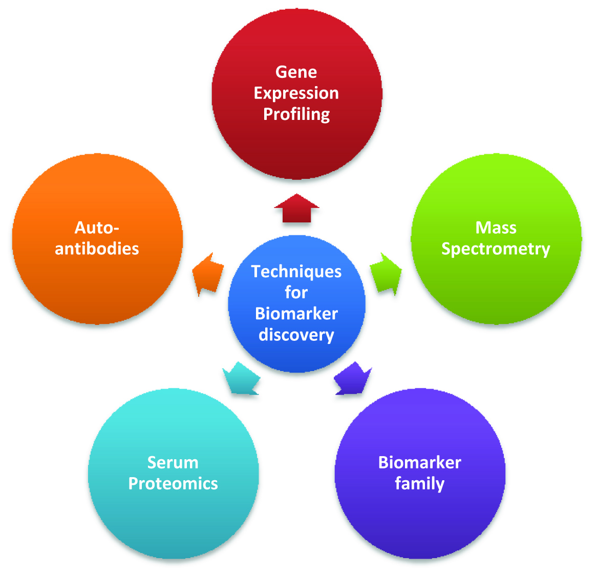
3.2 Techniques for Biomarker Discovery [Table/Fig-4]
Bioinformatics Tools for Biomarker Discovery
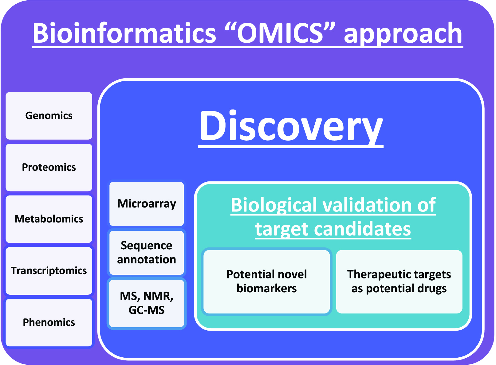
Various ‘omics’ tools and databases available online based on different types of data mining techniques such as Decision Trees, Clustering, Regression, Association Rules, Artificial Intelligence, Neural Networks, Genetic Algorithm, Nearest Neighbour method, Classification and other pattern based searches are being used as discovery tools for easier understanding of biological systems by incorporation of systems biology in the process of biomarker discovery [Table/Fig-4,5] [77,78]. Some of the major databases used by researchers have been described in Supplementary [Table/Fig-1].
Application of ‘omics’ data in Biomarker discovery.

Various techniques for biomarker discovery.

DNA and RNA microarray chips allow studying differentially expressed genes on a single chip-plate, however, similar development for providing conclusive multiple gene signatures to serve as biomarkers appearing in AD patients is still under progress [79,80].
Various techniques being used nowadays for biomarker discovery include, mass spectrometry imaging and profiling which basically explores the idea that miRNA might not provide complete information of the diseased state and the altered proteins could be assessed by mass spectrometry and used for diagnostic purposes by combining with mathematical algorithms. One of the most widely used technologies include SELDI-TOF-MS (Surface Enhanced Laser Desorption Ionization Time Of Flight Mass Spectrometry) which involves analysing small amount of unfractioned serum samples added on a protein chip. These patterns reflect the blood proteome without actual identity of proteins [81,82].
Another approach to biomarker discovery being explored is biomarker family approach, where in an assumption that a miRNA is already a member of biomarker family or have a potent target gene, then other miRNAs belonging to the same family or having same gene target might also be potential biomarkers for diagnosis is considered [83,84].
Serum proteome or low molecular weight plasma is another domain in which biomarkers are being investigated. Wijte et al., (2012) conducted a study on peptidome analysis of CSF from AD post-mortem brains and respective controls, and concluded with results of difference in profiles of endogenous peptides and protein bound peptide fractions [85]. The discriminating factors included VGF nerve growth factor inducible precursor, and complement C4 precursor, whereas the discriminating peptides in the protein-bound fraction were identified as VGF nerve growth factor inducible precursor, and alpha-2-HS-glycoprotein [86].
The role of auto-antibodies in pathology of neurodegenerative disorders by evaluating the changes in the spectrum of auto-antibodies in human sera is being carried out using High Throughput Protein Microarray Technology in most laboratories [87–89]. Researches show that in case of AD, early loss of pyramidal neurons may lead to breakdown of antigenic cellular products which then enter CSF and then enter into blood and lymph. This leads to production of auto-antibodies in the blood, which can then be analysed for their potential role as biomarkers for AD. This also can lead to identification of antigen targets as well as disease relevant pathways for further investigation [90].
Although various strategies are being discussed for biomarker discovery, however a major future challenge is defining a routine procedure [91,92]. These procedures should provide clear guidelines on
Collecting, transport, processing and storage of samples
Analysis of the samples
Interpretation and cut off values.
Conclusion
Till date, researchers vouch on amyloid beta, tau protein and phosphor tau as confirmed biomarkers for AD. However, with increase in knowledge of genomics, proteomics and systems biology a number of novel blood based biomarkers- circulatory miRNA and inflammatory biomarkers are being developed for better diagnosis by the research community for AD.
In the current scenario, research for biomarkers is not limited to diagnosis for neurodegenerative disorders. With new advances in technologies for testing and implementation of emerging therapeutic approaches, recognition of “at-risks” individuals also becomes crucial for clinical trials. AD patients have been known to show neuropathology in their brains for almost 10-20 years before the actual onset of disease. The new biomarkers should hence act as an asset for preclinical and early diagnosis of onset of AD should be sensitive, specific and reproducible biomarkers for detection of AD related neuropathological disorders. Thus, efforts are needed to be made in validating reliable, and inexpensive blood based methods for proper diagnosis, detection and monitoring of AD progression and estimation of therapeutic relevance.
[1]. Thies W, Bleiler L, Alzheimer’s disease facts and figuresAlzheimer’s & Dementia 2013 9(2):208-45. [Google Scholar]
[2]. Kumar P, Dezso Z, MacKenzie C, Circulating miRNA biomarkers for Alzheimer’s diseasePLoS One 2013 8(7):e69807 [Google Scholar]
[3]. Brookmeyer R, Gray S, Kawas C, Projections of Alzheimer’s disease in the United States and the public health impact of delaying disease onsetAmerican Journal of Public Health 1998 88(9):1337-42. [Google Scholar]
[4]. Barthel H, Gertz HJ, Dresel S, Peters O, Cerebral amyloid-β PET with florbetaben (18 F) in patients with Alzheimer’s disease and healthy controls: a multicentre phase 2 diagnostic studyThe Lancet Neurology 2011 10(5):424-35. [Google Scholar]
[5]. Humpel C, Identifying and validating biomarkers for Alzheimer’s diseaseTrends in Biotechnology 2011 29(1):26-32. [Google Scholar]
[6]. Sprott RL, Biomarkers of aging and disease: introduction and definitionsExp Gerontology 2010 45(1):2-4. [Google Scholar]
[7]. Blessed G, Tomlinson BE, Roth M, The association between quantitative measures of dementia and of senile change in the cerebral grey matter of elderly subjectsBr J Psychiatry 1968 114:797-811. [Google Scholar]
[8]. van der Vlies AE, Koedam EL, Pijnenburg YA, Twisk JW, Scheltens P, van der Flier WM, Most rapid cognitive decline in APOE epsilon4 negative Alzheimer’s disease with early onsetPsychol Med 2009 39:1907-11. [Google Scholar]
[9]. Nieuwenhuis-Mark RE, Diagnosing Alzheimer’s dementia in Down syndrome: problems and possible solutionsRes Dev Disabli 2009 30:827-38. [Google Scholar]
[10]. Rossor MN, Fox NC, Mummery CJ, Schott JM, Warren JD, The diagnosis of young-onset dementiaThe Lancet Neurology 2010 9(8):793-806. [Google Scholar]
[11]. WHO International Programme on Chemical Safety Biomarkers in Risk Assessment: Validity and Validation. 2001. Retrieved from http://www.inchem.org/documents/ehc/ehc/ehc222.htm. Accessed December 30, 2013 [Google Scholar]
[12]. Strimbu K, Tavel JA, What are biomarkers?Current opinion in HIV and AIDS 2010 5(6):463 [Google Scholar]
[13]. Gu Y, Schupf N, Cosentino SA, Nutrient intake and plasma β-amyloidNeurology 2012 78(23):1832-40. [Google Scholar]
[14]. Sunderland T, Mirza N, Putnam KT, Cerebrospinal fluid β-amyloid 1–42 and tau in control subjects at risk for Alzheimer’s disease: The effect of APOE ε4 alleleBiological Psychiatry 2004 56(9):670-76. [Google Scholar]
[15]. Zetterberg H, Blennow K, Hanse E, Amyloid β and APP as biomarkers for Alzheimer’s diseaseExperimental Gerontology 2010 45(1):23-29. [Google Scholar]
[16]. Blennow K, CSF biomarkers for Alzheimer’s disease: use in early diagnosis and evaluation of drug treatmentExpert Rev Mol Diagn 2005 5(5):661-72. [Google Scholar]
[17]. Pérez V, Sarasa L, Allue JA, Beta-amyloid-17 is a major beta-amyloid fragment isoform in cerebrospinal fluid and blood that shows diagnostic valueAlzheimer’s & Dementia 2012 8(4):P240 [Google Scholar]
[18]. Portelius E, Dean RA, Gustavsson MK, A novel Abeta isoform pattern in CSF reflects gamma-secretase inhibition in Alzheimer diseaseAlzheimers Res Ther 2010 2(7) [Google Scholar]
[19]. Pérez-Grijalba V, Pesini P, Monleón I, Several Direct and Calculated Biomarkers from the Amyloid-β Pool in Blood are Associated with an Increased Likelihood of Suffering from Mild Cognitive ImpairmentJournal of Alzheimer’s Disease 2013 36(1):211-19. [Google Scholar]
[20]. Blennow K, CSF biomarkers for mild cognitive impairmentJournal of Internal medicine 2004 256(3):224-34. [Google Scholar]
[21]. Andreasen N, Blennow K, CSF biomarkers for mild cognitive impairment and early Alzheimer’s diseaseClinical Neurology and Neurosurgery 2005 107(3):165-73. [Google Scholar]
[22]. Marksteiner J, Hinterhuber H, Humpel C, Cerebrospinal fluid biomarkers for diagnosis of Alzheimer’s disease: Beta-amyloid (l-42), tau, phospho-tau-181 and total proteinDrugs of Today 2007 43(6):423 [Google Scholar]
[23]. Blennow K, Hampel H, Weiner M, Zetterberg H, Cerebrospinal fluid and plasma biomarkers in Alzheimer diseaseNature Reviews Neurology 2010 6(3):131-44. [Google Scholar]
[24]. Ha M, Kim VN, Regulation of microRNA biogenesisNature Reviews Molecular Cell Biology 2014 15(8):509-24. [Google Scholar]
[25]. Singh AN, Sharma N, Developing novel miRNA biomarkers for early detection of Alzheimer’s disease 2015 Eposters.net: E23737. Retrieved from http://www.eposters.net/pdfs/developing-novel-mirna-biomarkers-for-early-detection-of-alzheimers-disease.pdf [Google Scholar]
[26]. Schipper HM, Maes OC, Chertkow HM, Wang E, MicroRNA expression in Alzheimer blood mononuclear cellsGene Regulation and Systems Biology 2007 1:263 [Google Scholar]
[27]. Krichevsky AM, King KS, Donahue CP, Khrapko K, Kosik KS, A microRNA array reveals extensive regulation of microRNAs during brain developmentRNA 2003 9:1274-81. [Google Scholar]
[28]. Leidinger P, Backes C, Deutscher S, A blood based 12-miRNA signature of Alzheimer disease patientsGenome Biology 2013 14:R78 [Google Scholar]
[29]. Nelson PT, Wang WX, MiR-107 is reduced in Alzheimer’s disease brain neocortex: validation studyJournal of the Alzheimer’s Disease 2010 21:75-79. [Google Scholar]
[30]. Goodall EF, Heath PR, Bandmann O, Neuronal dark matter: the emerging role of microRNAs in neurodegenerationFront. Cell. Neuroscience 2013 7:178 [Google Scholar]
[31]. Kumar P, Dezso Z, MacKenzie C, Circulating miRNA biomarkers for Alzheimer’s diseasePLoS One 2013 8:e69807 [Google Scholar]
[32]. Hebert SS, Horre K, Nicolai L, Loss of microRNA cluster miR-29a/b- 1 in sporadic Alzheimer’s disease correlates with increased BACE1/betasecretase expressionProc Natl Acad Sci 2008 105:6415-20. [Google Scholar]
[33]. Sala Frigerio C, Lau P, Salta E, Reduced expression of hsa-miR-27a-3p in CSF of patients with Alzheimer diseaseNeurology 2013 81:2103-06. [Google Scholar]
[34]. Wang X, Liu P, Zhu H, miR-34a, a microRNA up-regulated in a double transgenic mouse model of Alzheimer’s disease, inhibits bcl2 translationBrain Research Bulletin 2009 80(4):268-73. [Google Scholar]
[35]. Zovoilis A, Agbemenyah HY, Agis-Balboa RC, microRNA-34c is a novel target to treat dementiasEMBO J 2011 30:4299-308. [Google Scholar]
[36]. Cogswell JP, Ward J, Taylor IA, Identification of miRNA changes in Alzheimer’s disease brain and CSF yields putative biomarkers and insights into disease pathwaysJournal of Alzheimer’s Disease 2008 14:27-41. [Google Scholar]
[37]. Schonrock N, Matamales M, Ittner LM, Gotz J, MicroRNA networks surrounding APP and amyloid-beta metabolism— implications for Alzheimer’s diseaseExp Neurol 2012 235:447-54. [Google Scholar]
[38]. Lukiw WJ, Alexandrov PN, Zhao Y, Spreading of Alzheimer’s disease inflammatory signaling through soluble micro-RNANeuroreport 2012 23:621-26. [Google Scholar]
[39]. Kim J, Yoon H, Ramirez CM, MiR-106b impairs cholesterol efflux and increases Abeta levels by repressing ABCA1 expressionExp Neurol 2012 235:476-83. [Google Scholar]
[40]. Alexandrov PN, Dua P, Hill JM, microRNA (miRNA) speciation in Alzheimer’s disease (AD) cerebrospinal fluid (CSF) and extracellular fluid (ECF)Int J Biochem Mol Biol 2012 3:365-73. [Google Scholar]
[41]. Sethi P, Lukiw WJ, Micro-RNA abundance and stability in human brain: specific alterations in Alzheimer’s disease temporal lobe neocortexNeurosci. Lett 2009 459:100-04. [Google Scholar]
[42]. Lehmann SM, Krüger C, Park B, An unconventional role for miRNA: Let-7 activates Toll-like receptor 7 and causes neurodegenerationNat Neurosci 2012 15:827-35. [Google Scholar]
[43]. Bekris LM, Lutz F, Montine TJ, microRNA in Alzheimer’s disease: An exploratory study in brain, cerebrospinal fluid and plasmaBiomarkers 2013 18:455-66. [Google Scholar]
[44]. Bhatnagar S, Chertkow H, Schipper HM, Increased microRNA-34c abundance in Alzheimer’s disease circulating blood plasmaFront Mol Neurosci 2014 7:2 [Google Scholar]
[45]. Sheinerman KS, Tsivinsky VG, Crawford F, Plasma microRNA biomarkers for detection of mild cognitive impairmentAging 2012 4:590-605. [Google Scholar]
[46]. Grasso M, Piscopo P, Confaloni A, Denti MA, Circulating miRNAs as biomarkers for neurodegenerative disordersMolecules 2014 19:6891-910. [Google Scholar]
[47]. Geekiyanage H, Jicha GA, Nelson PT, Chan C, Blood serum miRNA: non-invasive biomarkers for Alzheimer’s DiseaseExp Neurology 2012 235(2):491-96. [Google Scholar]
[48]. Villa C, Ridolfi E, Fenoglio C, Expression of the transcription factor Sp1 and its regulatory hsa-miR-29b in peripheral blood mononuclear cells from patients with Alzheimer’s diseaseJournal of Alzheimer’s Disease 2013 35(3):487-94. [Google Scholar]
[49]. Bekris LM, Lutz F, Montine TJ, MicroRNA in Alzheimer’s disease: an exploratory study in brain, cerebrospinal fluid and plasmaBiomarkers 2013 18(5):455-66. [Google Scholar]
[50]. Koyama A, Okereke OI, Yang T, Plasma amyloid-β as a predictor of dementia and cognitive decline: a systematic review and meta-analysisArchives of Neurology 2012 69(7):824-31. [Google Scholar]
[51]. Pérez V, Sarasa L, Allue JA, Beta-amyloid-17 is a major beta-amyloid fragment isoform in cerebrospinal fluid and blood that shows diagnostic valueAlzheimer’s & Dementia 2012 8(4):240 [Google Scholar]
[52]. Oh ES, Mielke MM, Rosenberg PB, Comparison of conventional ELISA with electrochemiluminescence technology for detection of amyloid-β in plasmaJournal of Alzheimer’s Disease 2010 21(3):769-73. [Google Scholar]
[53]. Heneka MT, O’Banion MK, Terwel D, Kummer MP, Neuroinflammatory processes in Alzheimer’s diseaseJournal of Neural Transmission 2010 117(8):919-47. [Google Scholar]
[54]. Perry VH, Teeling J, Microglia and macrophages of the central nervous system: the contribution of microglia priming and systemic inflammation to chronic neurodegenerationSeminars in Immunopathology 2013 35(5):601-12. [Google Scholar]
[55]. Swardfager W, Lanctôt K, Rothenburg L, Wong A, Cappell J, Herrmann N, A meta-analysis of cytokines in Alzheimer’s diseaseBiological Psychiatry 2010 68(10):930-41. [Google Scholar]
[56]. Holmes C, Cunningham C, Zotova E, Systemic inflammation and disease progression in Alzheimer diseaseNeurology 2009 73(10):768-74. [Google Scholar]
[57]. Laske C, Schmohl M, Leyhe T, Immune profiling in blood identifies sTNF-R1 performing comparably well as biomarker panels for classification of Alzheimer’s disease patientsJournal of Alzheimer’s Disease 2013 34(2):367-75. [Google Scholar]
[58]. Ray S, Britschgi M, Herbert C, Classification and prediction of clinical Alzheimer’s diagnosis based on plasma signaling proteinsNature Medicine 2007 13(11):1359-62. [Google Scholar]
[59]. Fu H, Liu B, Frost JL, Hong S, Jin Complement component C3 and complement receptor type 3 contribute to the phagocytosis and clearance of fibrillar Aβ by microgliaGlia 2012 60(6):993-1003. [Google Scholar]
[60]. Weisman D, Hakimian E, Ho GJ, Interleukins, inflammation, and mechanisms of Alzheimer’s diseaseVitamins & Hormones 2006 74:505-30. [Google Scholar]
[61]. Mastrangelo MA, Sudol KL, Narrow WC, Bowers WJ, Interferon-γ differentially affects alzheimer’s disease pathologies and induces neurogenesis in triple transgenic-AD miceThe American Journal of Pathology 2009 175(5):2076-88. [Google Scholar]
[62]. Tobinick E, Tumour necrosis factor modulation for treatment of alzheimer’s diseaseCNS drugs 2009 23(9):713-25. [Google Scholar]
[63]. Das P, Golde T, Dysfunction of TGF-β signaling in Alzheimer’s diseaseJournal of Clinical Investigation 2006 116(11):2855 [Google Scholar]
[64]. O’Bryant SE, Waring SC, Hobson V, Decreased C-reactive protein levels in Alzheimer diseaseJournal of Geriatric Psychiatry and Neurology 2009 [Google Scholar]
[65]. Thambisetty M, Lovestone S, Blood-based biomarkers of Alzheimer’s disease: challenging but feasibleBiomarkers in Medicine 2010 4(1):65-79. [Google Scholar]
[66]. Mielke MM, Lyketsos CG, Alterations of the sphingolipid pathway in Alzheimer’s disease: new biomarkers and treatment targets?Neuromolecular Medicine 2010 12(4):331-40. [Google Scholar]
[67]. Mielke MM, Bandaru VVR, Haughey NJ, Serum sphingomyelins and ceramides are early predictors of memory impairmentNeurobiology of Aging 2010 31(1):17-24. [Google Scholar]
[68]. Zuliani G, Cavalieri M, Galvani M, Markers of endothelial dysfunction in older subjects with late onset Alzheimer’s disease or vascular dementiaJournal of the Neurological Sciences 2008 272(1):164-70. [Google Scholar]
[69]. Schindowski K, Peters J, Gorriz C, Apoptosis of CD+T and Natural Killer Cells in Alzheimer’s DiseasePharmacopsychiatry 2006 39(06):220-28. [Google Scholar]
[70]. Tacconi S, Perri R, Balestrieri E, Increased caspase activation in peripheral blood mononuclear cells of patients with Alzheimer’s diseaseExperimental Neurology 2004 190(1):254-62. [Google Scholar]
[71]. Sultana R, Butterfield DA, Role of oxidative stress in the progression of Alzheimer’s diseaseJournal of Alzheimer’s Disease 2010 19(1):341-53. [Google Scholar]
[72]. Stadtman ER, Levine RL, Free radical-mediated oxidation of free amino acids and amino acid residues in proteinsAmino Acids 2003 25(3-4):207-18. [Google Scholar]
[73]. Butterfield DA, BaderLange ML, Sultana R, Involvements of the lipid peroxidation product, HNE, in the pathogenesis and progression of Alzheimer’s diseaseBiochimica et Biophysica Acta (BBA)-Molecular and Cell Biology of Lipids 2010 1801(8):924-29. [Google Scholar]
[74]. Dattilo S, Mancuso C, Heat shock proteins and hormesis in the diagnosis and treatment of neurodegenerative diseasesImmunity & Ageing 2015 12(1):1 [Google Scholar]
[75]. Kulasingam V, Diamandis EP, Strategies for discovering novel cancer biomarkers through utilization of emerging technologiesNature Clinical Practice Oncology 2008 5(10):588-99. [Google Scholar]
[76]. Pepe MS, Etzioni R, Feng Z, Phases of biomarker development for early detection of cancerJournal of the National Cancer Institute 2001 93(14):1054-61. [Google Scholar]
[77]. Alawieh A, Zaraket FA, Li JL, Systems biology, bioinformatics, and biomarkers in neuropsychiatryFront Neurosci 2012 6:187 [Google Scholar]
[78]. Moore JH, Asselbergs FW, Williams SM, Bioinformatics challenges for genome-wide association studiesBioinformatics 2010 26(4):445-55. [Google Scholar]
[79]. Kong W, Mou X, Liu Q, Independent component analysis of Alzheimer’s DNA microarray gene expression dataMolecular Neurodegeneration 2009 4(1):1-14. [Google Scholar]
[80]. Panigrahi PP, Singh TR, Computational studies on Alzheimer’s disease associated pathways and regulatory patterns using microarray gene expression and network data: Revealed association with aging and other diseasesJournal of Theoretical Biology 2013 334:109-21. [Google Scholar]
[81]. Carrette O, Demalte I, Scherl A, A panel of cerebrospinal fluid potential biomarkers for the diagnosis of Alzheimer’s diseaseProteomics 2003 8:1486-94. [Google Scholar]
[82]. Rüetschi U, Zetterberg H, Podust VN, Identification of CSF biomarkers for frontotemporal dementia using SELDI-TOFExperimental neurology 2005 196(2):273-81. [Google Scholar]
[83]. Ho L, Fivecoat H, Wang J, Pasinetti GM, Alzheimer’s disease biomarker discovery in symptomatic and asymptomatic patients: experimental approaches and future clinical applicationsExperimental Gerontology 2010 45(1):15-22. [Google Scholar]
[84]. Wulfkuhle JD, Paweletz CP, Steeg PS, Petricoin III EF, Liotta L, Proteomic approaches to the diagnosis, treatment, and monitoring of cancerIn New Trends in Cancer for the 21st Century (pp. 59-68)USSpringer [Google Scholar]
[85]. Wijte D, McDonnell LA, Balog CI, A novel peptidomics approach to detect markers of Alzheimer’s disease in cerebrospinal fluidMethods 2012 56(4):500-07. [Google Scholar]
[86]. Ferrara P, The Unbiased Search of Biomarkers in Neurodegenerative DiseasesCurrent pharmaceutical biotechnology 2016 17(5):471-79. [Google Scholar]
[87]. Frenkel D, Kariv N, Solomon B, Generation of auto-antibodies towards Alzheimer’s disease vaccinationVaccine 2001 19(17):2615-19. [Google Scholar]
[88]. DeMarshall CA, Nagele EP, Sarkar A, Detection of Alzheimer’s disease at mild cognitive impairment and disease progression using autoantibodies as blood-based biomarkersAlzheimer’s & Dementia: Diagnosis, Assessment & Disease Monitoring 2016 :1-12. [Google Scholar]
[89]. Qu BX, Gong Y, Moore C, Beta-amyloid auto-antibodies are reduced in Alzheimer’s diseaseJournal of Neuroimmunology 2014 274(1):168-73. [Google Scholar]
[90]. McIntyre JA, Ramsey CJ, Gitter BD, Antiphospholipid autoantibodies as blood biomarkers for detection of early stage Alzheimer’s diseaseAutoimmunity 2015 48(5):344-51. [Google Scholar]
[91]. Kulasingam V, Diamandis EP, Strategies for discovering novel cancer biomarkers through utilization of emerging technologiesNature Clinical Practice Oncology 2008 5(10):588-99. [Google Scholar]
[92]. Jack Jr CR, Knopman DS, Jagust WJ, Shaw Hypothetical model of dynamic biomarkers of the Alzheimer’s pathological cascadeThe Lancet Neurology 2010 9(1):119-28. [Google Scholar]