Endogenous Panophthalmitis in a case of Multiple Myeloma and Diabetes Mellitus
Madhurima K. Nayak1, Neha Singh2
1 Senior Resident, Department of Ophthalmology, Kasturba Medical College, Manipal University, Mangalore, Karnataka, India.
2 Resident, Department of Ophthalmology, Kasturba Medical College, Manipal University, Mangalore, Karnataka, India.
NAME, ADDRESS, E-MAIL ID OF THE CORRESPONDING AUTHOR: Dr. Neha Singh, Resident, Department of Ophthalmology, Kasturba Medical College, Mangalore-575003, Karanataka, India.
E-mail: neha_kmc07@yahoo.com
Multiple myeloma cripples the human body in many ways, one of them being decreased immunity. Infections occurring spontaneously can increase the morbidity. We report a case of an elderly lady with multiple myeloma on treatment and uncontrolled diabetes, who developed loss of vision, swelling and redness of left eye of 4 days duration. There was no history of injury or entry of a foreign body. She also had left arm cellulitis. Ocular examination revealed visual acuity of 6/36 in right eye and no perception of light in left eye. Anterior segment of the right eye was insignificant while the left eye showed features suggestive of panophthalmitis. B scan revealed choroidal detachment and confirmed panophthalmitis. She underwent evisceration of the left eye. The cause of spontaneous infections is an immunocompromised state due to multiple myeloma and uncontrolled diabetes. This case report highlights the propensity of multiple myeloma to cause infections of the eye debilitating enough to cause severe visual morbidity.
B Scan, Evisceration, Perception of light negative, Sloughed cornea
Case Report
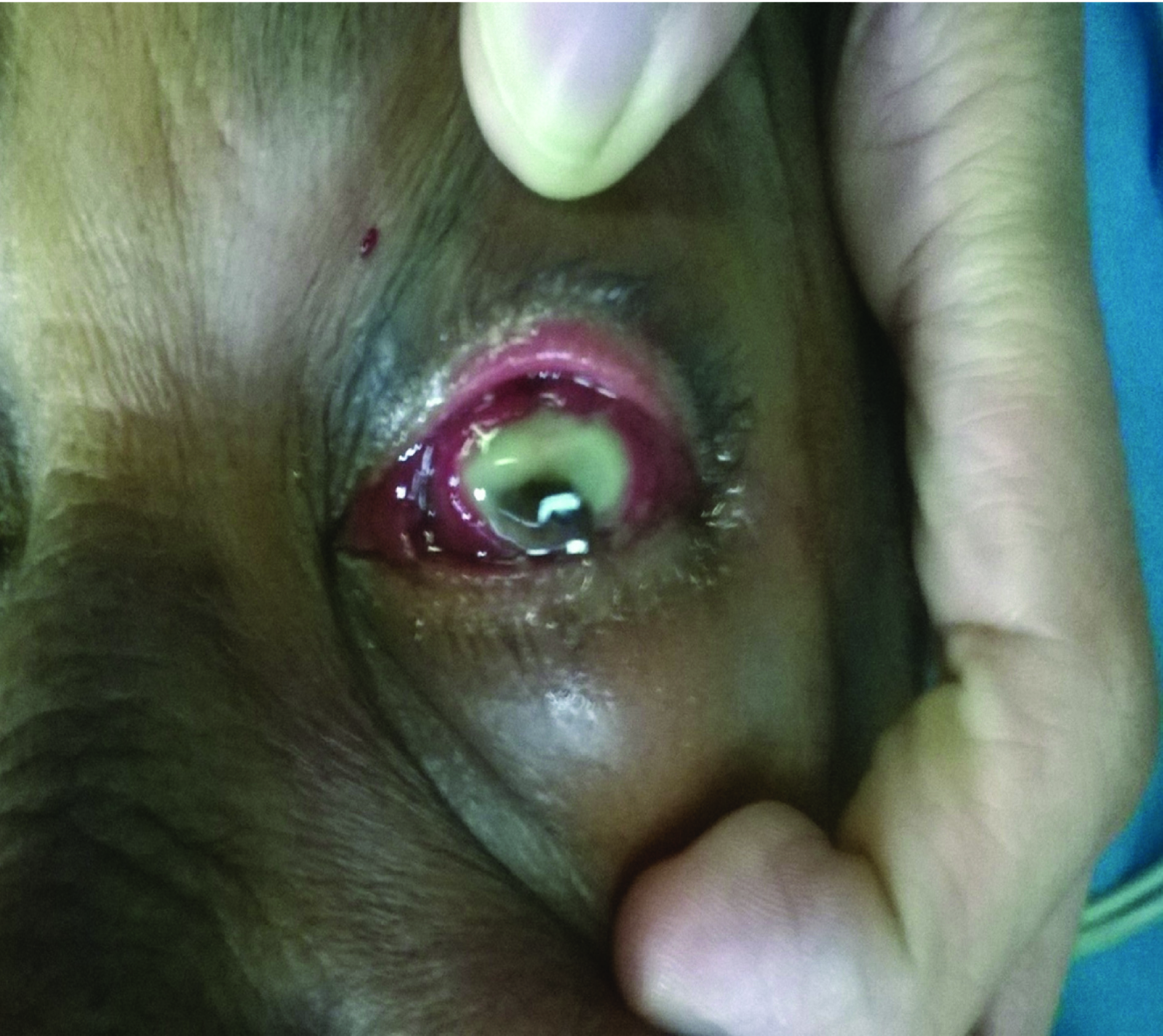
A 61-year-old lady reported with swelling and redness of left eye. The patient was a diagnosed case of multiple myeloma based on anaemia, neutropaenia and bone pain and confirmed with serum electrophpresis 2 years back, had completed three cycles of chaemotherapy with melphalan, dexamethasone and bortezomib and uncontrolled diabetes mellitus (HbA1C: 12.7) on insulin of 8 years duration and hypothyroidism presented with a four day history of rapid onset loss of vision, pain and redness of the left eye. There was no history of injury or entry of a foreign body. She also complained of pain and swelling in left arm and inability to move it. General examination revealed a pulse of 78/min, BP 140/80mm Hg and left arm cellulitis. Ocular examination showed visual acuity of 6/36 in right eye and no perception of light in left eye. Anterior segment of the right eye showed a clear cornea, briskly reacting pupil and an immature cataract, whereas in the left eye, ductions were restricted in all directions, lids were edematous, conjunctiva was congested and chemosed, and showed total corneal sloughing [Table/Fig-1]. Fundus was normal in the right eye. B scan of the left eye revealed 9X5mm serous choroidal detachment in the temporal aspect of the globe and confirmed panophthalmitis and ruled out uveal plasmacytomas. Blood investigations revealed Haemoglobin 11g%, a total count of 8,800/mm3, platelets of 1.6 lac and an ESR 99/hr. Differential count was suggestive of neutrophilia with toxic granules.
Left eye & ductions were restricted in all directions, lids were edematous, conjunctiva was congested and chemosed showing total corneal sloughing.
She was started on intravenous Clindamycin and Metronidazole. Ultrasound of the wrist joint showed only soft tissue swelling ruling out septic arthritis. As the patient did not consent evisceration at this stage, surgery was postponed. The next day the cornea perforated in the central two thirds and showed iris prolapse. Then she underwent evisceration of the left eye. Centre of the cornea showed a 5mm long horizontal perforation with iris prolapse. Lens was found dislocated in the vitreous cavity. Vitreous culture and blood culture did not show any growth. The cellulitis of the arm was controlled. The patient was followed up regularly till 3 months and has been fitted with a prosthesis.
Discussion
Multiple myeloma is a malignant proliferation of plasma cells originating from a single clone. It is caused by infiltration of bone marrow by bone marrow-derived plasma cells followed by the production of monoclonal Ig by these cells. The characteristic signs of myeloma include bone pain, hypercalcaemia, anaemia, renal failure and hyperviscosity syndrome. Symptomatic hyperviscosity is observed in 2–6% of patients with multiple myeloma [1]. Multiple myeloma causes morbidity by various mechanisms: abnormal immunoglobulins leading to decreased immunity, pancytopaenia and susceptibility to infections and bleeding tendencies, plasmacytomas and metatstases and pathological fractures.
Decreased synthesis and increased catabolism of normal immunoglobulins leads to hypogammaglobulinaemia. As a result, these patients are prone to bacterial infections, especially the Pneumococcus, Klebsiella pneumoniae, and Staphylococcus aureus affecting the lung and Escherichia coli and other gram-negative pathogens affecting the urinary tract. The infections; caused by bacteria, fungi and viruses; have been described in patients with multiple myeloma are pneumonia, sinusitis, discitis, osteomyelitis, septic arthritis, cellulitis, septic thrombophlebitis, colitis, abdominal abscess and periodontal abscess [2].
Manifestations of multiple myeloma can be seen in all structures of the eye, including both anterior and posterior segment. Bilateral eyelid ecchymoses was one of the first signs in detecting multiple myeloma, as described in a patient [3]. Multiple myeloma has also presented initially with recurrent subconjunctival haemorrhages [4]. Deposition of amyloid crystals in all layers of cornea has been reported [3].
Multiple myeloma has posterior segment manifestations too. Bilateral central retinal venous occlusions [5], Purtscher’s retinopathy [6] have been reported as initial manifestation of multiple myeloma. Endogenous endophthalmitis due to Pneumococcus has been reported in multiple myeloma [7]. Panophthalmitis is inflammation of all the coats of the eyeball. It is characterized by loss of vision, restricted eye movements and proptosis. The most important cause is entry of an exogenous infective etiology, usually following trauma. However, endogenous endophthalmitis and panophthalmitis do occur in immunocompromised state. This patient had cellulitis of the left upper arm concurrent with panophthalmitis. However, blood culture and vitreous culture were inconclusive, probably as the patient had already been started on intravenous antibiotics prior to being referred to us. Spontaneous panophthalmitis has been reported in uncontrolled insulin dependent diabetes [8] and urosepsis [9]. Panophthalmitis has a poor prognosis, and is one of the few indications of evisceration. The immune status of the patient was crippled by two morbidities- Multiple myeloma and diabetes mellitus, which was accentuated by steroids. The extent of contribution of each in causing panophthalmitis cannot be commented upon.
Conclusion
Multiple myeloma predisposes the patients to a wide spectrum of infections, which in this case was further exaggerated by uncontrolled diabetes mellitus. This case report highlights the propensity of ocular damage that can come across in Multiple myeloma.
[1]. Mehta J, Singhal S, Hyperviscosity syndrome in plasma cell dyscrasiasSemin Thromb Haemost 2003 29(5):467-71. [Google Scholar]
[2]. Mahfouz T, Miceli MH, Saghafifar F, Stroud S, Jones-Jackson L, Walker R, 18F-Fluorodeoxyglucose positron emission tomography contributes to the diagnosis and management of infections in patients with multiple myeloma: a study of 165 infectious episodesJ Clin Oncol 2005 23:7857-63. [Google Scholar]
[3]. Goldstein DA, Schteingart MT, Birnbaum AD, Tessler HH, Bilateral eyelid ecchymoses and corneal crystals: an unusual presentation of multiple myelomaCornea 2005 24(6):757-58. [Google Scholar]
[4]. Felipe AF, Nottage JM, Rapuano CJ, Recurrent bilateral subconjunctival haemorrhage as an initial presentation of multiple myelomaOman Journal of Ophthalmology 2012 5(2):133-34. [Google Scholar]
[5]. Kamath SJ, Kamath GM, Roopashree Bilateral simultaneous central retinal vein occlusion presenting as initial manifestation of multiple myelomaOnline J Health Allied Scs 2014 13(2):9Available at URL: http://www.ojhas.org/issue50/2014-2-9.html [Google Scholar]
[6]. Nautiyal A, Amescua G, Jameson A, Gradowski JF, Hong F, Doft B, Sudden loss of vision: Purtscher retinopathy in multiple myelomaCan med acad J 2009 181(12) [Google Scholar]
[7]. Baker TR, Spencer WH, Ocular findings in multiple myeloma: a report of two CasesArch Ophthalmol 1974 91(2):110-13. [Google Scholar]
[8]. Hassan NA, Al-Baqsomi A, Reddy MA, Endogenous Staphylococcus aureus panophthalmitisMiddle East Afr J Ophthalmol 2007 14:32-4. [Google Scholar]
[9]. Murthy TA, Rangappa P, Rao S, Rao K, ESBL E. coli Urosepsis resulting in endogenous panophthalmitis requiring evisceration of the eye in a diabetic patientCase Reports in Infectious Diseases 2015 2015:897245 [Google Scholar]