Success of any endodontic treatment depends on strict adherence to ‘endodontic triad’. Preparation of root canal system is recognized as being one of the most important stages in the root canal treatment [1]. Thus, successful endodontic treatment requires predictably shaped root canals to facilitate three dimensional obturation.
At times we inevitably end up damaging the root dentin which becomes Gateway for infections like perforation, zipping, dentinal cracks and minute intricate fractures, thereby resulting in failure of treatment.
The development of nickel–titanium alloys for the manufacture of manual instruments initially and then rotary endodontic instruments, has brought about a complete revolution in endodontics over the last few years. These systems differ from each other in the design of cutting blades, body taper, metallurgy, tip configuration. Despite the obvious advantages of these instruments over hand instrumentation, these NiTi have tendency to fracture without any warning [2].
A new NiTi rotary instrument ProTaper Next (PTN) is machined from a wire (termed M-wire) by subjecting it to a proprietary novel thermo- mechanical processing. It is said that this new M-wire instrument has considerably improved flexibility compared to conventional rotary instruments that are machined from super elastic (SE) austenitic NiTi wire – ProTaper Universal (PTU) [3].
Also, till date, not many studies have reported the relationship between instrumentation length and development of apical root cracks.
The aim of the study was to evaluate and compare the incidence of apical root crack formation after root canal preparation with hand and newer rotary files and to evaluate the potential effect of various endodontic instrumentation lengths on crack formation in apical root dentin.
Materials and Methods
The present in vitro study was carried out in the Department of Conservative Dentistry and Endodontics at JSS Dental College, Mysuru from April 2015 to September 2015 after the approval by the Ethical Committee of the institution.
A) Selection of teeth
A total of 70 freshly extracted premolars with complete apices and single, straight root and root canal were selected and stored in 0.1% thymol. Pre operative radiographs using RVG was taken to exclude teeth with curved roots and anatomic irregularities.
B) Tooth preparation
i) The roots were wrapped with a single layer of aluminium foil and embedded in auto polymerizing resin set in an aluminium hollow block. Aluminium foil was then peeled off. The prepared acrylic blocks were cut towards the apical end such that 1-2mm of apical root portion was exposed.
ii) The root surface and acrylic “socket” were coated with a hydrophilic vinyl polysiloxane impression material, and the root was repositioned immediately. The teeth were decoronated to ensure straight line access, to provide a reference plane and uniformity of root lengths of 16mm.
iii) The root tip was cut approximately 1mm and stained with methylene blue for better visualization under the stereomicroscope (X20). Initial photographs of cut apex were taken using stereomicroscope (X20) connected to a computer. The roots were examined for cracks and discarded if many cracks were present. In case of few cracks, the teeth were included in the study and the position of cracks were noted.
iv) The teeth were randomly distributed into six experimental and a control group of 10 teeth per group. A 10 size K file was inserted into the canal until the tip of the file was visible at resected root tip. The distance between the reference point and the tip of the file was defined as Root Canal Length (RCL).
C) Root canal preparation
Teeth in control group were left unprepared. Teeth in sub groups A and B were instrumented using Stainless Steel (SS) files up to RCL and (RCL-1mm) respectively. Teeth in sub groups C and D were instrumented using ProTaper Universal (NiTi) rotary files up to RCL and (RCL-1mm) respectively. Teeth in sub groups E and F were instrumented using ProTaper Next (M wire) rotary files up to RCL and (RCL-1mm) respectively [Table/Fig-1]. All the samples were examined under stereomicroscope with 20X magnification and photographs of samples taken both pre–operative and post–operative at incremental sequence of file sizes.
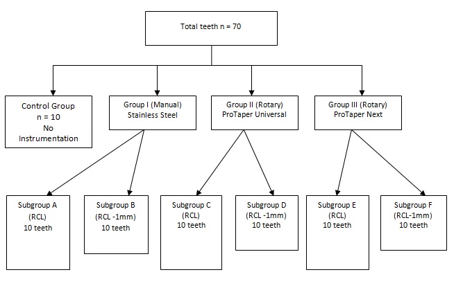
Grouping: Total teeth (n) = 70
D) Evaluation
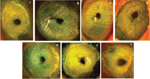
Once the instrumentation length was reached, teeth were stained with methylene blue and viewed under stereomicroscope (X20) and photographs were taken after each file change [Table/Fig-2a–2g]. Slide show was prepared and photographs were compared with preceding photographs and a note of cracks was made regarding presence or absence, number of cracks, originating at resected root tip and the file size inducing cracks was made. Roots were classified as ‘no crack formation’, ‘incomplete root crack formation – when the crack line extends from canal wall into dentin without reaching outer root surface; ‘propagation of existing crack – when there is crack visibly longer compared to previous image; ‘complete crack formation – when the crack extending from canal wall reaches the outer root surface. Samples were considered defected when any of the above mentioned condition was noted.
Crack formation on instrumentation. 2a: Control group, 2b: Sub group A, 2c: Sub group B, 2d: Sub group C, 2e: Sub group D, 2f: Sub group E, 2g: Sub group F
Statistical Analysis
The data was analysed statistically using descriptive analysis by ‘Phi’ and ‘Cramers’ (since data is categorical-crack present or absent) test to find out statistical significance between the groups and one-way analysis of variance (since data is continuous and more than two groups are present) was used to compare the number of crack formation between hand and rotary instrumentation methods and instrumentation lengths. All statistical analyses were performed at a 0.05% significance level.
Results
Control group showed no crack formation while experimental groups showed crack formation.
In group I, the samples were instrumented using stainless steel hand K files. Sub group ‘A’ which was instrumented up to RCL showed cracks in five out of 10 samples. Whereas, sub group ‘B’ which was instrumented 1mm short of RCL showed cracks in three out of 10 samples.
In group II, the samples were instrumented using ProTaper Universal rotary files. Sub group ‘C’ which was instrumented up to RCL showed cracks in three out of 10 samples. Whereas, sub group ‘D’ which was instrumented 1mm short of RCL showed cracks in three out of 10 samples (the results of sub group C were tabulated as 2 cracks since crack formed by file S2 was eliminated in order to standardise the file size amongst the experimental groups) [Table/Fig-2].
In group III, the samples were instrumented using ProTaper Next rotary files. Sub group ‘E’ which was instrumented up to RCL showed cracks in four out of 10 samples. Whereas, sub group ‘F’ which was instrumented 1mm short of RCL showed cracks in two out of 10 samples.
Stainless steel group showed the highest number of cracks followed by ProTaper Universal and ProTaper Next. But the statistical analysis showed no significant difference between the groups.
In all the groups, samples instrumented 1mm short of the Root Canal Length showed lesser number of cracks than samples instrumented up to RCL, although there was no statistical significance between the sub groups. Greater number of cracks was seen in samples instrumented with size #30 file compared to instrumentation done using #25 and #20 size files [Table/Fig-3].
File size producing crack in the affected teeth and total.
| Sub Groups | #20 | #25 | #30 | Total |
|---|
| A | - | 3 | 2 | 5 |
| B | 1 | 1 | 1 | 3 |
| C | - | 2 | 1 | 3 |
| D | - | - | 2 | 2 |
| E | - | 1 | 3 | 4 |
| F | - | - | 2 | 2 |
| Total | 1 | 7 | 11 | |
[Table/Fig-4] summarizes the mean number of teeth with apical cracks in relation to instrumentation length, although there was no statistically significant difference between the groups.
Mean number of teeth with apical crack in relation to instrumentation length.
| N | Mean | Std. Deviation | Minimum | Maximum | F value | p value* |
|---|
| Group A | 10 | .50 | .972 | 0 | 3 | 0.298 | 0.912 (NS) |
| Group B | 10 | .30 | .483 | 0 | 1 |
| Group C | 10 | .30 | .483 | 0 | 1 |
| Group D | 10 | .30 | .483 | 0 | 1 |
| Group E | 10 | .40 | .966 | 0 | 3 |
| Group F | 10 | .20 | .632 | 0 | 2 |
| Total | 60 | .33 | .681 | 0 | 3 |
* Test of Significance – ANOVA, NS: Non-significant
One way ANOVA showed no significant effect of hand and rotary instrumentation methods and instrumentation length on crack formation [Table/Fig-4].
Discussion
Endodontic treatment of an inflamed or infected tooth is beneficial in creating a healthy environment that is conducive to the tooth’s continual performance as a functional member of the masticatory apparatus. However it is also important to ensure that iatrogenic harm to the root dentin be minimized in order that the tooth is sufficiently strong for a long term function.
Studies have shown that the alloy from which the material is manufactured is a more important factor in determining the dentin damaging potential of single-file instruments than the motion of instrumentation [4]. Hence in the present study instruments made of different alloys – Stainless steel (K file), and two types of NiTi instruments – conventional NiTi (ProTaper Universal) and thermally treated NiTi (ProTaper Next) were investigated for their potential to cause crack formation.
Stainless steel K files are manufactured by twisting square or triangular metal blanks along their long axis, producing partly horizontal cutting blades and a negative rake angle. ISO standardized K files are available in lengths 21, 25 & 31mm and a standard taper of 0.32 over 16mm of cutting blades. The tip size increases by 0.05mm for file sizes #10 to #60; for sizes #60 to #140, the absolute increase is 0.1mm [5].
The ProTaper Universal instrument having tip diameters of S1–0.185mm, S2–0.2mm, F1- 0.2mm, F2-0.25mm, F3-0.3mm, F4-0.4mm, F5-0.5mm respectively, and apical taper of 0.02%, 0.04%, 0.07%, 0.08%, 0.09%, 0.06% and 0.05% respectively also has a convex triangular cross sectional design, a non-cutting safety tip and a negative rake angle.
ProTaper Next (PTN) has an off–centred rectangular cross section which gives it a snake like swaggering movement. They have a negative rake angle and are available in different sizes, respectively. They have variable taper along the instruments’ long axes [6].
Canal preparation was standardized to master apical file size #30 in all the groups as the apical region was the prime area of focus for observing the crack formation. External reinforcement was avoided using a thin layer of silicone to simulate periodontal ligament [7]. Because an “exposed” apex is not uncommon in teeth with chronic apical periodontitis or periapical cysts [7], the apical 2mm to 3mm portion of the root was exposed to allow for intraoperative image recordings. Approximately 1mm of the apical tip was resected in order to gain flat surface and better visualization under stereomicroscope. Also flattening of the apex facilitates exposure of dentin tissues where in crack formation could be appreciated better and the working length determined accurately.
Higher concentrations of NaOCl solution significantly decrease the elastic modulus and flexural strength of human dentin compared with physiologic saline, and solutions of lower concentrations [8]. Hence, in the present study, a 1% NaOCl solution was used for irrigation.
The molecular size of methylene blue is 120nm which is much smaller than the size of a bacterium. Since methylene blue has a low molecular weight (318.85) which is even lower than basic fuschin (323.45), it penetrates more deeply than other dyes [5]. Hence, in the present study, methylene blue was used for staining the specimens.
The results of the present study [Table/Fig-3] showed that, stainless steel hand K files showed the highest number of crack formation followed by ProTaper Universal and ProTaper Next. Although there was no statistically significant difference between the groups, the increase in the incidence of crack formation in the stainless steel hand group may be attributed to the rigidity of the instrument in contrast to the other two instruments ProTaper Universal and ProTaper Next, which are more flexible. A recent finite element analysis study concluded that stiffer file designs generate higher stress concentration in the apical root dentin, which could lead to higher risk of crack initiation [9]. The crack formation using ProTaper Universal and ProTaper Next did not show any difference which is in contrast to other studies which showed that ProTaper Next resulted in fewer cracks compared to ProTaper Universal [10]. A probable reason could be the variation in the study model between the two studies where they evaluated the crack formation at 3mm, 6mm, and 9mm of the roots but in the present study only the apical resected portion was examined. Also, despite having the advantage of M–wire technology and lesser taper compared to ProTaper Universal. ProTaper Next has an offset mass of rotation which generates a mechanical wave of motion analogous to the oscillation noted along a sinusoidal wave. As a result of this design, ProTaper Next file cuts a bigger envelope of motion compared to a similarly-sized file with a symmetrical mass and axis of rotation [11].
The results [Table/Fig-4] of this study indicate that the crack is more likely to appear when the working length was Root Canal Length (RCL) than 1mm short of Root Canal Length (RCL- 1mm). Conservation of the dentin adjacent to the apical root canal is crucial to maintain strength and fracture resistance of the tooth structure. Furthermore, because of the proximity to the apical foramen, the file tips reaching RCL has caused cracks. On the other hand, the file tips reaching (RCL-1mm) had a sufficient amount of dentin around the file tip to resist the formation of cracks, although cracks were found in few samples [10].
In the present study, the incidence of crack formation increased with increasing size of the instrument. Since file size #15 is not present in the rotary file groups, file size #15 is eliminated while tabulating the results in [Table/Fig-3] in order to standardize the file sizes between the experimental groups. Samples instrumented with size #30 file showed more number of cracks when compared to samples instrumented with #25 and #20 size files [Table/Fig-3]. This could be explained by the fact that as the size of the instrument increases, the rigidity of the instrument increases, which causes increased crack formation [11].
The absence of coverage of the apex during instrumentation procedures because the periodontal ligament might “protect” the apex against crack initiation. Also, the thickness of the cellular cementum increases with age [7]. Additionally, compensatory cementum deposition, which occurs in the apical area to counter balance occlusal attrition, might cover the crack and it’s unknown whether or not it might limit its progression [7]. Methylene blue stain could not be washed away completely from the samples in between subsequent staining using alcohol and water which may lead to misinterpretation as additional crack.
Conclusion
Within the limitations of the study, the samples of all the experimental groups showed crack formation irrespective of hand or rotary instrumentation technique used. Samples instrumented with stainless steel hand files showed highest number of cracks followed by ProTaper Universal and ProTaper Next. Although lesser number of cracks was anticipated in samples instrumented with ProTaper Next - a thermally treated unique NiTi compared to ProTaper Universal - a conventional NiTi instrument, the present study showed equal number of cracks on instrumentation with ProTaper Universal and ProTaper Next. Samples instrumented upto 1mm short of root canal length showed less number of cracks compared to samples instrumented upto root canal length. Increase in the number of crack formation was seen when the samples were instrumented with size #30 file compared to #25 and #20 size files.
* Test of Significance – ANOVA, NS: Non-significant