Unilateral Existence of Chondro-epitrochlearis: Its Embryological Perspectives and Clinical Implications
Abhinitha Padavinangadi1, Naveen Kumar2, Mohandas KG Rao3, Satheesha B Nayak4
1 Lecturer, Department of Anatomy, Melaka Manipal Medical College, Manipal Campus, Manipal University, Manipal, India.
2 Assistant Professor, Department of Anatomy, Melaka Manipal Medical College, Manipal Campus, Manipal University, Manipal, India.
3 Professor, Department of Anatomy, Melaka Manipal Medical College, Manipal Campus, Manipal University, Manipal, India.
4 Professor, Department of Anatomy, Melaka Manipal Medical College, Manipal Campus, Manipal University, Manipal, India.
NAME, ADDRESS, E-MAIL ID OF THE CORRESPONDING AUTHOR: Dr. Naveen Kumar, Assistant Professor, Department of Anatomy, Melaka Manipal Medical College, Manipal Campus, Manipal University, Manipal, India.
E-mail: naveentonse@gmail.com
Chondroepitrochlearis (CET) is an anomalous muscular slip that originates from the pectoralis major muscle and inserts into epicondyle of the humerus. The morphology of this variant form of pectoralis major can vary from slender to strong musculo-tendinous. In its course, it usually crosses the neurovascular structures of arm; their compression is a major complication that could be manifested by its persistence. In the present case, potentially anomalous CET muscle with the slender slip of origin, but strong tendinous insertion to the medial epicondyle of the humerus was found unilaterally. This musculo-tendinous structure was found to be compressing the brachial artery and median nerve in the arm. Detailed embryological and clinical perspective of such variant muscular slip helps the physiotherapists, orthopaedicians in their treatment strategy in complain of restricted shoulder movement. It may also help the neurologists, radiologists in their diagnostic approach of ulnar neuropathy.
Pectoralis major, Shoulder movement, Ulnar neuropathy
Case Report
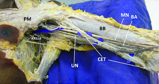
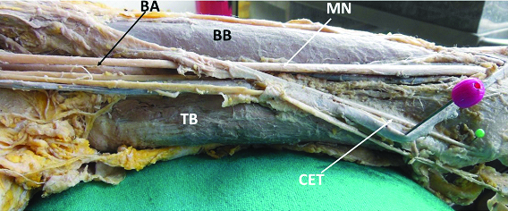
During routine cadaveric dissection for undergraduate medical students we observed a variant muscle slip known as chondroepitrochlearis on the left upper limb in a male cadaver aged about 60 years. It was arising from the lower border of the pectoralis major muscle near its insertion. This muscular slip then descended downwards and obliquely in relation to medial side of the arm and superficial to biceps brachii and corachobrachialis muscles [Table/Fig-1]. On its way to medial epicondyle, it crossed the median nerve, brachial artery and ulnar nerve superficially from lateral to medial side. In the middle of the medial side of the arm it formed a strong tendon and covered by the deep fascia of the arm with the enclosure of medial cutaneous nerve of the forearm. The tendon was inserted to medial epicondyle of the humerus near the common origin of flexors of forearm [Table/Fig-2].
Dissection of the flexor compartment of the left arm showing the morphology of chondroepitrochlearis (CET) muscle. PM- Pectoralis Major Muscle, BB- Biceps brachii, MN –Median Nerve, BA- Brachial artery, UN- Ulnar Nerve.
Showing the insertion of Chondroepitrochlearis (CET). BA- Brachial artery, BB- Biceps brachii, MN – Median Nerve, TB- Triceps brachii.
Discussion
Chondroepitrochlearis (CET), as described by Bergmann is an anomalous muscular slip arising from one or more ribs, crossing the axilla and inserting into medial humeral epicondyle or medial intermuscular septum [1]. There have been cases reported both of unilateral [2,3] and bilateral [4–6] occurrence of the CET with, and without, the presence of an axillary arch muscle. CET reported in a small number of cases as an unusual muscular variation. The muscle is related phylogenetically to the patagii muscle in birds and abdomino humeralis or to the xiphihumeralis in quadrupeds [7]. Presence of CET has also been linked with chromosomal defects namely trisomy 13 with a DD translocation as evidenced by the autopsy study done on infants [8].
During embryogenesis, the genetic code for such atavistic muscle is normally suppressed as postulated by Barash et al., [9]. In about fifth week of intrauterine life, the pectoral musculature is derived from the ventral limb bud masses from the myoblasts. These myoblasts eventually migrate out of the last five cervical and the first thoracic myotomes into the developing limb buds. Through a combination of migration, fusion and apoptosis of the muscle cell precursors, the pectoral muscles assume their final forms. The persistence of a muscle slip in the form of the CET results due to the failure of apoptosis to occur at the right time in the myoblasts [10]. It has also been considered that, CET muscle slip is the result of absence of the twisted insertion of the pectoralis major [7].
Anomalous CET having two heads, one ventral and one dorsal head has been reported by Nakajima et al., [3]. In this case, the greater part of the dorsal head and the ventral head was innervated by the pectoral nerves, whereas, small part of the dorsal head was innervated by the intercosto-brachial nerve. Due to its unusual double head, the ventral head was considered to be the CET muscle, while the dorsal head was an aberrant type of the muscular arch because of its similar attachment and innervations as ventral head [3]. Bilateral presence of CET with the prevalence of 2% of population has been reported [7]. Presence of CET could restrict the abduction of the humerus and might result in ulnar nerve entrapment as reported by Levent et al., [11] and it can even cause axillary vein thrombosis and lymphedema [12]. Voto and Weiner reported a clinical scenario with contracture of the CET in an infant [13]. Trobs et al., reported a case of CET as a persistence of phylogenetic remnant in an infant causing abnormal shoulder shape [14]. Therefore, detailed information on CET is not only imperative in the interpretation of magnetic resonance but also during reconstructive surgeries of the muscles.
Conclusion
In the present case, though the origin of CET muscle as a slender muscular slip, as it proceeded further towards its distal attachment it became more muscular and tendinous. Having such morphological transformation from its origin to insertion site will have all possibilities of restricted movements at shoulder joint as well as ulnar neuropathy. Therefore, reporting this unique muscular variation helps the physiotherapists, orthopedicians in their treatment strategy in complain of restricted shoulder movement. It may also help the neurologists, radiologists in their diagnostic approach of ulnar neuropathy.
[1]. Bergman RA, Thompson SA, Afifi AK, Saadah FA, Compendium of human anatomic variation 1988 BaltimoreUrban and Schwarzenberg:264-66. [Google Scholar]
[2]. Chiba S, Suzuki T, Kasai T, A rare anomaly of the pectoralis major chondroepitrochlearisOkajimas Folia Anat Jpn 1983 60(2-3):175-85. [Google Scholar]
[3]. Nakajima K, Chiba S, Kobayashi K, Wakatuki E, Kumaki K, Hoshino T, A rare muscular anomaly in the upper arm–the chondroepitrochlearis muscle with an aberrant type of muscular arch of axillaActa Anat Nippon 1999 74:209-13. [Google Scholar]
[4]. Di Gennaro GL, Soncini G, Andrisano A, Valdiserri, The chondroepitrochlearis muscle: case reportChir Organ Mov 1998 83:419-23. [Google Scholar]
[5]. Flaherty G, O’Neill MN, Folan-Curran J, Case report: Bilateral occurrence of a chondroepitrochlearis muscleJ Anat 1999 194:313-15. [Google Scholar]
[6]. Sarikcioglu L, Fatos BY, Chiba S, Unilateral occurrence of chondroepitrochlearisMuscle Clin Anat 2004 17:272-75. [Google Scholar]
[7]. Aruna S, Hannah Sugirthabai Rajila R, Vaithianathan G, Bilateral chondroepitrochlearis muscle: incidence, phylogenetic and clinical significanceJCDR 2011 5(1):31-34. [Google Scholar]
[8]. Aziz MA, Anatomical defects in a case of trisomy 13 with a DD translocationTeratology 1980 22:217-27. [Google Scholar]
[9]. Barash BA, Freedman L, Opitz JM, Anatomical studies in the 18-trisomy syndromeBirth Defects, Original Article Series 1970 V1(4):3-15. [Google Scholar]
[10]. Sweeney LJ, Basic concepts in embryology 1998 McGraw-Hill:136-38. [Google Scholar]
[11]. Sarikcioglu L, Fatos Belgin Y, Shoji C, Unilateral occurrence of a chondroepitrochlearis muscleClinical Anatomy 2004 17(3):272-75. [Google Scholar]
[12]. Thomet C, Vankerckhove S, Belgrado JP, Vandermeeren L, The chondroepitrochlearis muscle: a rare cause of axillary vein thrombosis and lymphedemaLymphology 2015 available at http://hdl.handle.net/2013/ULB-DIPOT:oai:dipot.ulb.ac.be:2013/217683 [Google Scholar]
[13]. Voto SJ, Weiner Ds, The chondro-epitrochlearis muscleJournal of Pediatric Orthopedics 1987 7:213-14. [Google Scholar]
[14]. Trobs RB, Gharavi B, Neid M, Cernaianu G, Chondroepitrochlearis muscle – A phylogenetic remnant with clinical importanceKlin Padiatr 2015 227(4):243-46. [Google Scholar]