One of the major causes for mass morbidity and mortality throughout the world is due to major fire accidents and identification of burnt bodies during these accidents becomes a difficult task [1]. In such instances investigation of the burnt bodies for the identification of the victims starts with the remnant objects found at the site of investigation. As the soft tissue destruction is more evident, victim identification becomes nearly impossible in majority of fire accidents. In these circumstances hard tissue remnants like skeletal and dental evidences may provide a clue in victim identification. In the human body teeth have highest resistance to severe environmental effects like fire, desiccation and decomposition, hence considered as most indestructible components. A tooth serves as a tool for positive personal identification, as they survive in most of natural disasters where victim bodies are in unrecognizable state [2].
In forensic context precise knowledge on physical and histological changes of teeth subjected to high temperatures is of great importance. In crime and forensic investigation, the type and severity of structural damage provides valuable clues, especially when only a dental remnant serves as evidence. Most of the damaged features of the oral tissues and dental restorations can be observed directly by the naked eye, but electron microscopic investigation can be very useful mainly in two ways: firstly to identify the changes that the dental tissues have undergone so that temperatures they were exposed to can be estimated and; secondly different types of dental treatments can be characterized [3]. A very few studies have been found in literature regarding the physical, histological and ultra-structural changes in teeth subjected to high temperatures [4,5]. In a short study performed previously [4], histologic findings were correlated to the temperature and as an extension to it the present study was undertaken to investigate structural damage in freshly extracted teeth to heating, at different temperatures for a certain length of time in laboratory and; physical and ultra-structural findings were correlated to the temperature.
Materials and Methods
Freshly extracted 60 permanent teeth were collected from an age group of individuals between 20-30 years. This particular age group was taken because the rate of dental caries and periodontal pathology is less in this age group compared to elderly age group. The physically and anatomically normal teeth were included, and grossly decayed, restored and endodontically treated teeth were not included in the study.
The extracted teeth were debrided with 3% hydrogen peroxide solution, rinsed thoroughly with tap water and fixed in 10% formalin. The pre SEM analysis tooth preparation like debridement and fixation was done in histopathology laboratory where as heating teeth to different temperatures was done in the Prosthodontics laboratory, Kamineni Institute of Dental Sciences, Telangana, India and final SEM analysis was performed in National Institution of Nutrition (NIN), Hyderabad.
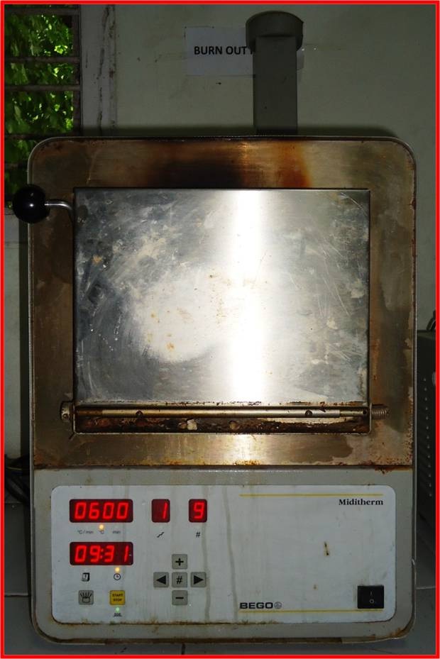
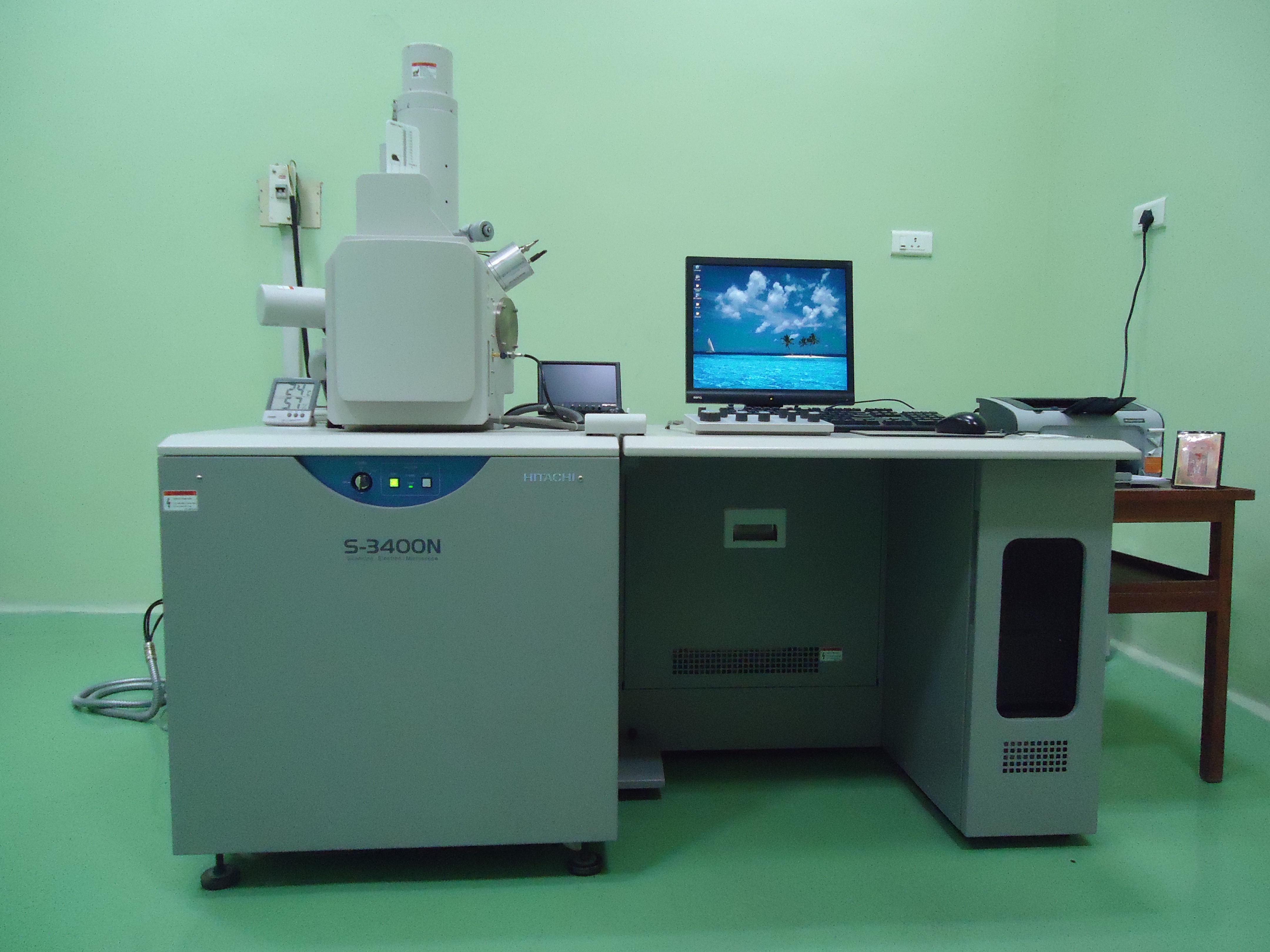
The collected teeth were categorized into four groups, Group A comprised of control group which included teeth that were not subjected to heat whereas Group B, C and D (study groups) comprised of teeth that were subjected to different temperatures i.e., 100oC, 300oC and 600oC respectively for a time of fifteen minutes in laboratory furnace (Model-BEGO- MIDITHERM) [Table/Fig-1], after which they were processed for SEM examination. Each group included 15 teeth; 5 anteriors, 5 premolars and 5 molars. The SEM (Model-HITACHI S-3400N) [Table/Fig-2] analysis was done at NIN, Hyderabad, India over a period of three months.
Laboratory furnace (Model- BEGO@MIDITHERM) used in the present study.
Scanning electron microscope (Model-HITACHI S-3400N) used in the present study.
During heating of teeth to different temperatures, the heat should be equally distributed to all the surfaces of the teeth. In order to achieve this, a custom made steel tray was designed in such a way that it could with stand high temperatures, and loops of tray holds the teeth in upright position in the furnace which renders equal distribution of heat around teeth. The tray holding the teeth in upright position was placed in furnace, where the heating is done gradually to different temperatures as specified above. After specified temperature is reached, it is held for fifteen minutes, followed by gradual cooling to room temperature [6].
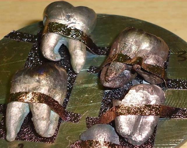
The physical changes like color, contour, texture and morphology of burnt teeth was examined after they were removed from furnace. After thorough physical examination each burnt tooth sample of study groups and tooth of control group was kept in vacuum desiccator for removal of moisture remnants and coated with electrically conductive material namely gold of 600 Ao thickness by low vacuum ion sputter technique for scanning electron microscopy analysis [Table/Fig-3]. The teeth samples were examined by SEM under magnifications of 9X, 500X, 1.2KX, 2.5KX for the evaluation of enamel surface (crown); and 9X, 250X, 500X, 1KX for the evaluation of cementum surface (root).
Results
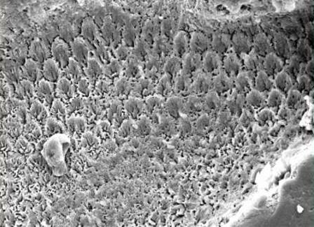
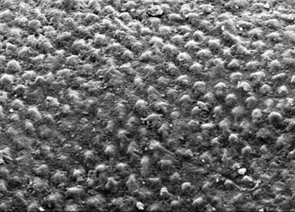
Enamel: The enamel surface of teeth unexposed to heat (Group A) when examined under SEM showed characteristic fish scale/keyhole pattern of enamel rods [Table/Fig-4]. At 1000C, there were no significant changes in the morphology of the teeth (Group B). On examination under SEM the characteristic fish scale pattern of enamel rods was lost and instead they appeared as pebbles on enamel surface [Table/Fig-5].
Incinerated teeth coated with gold of 600 Ao thickness by low vacuum sputter technique for scanning electron microscopy.
The enamel surface of teeth unexposed to heat (Group A) showing characteristic fish scale/keyhole pattern when examined under SEM (1.2 KX).
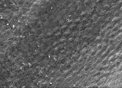
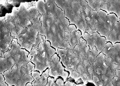
At 300oC, there were significant changes in the morphology of teeth (Group C) where the color of crown has changed to creamish yellow. Under SEM enamel rods on enamel surface appeared as granules [Table/Fig-6]. At 600oC, the color of the teeth (Group D) appeared black like coal, enamel completely came out like a cap and became very fragile. Under SEM surface of enamel showed cracks and the enamel rods appeared as tiny dots [Table/Fig-7]. With increase in temperature the enamel fragments became consistently smaller.
The enamel surface of teeth exposed at 100oC (Group B) shows enamel rods as pebbles on enamel surface when examined under SEM (1.2 KX).
The enamel surface of teeth exposed at 300oC (Group C) shows enamel rods on enamel surface as granules when examined under SEM (1.2 KX).
The enamel surface of teeth exposed at 600oC (Group D) shows the enamel rods as tiny dots when examined under SEM (1.2 KX).
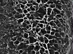
Cementum: The root surface of teeth unexposed to heat (Group A) when examined under SEM showed characteristic coral pattern [Table/Fig-8]. At 100oC, there were no significant changes in the morphology of the teeth (Group B) and on examination under SEM, cementum surface showed the characteristic soap bubble pattern [Table/Fig-9].
The root surface of teeth unexposed to heat (Group A) showing characteristic coral pattern when examined under SEM (1.0 KX).
The root surface of teeth exposed at 100oC (Group B) shows characteristic soap bubble pattern when examined under SEM (1.0 KX).
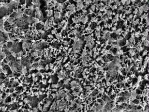
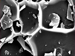
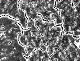
At 300oC, there were significant changes in the morphology of the teeth (Group C) where the root colour changed to brownish black and under SEM, cementum surface showed the characteristic honey comb pattern [Table/Fig-10]. At 600oC, the color of the teeth (group D) appeared black like a coal and under SEM surface of cementum showed cracks and characteristic snail track pattern [Table/Fig-11].
The root surface of teeth exposed at 300oC (Group C) shows characteristic honey comb pattern when examined under SEM (1.0 KX).
The root surface of teeth exposed at 600oC (Group D) shows characteristic snail track pattern when examined under SEM (1.0 KX).
Discussion
Identification of victims in mass fire disasters is particularly based on hard tissue remnants like teeth and bone. The destruction of bodies is temperature dependent [1]. The temperature of fire accidents varies according to site involved i.e., in closed environments temperature is more compared to open environments. It is also dependent on the oxidant involved, duration of exposure to fire and the burning atmosphere that may have a considerable effect on teeth. For example a house hold fire reaches to a maximum temperature of 1200°F (649°C), whereas cremation occurs at temperatures between 871-982°C and combustion of petrol occurs at a temperature of 1600-1800°F (800-1100°C) [7,8]. The present study was carried out on freshly extracted permanent teeth to understand the ultra-structural changes that occur in the teeth when subjected to different temperatures. Only permanent teeth were included in the present study because deciduous teeth will not be available with anatomically and morphologically sound structure as they have a tendency for root resorption and the study of root morphology becomes difficult.
In the human body teeth have highest resistance to severe environmental effects like fire, dessication and decomposition, hence considered as most indestructible components; this is because of their complex structure and composition. Enamel has 96% inorganic and 4% organic content, where as cementum contains 45% to 50% inorganic material and 50% to 55% organic content. The chief inorganic content is hydroxyappetite (Ca10(PO4)6(OH)2) [9]. Hydroxyapatite (HAP) has two types of water in its structure adsorbed and lattice water [10]. Adsorbed water shows characterized reversibility, thermal instability from 250C to 2000C, and weight loss without any effect on lattice parameters. Lattice water is irreversibly lost at the temperature of 2000C to 4000C, which causes a contraction in the α-lattice dimension during heating. At higher temperature, the hydroxyapatite gradually dehydrates. Complete dehydration of HAP cause lattice destruction, giving rise to a mixture of Tricalcium phosphate and Tetracalcium phosphate [11].
Examination of incinerated restored dental hard tissues can be performed by normal simple microscope, but SEM reveals distinct surface changes, grooves and striations that were formed from the use of a dental drill [12]. Linear and cross-sectional views of dentinal tubules have been identified by SEM in almost totally destroyed human dental remains, confirming that the residual fragments were teeth [13]. Considering the above facts the present study was aimed to investigate structural damage in freshly extracted teeth to heating, at different temperatures for a certain length of time in laboratory using SEM.
In the present study as the teeth were exposed to different temperatures such as 100oC, 300oC and 600oC, the normal fish scale/keyhole pattern of enamel rods changed to pebbled, granular appearance and finally became a tiny dot in appearance along with surface cracks. This change of appearance is due to loss of water from the hydroxyapetite because of which there is shrinkage of enamel rods. In case of cementum there is loss of collagenous matrix as well as loss of water from hydroxyapetite crystals leading to destruction of cementum layer and exposure of dentinal tubules. The soap bubble pattern in cementum is due to destruction of cementum layer by incineration leading to exposure of dentinal tubules. On further more exposure to heat, there was increase in the loss of lattice water leading to expansion of exposed dentinal tubules giving a characteristic honey comb appearance. When exposed to further higher temperatures the surface cracks are seen with loss of dentinal tubular structure showing snail track pattern in cementum which is attributed to complete loss of lattice water from HAP and formation of Tricalcium phosphate and Tetracalcium phosphate. Similar characteristic honey comb pattern of cementum was reported by Chetan A Pol et al., in their study [5].
It is evident from the present study that at specific temperatures specific patterns were evident which can be correlated easily to estimate fire temperature ranges during investigative procedures. The present study signifies specific pattern of ultra structural changes, exhibited by enamel and cementum upon incinerating the teeth, helping in estimation of temperatures with a minimum margin of error. In most of fire accidents burnt bodies does not give any clue of soft tissue remnants but in these cases teeth are the only recognizable structures as they are well mineralized and with stand the thermal effect, thus teeth have great value in fire investigations and provide clue to understand the chain of events that occurred during fire accidents. Macroscopic examination of teeth found at site of incidence and further SEM analysis of the same makes easy in identification of victims. SEM as a whole has additional benefits in investigations as it is not associated with complex procedural skills, direct examination of evidence and accuracy in its identification are trust worthy factors in considering SEM as positive tool for investigative procedures. As SEM is more sensitive in predicting the results small remnants of debris over the sample may give false positive results. Care should be taken to avoid moisture contamination of the sample before it is going for SEM analysis. Gold, platinum or palladium used for ion sputtering of the sample were proved to be costly and technique sensitive. Though handling and preparatory procedures for SEM pose difficulties but results obtained degrades these challenges and enlightens SEM as a useful tool in identifying humans in a mass disaster involving fire [5].
Conclusion
Mass disasters cause massive morbidity and mortality for human beings. It is important to identify the victims correctly in shorter period of time which helps in narrowing down the investigation in these cases. There are several methods of identification, which ranges from simple physical comparisons to complex DNA analysis. The present study provides a valuable clue that in fire investigations SEM analysis of incinerated teeth provides more precise information about the temperature range at which fire accidents occurred. Further studies on larger sample and more temperature ranges, with variables like caries, restorations, and endodontically treated teeth will provide more reliable data for practical implications.