Colloidion Baby: A Rare Clinical Entity
Rakesh Kumar1, Gulnaz Nadri2, Vineeta Wadhwa3, Rajlaxmi Mundhra4
1 Senior Resident, Department of Pediatrics, Dr. Baba Saheb Ambedkar Hospital, Delhi, India.
2 DNB Trainee, Department of Pediatrics, Dr. Baba Saheb Ambedkar Hospital, Delhi, India.
3 CMO, Department of Pediatrics, Dr. Baba Saheb Ambedkar Hospital, Delhi, India.
4 Senior Resident, Department of Obstetrics and Gynecology, AIIMSRishikesh, Delhi, India.
NAME, ADDRESS, E-MAIL ID OF THE CORRESPONDING AUTHOR: Dr. Gulnaz Nadri, Doctors Hostel, Baba Saheb Ambedkar Hospital, Rohini Sector 6, Delhi-110095, India.
E-mail: Gulnaz.nadri@gmail.com
Conjunctivitis, Fissures, Harlequin, Ichthyosis
Colloidion baby is a rare clinical entity. Herein the neonate is born with a shiny, yellowish parchment like transparent membrane stretched over the skin. It was Hallopeau and Watelet in 1892, who first used this term and since then literature has a record of nearly 270 colloidian babies [1]. Eventually, these children will develop signs of one of the several types of ichthyosis, giving the skin ‘fish scales’ appearance [2]. We herein report one such case owing to its rarity.
A primi gravida presented at 32 weeks of gestation according to her last menstrual period with complaint of leaking per vaginum for two days prior to presentation. There was no personal or family history of diabetes, any exposure to teratogenic drug, no history of any chronic medical disorders. Mother was diagnosed as pregnancy induced hypertension but was not on any treatment. Her present pregnancy was followed in a local hospital with regular follow-up with ultrasonography examination done one week prior to delivery showing oligohydramnios (decreased amniotic fluid index=4). There was no comment on any congenital malformations.
On admission patient was haemodynamically stable. On abdominal examination, fundal height corresponded to 32 weeks of gestation with breech presentation with normal fetal heart rate pattern. On per vaginal examination cervix was closed and not effaced with high breech with clear dribbling liquid. She was started on intravenous antibiotics and betamethasone was given.
Caessarean section was planned after 48 hours and a 1900 grams female baby was born with following morphological features [Table/Fig-1].
Appearance of baby on day 1 of life.
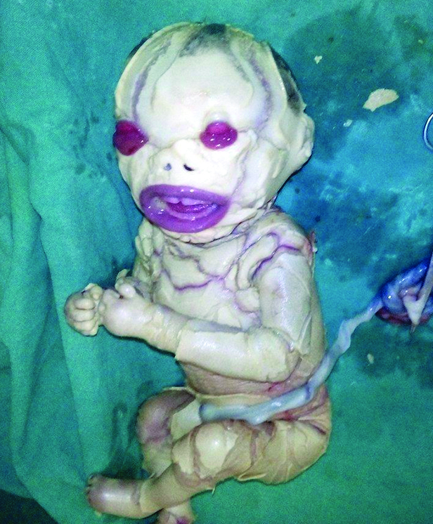
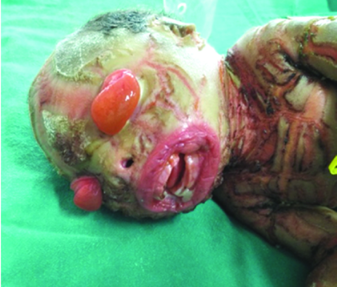
Thick shield like taut skin all over body with cracked pinkish areas in between, scanty hair, malformed ears, everted eyelids with exposed swollen conjunctiva, slit like nostril, open mouth, flexed limbs with malformed palms, oedematous feet were present. Baby did not need active resuscitation and was shifted to neonatal intensive care unit. Dermatological opinion was taken and a diagnosis of Harlequin ichthyosis was made. Baby was managed with intravenous fluids, intravenous antibiotics and topical emollients. Baby remained haemodynamically stable with cracks in the skin increasing [Table/Fig-2]. Baby was taken to home against medical advice on fifth day of life and lost to follow-up.
Appearance of baby on day 4 of life.
Approximately 75% of collodion baby develops a type of autosomal recessive congenital ichthyosis, either lamellar ichthyosis or congenital ichthyosiform erythrodema) [3]. The third type of autosomal recessive congenital ichthyosis, Harlequin ichthyosis, is the most rare and severe form. Other rare causes of colloidion baby include-Epidermolytic hyperkeratosis (bullous congenital ichthyosiform erythroderma), Gaucher’s disease and Sjögren-Larsson syndrome. Although, the colloidian membrane develops due to an epidermal cornification disorder, keratinocyte protein and lipid metabolism defects resulting from autosomal recessive genetic mutations have also been notified as important cofactors [4]. Transglutaminase 1 gene mutation localized on the 14q11 have been implicated in congenital autosomal recessive ichthyosis [5].
Owing to impairment of the skin barrier function, collodion babies prone for a number of complications like hypernatremic dehydration, hypothermia, skin infections, fissures, conjunctivitis, sepsis, dehydration and constrictive bands of the extremities resulting in vascular compromise and oedema [1,2].
They require proper fluid and electrolyte balance, emollients and lubricants for hydrating the membrane. Suitable eye care and pain control should be carried out for the collodion babies with ectropion. Prophylactic antibiotics should be used to prevent sepsis. In the present scenario, owing to intensive care, the survival of neonates has increased. Humidified incubators and water dressings followed by emollient agents are important aspects in its management.
[1]. van Gysel D, Lijnen RLP, Moekti SS, De Laat PCJ, Oranje AP, Collodion baby: a follow-up study of 17 casesJournal of the European Academy of Dermatology and Venereology 2002 16(5):472-75. [Google Scholar]
[2]. Roberts JB, Adelson D, Case report: prolonged collodion membrane causing constrictive bands of the digits and treatmentDermatology Online Journal 2010 16(1):15 [Google Scholar]
[3]. Dermatology at the Millennium. By Delwyn Dyall-Smith, Robin Marks, Page 586, Published by Informa Health Care, 1999, ISBN [Google Scholar]
[4]. Judge MR, Collodion baby and Harlequin ichthyosis. Harper J, Oranje A, Prose NTextbook of Pediatric Dermatology 2006 Second editionMaldenBlackwell Publishing:118-125. [Google Scholar]
[5]. Shwayder T, Akland T, Neonatal skin barrier: structure, function and disordersDermatol Therapy 2005 18:87-103.PMID: 15953139 [Google Scholar]