Cadaveric Study of Berretini Communications in North Indian Population
Neelamjit Kaur1, Rajan Kumar Singla2, Jagdev Singh Kullar3
1 Associate Professor, Department of Anatomy, Maharishi Markandeshwar Medical College and Hospital, Solan, Himachal Pradesh, India.
2 Professor, Department of Anatomy, Government Medical College, Patiala, Punjab, India.
3 Additional Professor, Department of Anatomy, Government Medical College, Punjab, India.
NAME, ADDRESS, E-MAIL ID OF THE CORRESPONDING AUTHOR: Dr. Neelamjit Kaur, 1083, Phase 9, Mohali, Punjab, India.
E-mail: neelamjit@yahoo.co.in
Introduction
Intercommunication between peripheral nerves deserves special attention in view of their clinical significance. Superficial palmar communication between the median nerve and ulnar nerve is referred to as Berrettini Anastomosis. The presence or absence of this communicating branch varies between individuals. Earlier, incidence of Berretini communication reported varied significantly (4-94%).
Aim
The aim of this study was to find out the frequency with which Berretini communication is found in North Indians.
Materials and Methods
The present study was conducted on 60 upper limbs of 30 cadavers at the Government Medical College, Amritsar. The whole course of the median nerve and the ulnar nerve was exposed. Communicating rami in the hand were identified, cleaned and photographed.
Results
In all the six (10%) variant limbs, the communicating branch originated from lateral common palmar digital branch of ulnar nerve and joined medial common palmar digital branch of median nerve. Single communication with oblique course was seen in all the variants. Further, its ontogeny and clinical implications have been discussed in detail.
Conclusion
The Berretini anastomosis was seen in 10% upper limbs of the present study. Damage to the communicating branch or the severing of the branch might result in sensory loss which may be difficult to diagnose owing to the large number of variations in the origin of the communicating branch. The patterns of sensory impairment may vary depending upon the branch of median and ulnar nerve it is seen connecting.
Anastomosis, Carpal tunnel, Communicating rami, Median, Ulnar, Variations in hand
Introduction
Berretini’s anatomic drawings from 1741 are the earliest illustrations of communicating branches [1]. These communications between common digital nerves that arise from the ulnar and median nerves in the palmar surface of hand are called Berretini communications or ‘ramus communicans cum nervi ulnari’ in Terminologia Anatomica [2]. The probable reason for the occurrence of such communicating branches may be due to the fact that peripheral processes of both the sensory and motor axons develop in different directions with the aid of various chemoattractants. However, the fibres which have to supply its assigned region end by supplying it after coursing over for a short distance following which it joins its parent nerve. This sort of travelling and shifting of some nerve fibres from adjacent nerves to the parent nerve may result in the formation of communicating branches [3].
According to literature, the first report was published by Meals and Shaner [4] on definition of the Berretini anastomosis and then their classification was modified by other authors [1,5,6]. The knowledge of such communications is significant to interpret any abnormal functioning of these nerves due to communications, and to avoid injury to the communicating branch while performing surgeries. As there are a large number of variations in the origin of the communicating branch so the surgeons should be aware of these variations while operating.
The incidence of Berretini anastomosis reported in earlier studies [1,5,7,8] varied significantly (4-94%). So the aim of this study was to find out the frequency with which Berretini communication is found in North Indians.
Materials and Methods
The present study was conducted on 60 upper limbs which belonged to 30 formalized and preserved cadavers (M:F::28:2) obtained from the Department of Anatomy, Government Medical College, Amritsar, Punjab, India. The limbs were dissected during the period 2007-2009 as per the dissection guidelines which were given by the Cunningham’s manual of Practical Anatomy [9], to expose the median nerve and its whole course from its formation till its termination. Similiarly, the ulnar nerve was traced till its termination. Communicating rami between these two nerves were exposed, photographed and studied. Out of these Berretini communications are being presented here.
Results
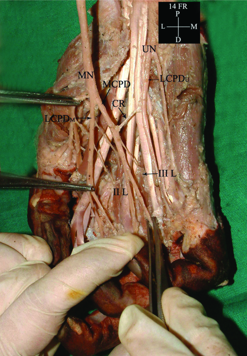
Out of the 60 upper limbs, Berritini communications i.e. communication between superficial branches of median nerve and ulnar nerve were observed in 6(10%) limbs. Five out of these belonged to male sex and one to female. Right to left ratio being 4:2. In one cadaver this variation was bilateral. In all the limbs, the communicating ramus originated from lateral common palmar digital branch of ulnar nerve to join medial common palmar digital branch of median nerve [Table/Fig-1]. The origin of communicating ramus was distal to flexor retinaculum and the course was oblique. All the variant limbs showed only one communicating branch.
Berretini communication between median nerve and ulnar nerve in hand (MN-Median nerve, UN-Ulnar nerve, MCPD-Medial common palmar digital nerve. LCPD-Lateral common palmar digital nerve, II L-Second lumbrical, III L-Third lumbrical).
Discussion
During this study two types of communications were seen between median nerve and ulnar nerve i.e., Martin-Gruber anastomosis and Berretini communications. The former have been already published by the author [10] while the later is being presented here. Berrettini communications i.e. the communication between common digital branch of median nerve and ulnar nerve in the hand, have been classified by Meals and Shaner [4] and later modified by Bas and Kleinert [11] into four types as follows:
Type I-The communication that originates from the ulnar nerve proximally and proceed distally to join the median nerve;
Type II-The communication that originates proximally from the median nerve and extends distally to join the ulnar nerve;
Type III-The perpendicular transverse communication between the median and ulnar nerves, however, it’s not possible to determine which nerve corresponds to the point of origin;
Type IV-Multiple communicating branches, anatomically complex, from both the median and ulnar nerves.
According to this classification all the branches seen in the present study fall in Type I of this classification. [Table/Fig-2] compares the incidence of different types of Berretini communications as observed by previous workers.
Comparison of Incidence of Berretini Communications in Different Studies
| Sl. No. | Authors (Year) | Incidence of Berrettini Communications |
|---|
| Type I | Type II | Type III | Type IV |
|---|
| 1 | Meals & Shaner [4] 1983 | 76% | 2% | 0% | 0% |
| 2 | Bas & Kleinert [11] 1999 | 37% | 13% | 0% | 17% |
| 3 | Stancic et al., [12] 2006 | 65% | 0% | 16% | 0% |
| 4 | Don Griot et al., [1] 2000 | 83% | 3.50% | 7.50% | 0% |
| 5 | Tagil et al., [8] 2007 | 40% | 3.30% | 6.70% | 10% |
| 6 | Zolin et al., [13]2014 | 33.34% | 0% | 3.40% | 6.70% |
| 7 | Present Study(2016) | 10% | 0% | 0% | 0% |
Ontogeny
The development of forelimb takes place during the fifth week of embryonic life. The mesenchyme of the paraxial mesoderm gives rise to the muscles of the upper limb. The upper limb nerves develop from the peripheral processes of spinal nerves. Within the mesenchyme, the peripheral processes of both the motor and sensory axons develop in different directions with the aid of various chemoattractants. The route taken up by the axons may vary while they ultimately reach the main trunk to which they belong. The developmental changes present during the formation of the nerve persist even after its formation and this result in communicating branches in adults. Many chemoattractants and chemorepulsants are involved in co-ordinating the development of a nerve. Some circulatory factors are also believed to be involved in its formation. Differences in the signaling system maybe the causative factor for the abnormal development of nerves which ultimately results in variations [14].
Clinical Significance
Knowledge of this variation is necessary to avoid injury to the communicating branch while performing release of the recurrent motor branch of median nerve in case of carpal tunnel syndrome. Though the communication was seen only in 10% of specimens, the presence of the communicating branch between the two nerves may compromise the sensory loss occurring due to lesion in median nerve to a certain extent. This variation can also be of significant use to neurologists and orthopedicians while examining cases with sensory loss over the distal part of the upper limb. Care should also be taken to prevent injury of the nerve while performing endoscopic release of carpal tunnel for carpal tunnel syndrome by looking out for the communicating branch prior to the surgery. Ferrari and Gilbert [7] described a danger zone, defined as an area that extends from the half of the hypothenar eminence limited distally by the transverse crease of the carpal on the palmar region and radially by a longitudinal fold between the thenar and hypothenar eminence, in which there is greater possibility of iatrogenic lesions in the approach to the carpal tunnel. According to Agge et al., and Jimenez et al., these lesions are more frequent after endoscopic carpal tunnel release, although this technique has lower risk of complications in general, and provides quicker return to functional activities [15,16].
Conclusion
To conclude, the Berretini anastomosis was seen in 10% upper limbs of the present study. Its formation can be explained ontogenically. Such communications have got a great clinical implication. Care should be taken to avoid injury to the communicating branch while performing release of the recurrent motor branch of median nerve in case of carpal tunnel syndrome.
[1]. Don Griot JPWD, Zuidam JM, Kooten EOV, Prose LP, Hage JJ, Anatomic study of the ramus communicans between the ulnar and median nervesJ Hand Surg 2000 25A:948-54. [Google Scholar]
[2]. [No authors listed]. Federative committe on Anatomical Technology. Terminologia Anatomica. Stuttgart, Thieme. 1998; 138. Cited by: Dogan NU, Uysal II, Seker M. The communications between the ulnar and median nerves in upper limb. Neuroanat. 2009;8:15–19 [Google Scholar]
[3]. Janani Y, Sugirthabai H, Rajendran R, Balaji T, Saran RS, Mounissamy B, A study on the communications of median nerve with musculocutanous and ulnar nervesInt J Reas Hlth Sci 2014 2(2):480-87. [Google Scholar]
[4]. Meals RA, Shaner M, Variations in digital sensory patterns: a study of the ulnar nerve-median nerve palmar communicating branchJ Hand Surg. [Am] 1983 8:411-14. [Google Scholar]
[5]. Loukas M, Louis ZR Jr, Stewart L, Hallner B, Deluca T, Morgan W, The surgical anatomy of ulnar and median nerve communications in the palmar surface of the handJ. Neurosurg 2007 106:887-93. [Google Scholar]
[6]. Kawashima T, Sato K, Sasaki H, Stratification of the flexor retinaculum and the course and distribution of the ulnar, median and palmar digital nerves: An anatomical studyClin. Anat 2004 17:643-50. [Google Scholar]
[7]. Ferrari GP, Gilbert A, The superficial anastomosis on the palm of the hand between the ulnar and median nervesJ. Hand Surg 1991 16B:511-14. [Google Scholar]
[8]. Tagil SM, Bozkurt MC, Ozcakar L, Ersoy M, Tekdemir I, Elhan A, Superficial palmar communications between the ulnar ve median nerves in Turkish cadaversClin. Anat 2007 20:795-98. [Google Scholar]
[9]. Romanes GJ, The pectoral region and the axilla, the arm and the forearm and the handIn: Cunninghams Manual of Practical Anatomy 1986 115th EditionEdinburgh, LondonThe English Language Book Society and Oxford University Press:28-89. [Google Scholar]
[10]. Kaur N, Singla RK, Kullar JS, Martin–Gruber Anastomosis- A cadaveric study in north indian populationJour Clin Diag Res 2016 10(2):AC09-AC11. [Google Scholar]
[11]. Bas H, Kleinert JM, Anatomic variations in sensory innervation of the hand and digitsJ Hand Surg Am 1999 24(6):1171-84. [Google Scholar]
[12]. Stancić MF, Burgić N, Mićović V, Marinacci communication. Case reportJ Neurosurg 2000 92(5):860-62. [Google Scholar]
[13]. Zolin Anatomical study of sensory anastomoses in the handActa Orthop Bras 2014 22(1):34-37. [Google Scholar]
[14]. Collins P, Development of the limbs and Development of the pectoral girdle and upper limb. 40th edIn: Gray’s anatomy: The anatomical basis of clinical practice 2008 Churchill Livingstone:899-906.Standring S, Editor [Google Scholar]
[15]. Agee JM, Peimer CA, Pyrek JD, Walsh WE, Endoscopic carpal tunnel release: a prospective study of complications and surgical experienceJ Hand Surg Am 1995 20(2):165-71. [Google Scholar]
[16]. Jimenez DF, Gibbs SR, Clapper AT, Endoscopic treatment of carpal tunnel syndrome: a critical reviewJ Neurosurg 1998 88(5):817-26. [Google Scholar]