Cadaveric Study on Morphology of Dorsal Interossei of Hand and its Anatomical Variation
Vanishri S Nayak1, Abhilasha Priya2, Nandini Bhat3, Sunil S Nayak4, Antony Sylvan D’Souza5, Hemalatha Bangera6, Suhani Sumalatha7
1 Senior Grade Lecturer, Department of Anatomy, K.M.C. Manipal, Manipal University, Udupi, Karnataka, India.
2 Assistant Professor, Department of Anatomy, School of Medical Sciences and Research, Sharda University, Greater Noida, Uttar Pradesh, India.
3 Tutor, Department of Anatomy, K.M.C. Manipal, Manipal University, Udupi, Karnataka, India.
4 Reader, Department of Oral and Maxillofacial surgery, Srinivas Institute of Dental Sciences, Mukka, Suratkal, Rajiv Gandhi University of Health Sciences (RGUHS), Karnataka, India.
5 Professor and Associate Dean, Department of Anatomy, K.M.C. Manipal, Manipal University, Udupi, Karnataka, India.
6 Junior Research Fellow, Department of Anatomy, K.M.C. Manipal, Manipal University, Udupi, Karnataka, India.
7 Senior Grade Lecturer, Department of Anatomy, K.M.C. Manipal, Manipal University, Udupi, Karnataka, India.
NAME, ADDRESS, E-MAIL ID OF THE CORRESPONDING AUTHOR: Dr. Vanishri S Nayak, Senior Grade Lecturer, Department of Anatomy, K.M.C. Manipal, Manipal University, Karnataka-576104, India.
E-mail: vanishri_nayak@yahoo.co.in
Introduction
The dorsal interossei are the abductors of the fingers and the knowledge of its variation help the surgeon in treatment of fractures, claw hand and compartment syndromes.
Aim
To note the origin, insertion, pattern of muscle fibres and tendon length of all the dorsal interossei of hand.
Materials and Methods
Routinely dissected 30 formalin fixed hand in the Department of Anatomy, KMC, Manipal, were observed for origin, insertion and tendon length and muscle pattern of dorsal interossei.
Results
Out of 30 hands, presence of supernumerary muscle was observed in three hands. Presence of three heads of dorsal interossei was noted in one hand.
Conclusion
The finding of present study is of importance to surgeons and orthopaedicians during conservative and surgical management of hand deformity. Adequate knowledge of these muscular variations is also important in treatment of fractures, stiff hand, claw hand or tendon transfer.
Muscular variation, Supernumerary muscle, Abductors
Introduction
Dorsal interossei muscle are the bipennate intrinsic muscles of the hand arising from the two adjacent metacarpal bones, their tendon pass to the lateral sides of the index and middle fingers. Part of the tendon passes deep to the extensor expansion into the base of proximal phalanx; the remainder joins the corresponding margin of extensor expansion. They abduct these fingers from the line of the middle finger, extend the interphalangeal joints, and play a part in flexion of the metacarpophalengeal joints [1,2]. The act of gripping and holding objects with the palm of the hand is usually carried out by extension of wrist, fingers and thumb with the help of long extensor tendons and intrinsic muscles of the hand which insert into the wing tendons. The action is initiated by extrinsic flexors and sustained by interossei muscles that are inserted into the phalanges [3].
The interosseous muscle of hand both dorsal and palmar are considered as key to the functioning of the hand and offer a foundation to all the movements such as finger balance, clutch, clasp and pinch functions [4].
A study on morphology and functional anatomy of the first dorsal interossei showed that the muscle had 2 heads superficial and deep. The superficial head mainly causes abduction and the deep head of the muscle causes flexion of thumb and index fingers to form pinch [5].
The interosseous muscles have a high fibre to muscle length ratio indicating that they are susceptible to instability resulting from change in the surrounding bony framework. These muscles exhibit decreased power for every millimetre reduced metacarpal length [6]. Interossei muscles shortening is called Intrinsic Tightness (IT), may be initiated by an injury resulting in flow of events. As interossei muscles are located in tight compartments any inflammatory swelling will cause increase in pressure in the compartment leading to decreased blood supply, subsequent fibrosis and muscle shortening. Rheumatoid arthritis also causes shortening of the muscle following a different pathology [7].
Dorsal interossei is known to be used for musculocutaneous flaps by plastic surgeons to correct unusual small defects of the hand, it is the most preferred muscle as it can be easily accessed through the dorsum. So any additional heads can be used following MRI evaluation [8].
Awareness of the anatomy and variations of the intrinsic muscles on the dorsum of the hand is indispensable when assessing the injured or diseased hand and when considering tendons for repair or transfer. A complete quantitative documentation of the dorsal interossei is deficient in literature.
Materials and Methods
This is a prospective study done in the Department of Anatomy, Kasturba Medical College, Manipal from August 2013 to December 2015. Thirty embalmed upper limbs of 15 adult human cadavers (without gender predilection) of south Indian origin, with the age range of 30-70 years were selected for this study.
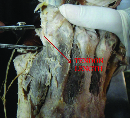
Palms were dissected with care to identify the dorsal interossei. Their proximal and distal attachment, tendon length and muscle pattern were noted along with the available variations. All measurements were recorded by a single author to minimize the inter-observer variations. The tendon length is measured from distal end of the muscle belly to its point of insertion using digital vernier calliper [Table/Fig-1].
Dorsal Interossei of hand.
Results
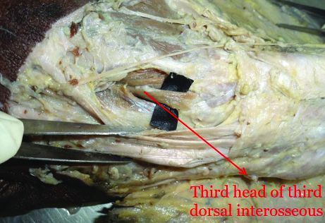
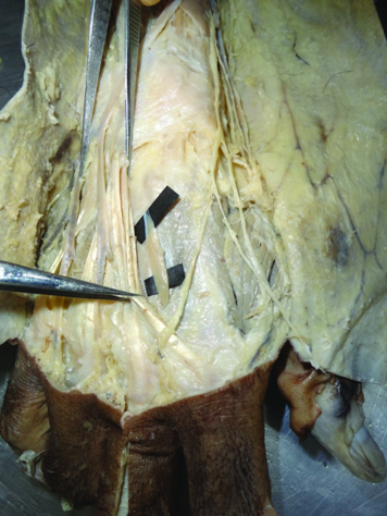
When we observed these muscles except 3 hands all other hands had normal origin of muscle from adjacent metacarpal bones. These muscles are inserted into the base of proximal phalanx and then into the dorsal digital expansion. There is negligible variation in the tendon length, which varied between1.2 to 1.5cm. All muscles had bipennate muscle fibre except one which had 3 heads in third dorsal interossei [Table/Fig-2].
Picture showing third head of third dorsal interosseous.
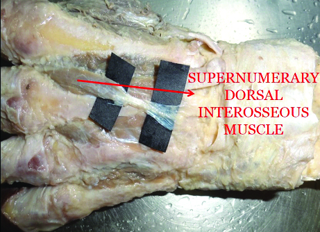
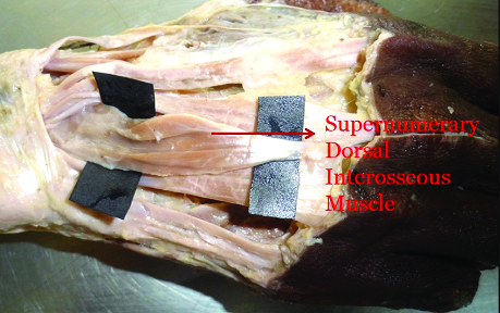
But in 3 out of 30 dissected hand had supernumerary muscle. One supernumerary dorsal interosseous was originated from third metacarpal bone and gets inserted into radial side of dorsal digital expansion of third finger [Table/Fig-3]. In one more case a supernumerary dorsal interosseous originating from third metacarpal bone and getting inserted into ulnar side of dorsal digital expansion of third finger was observed [Table/Fig-4]. In one more case there was similar origin where the supernumerary dorsal interosseous has its origin from the third metacarpal bone but it gets inserted into the ulnar side of the dorsal digital expansion of the second finger [Table/Fig-5].
Picture showing supernumerary dorsal interosseous arising from third metacarpal bone and inserting into radial side of dorsal digital expansion.
Picture showing supernumerary dorsal interosseous muscle arising from third metacarpal bone and inserting into ulnar side of dorsal digital expansion of third finger.
Picture showing supernumerary dorsal interosseous arising from third metacarpal bone and inserting into the ulnar side of dorsal digital expansion of second finger.
Discussion
Dorsal interossei are responsible for complex movements of hand. Anatomical variations in the morphology of the dorsal interossei muscle are reported in literature since 18th century. Bergman et al., have reported of absence of these muscles in one or more spaces, muscles with three heads and also double muscle in each space [9]. Eladoumikdachi et al., reported three heads of dorsal interossei muscle, each with different distal attachments [10]. In the present study one case of dorsal interossei with three heads was noted.
Natis et al., reported one supernumerary muscle on the right hand lying superficial to normal fourth dorsal interossei muscle, proximally attached to the dorsal surface of fourth metacarpal base, coursing obliquely and distally attached to head of fifth metacarpal [11]. In the present study 3 out of 30 hands had presence of supernumerary muscle. Among them one was second supernumerary muscle and two was third supernumerary muscle.
Functions of skeletal muscles depend on its architecture. Dorsal interossei muscles have short fibres for better function and estimated to provide 40-90% of grip strength [12]. Additional head of muscle as present in the study will contribute to additional grip strength.
Presence of additional head of dorsal interosseus muscle and supernumerary muscle in hand is mostly asymptomatic. However since they are occupying different compartment in hand, additional muscle head or supernumerary muscles can result in chronic compartment syndrome due to increase in intracompartmental pressure locally. Exercise induced chronic compartment syndrome in the first dorsal compartment is reported in literature that showed signs of improvement with surgical fasciotomy [13,14].
Bharambe et al., observed additional heads of dorsal interosseous muscle in four cases out of 25 dissected hands [15]. In the present study additional head of dorsal interosseous muscle is observed in one case out of 30.
A study conducted by Pridgen et al., showed that ultrasound is useful for measurement of hand muscles. A significant decrease in cross-sectional area of the first dorsal interossei was observed with the help of an ultrasound study in osteoarthritis of first carpometacarpal joint [16].
Limitations
The sample size is less, also the differentiation between the variation in male and female hands were not done because of the lack of availability of female specimens in good number. The innervation of the dorsal interossei was not observed.
Conclusion
Muscular variations are always important to a clinician in diagnosis and treatment. Knowledge of these variations in the morphology of dorsal interossei in the hand is important for orthopaedicians and hand surgeons in treatment of fractures, claw hand, stiff hand and compartment syndromes. The above described supernumerary muscle and additional head of the dorsal interossei will add extra data to the existing literature.
[1]. Gray H, Standring S, Ellis H, Berkovitz B, Gray’s Anatomy 2008 40th edSpainChurchill Livingstone Elsevier:811 [Google Scholar]
[2]. Cunningham D, Romanes GJ, Cunningham’s Manual of Practical Anatomy 1986 Vol 115th edLondonOxford University Press:16-18. [Google Scholar]
[3]. Biomechanics of the Hand: A Basic Research Study by E Y S Chao, K-N An, W P Cooney, RL LinscheidWorld Scientific 1989 Apr :35 [Google Scholar]
[4]. Liss F, The interosseous muscles: The foundation of hand functionHand Clinics 2012 28(1):9-12. [Google Scholar]
[5]. Masquelet AC, Salama J, Outrequin G, Serrault M, Chevrel JP, Morphology and functional anatomy of the first dorsal interosseous muscle of the handSurg Radiol Anat 1986 8(1):19-28. [Google Scholar]
[6]. Meunier MJ, Hentzen E, Ryan M, Shin AY, Lieber RL, Predicted effects of metacarpal shortening on interosseous muscle functionThe Journal of Hand Surgery 2004 29(4):689-93. [Google Scholar]
[7]. Schreuders TA, Selles RW, Roebroeck ME, Stam HJ, Strength measurements of the intrinsic hand muscles: A review of the Development and Evaluation of the Rotterdam Intrinsic Hand myometerJournal of Hand Therapy 2006 19(4):393-402. [Google Scholar]
[8]. Tang M, Sun H, Morris SF, Anatomic basis and clinical application of the interosseous muscle flap in the handJ Bone Joint Surg Br 2009 91-B:242 [Google Scholar]
[9]. Bergman RA, Thompson SA, Afifi AK, Saadeh FA, Compendium of human anatomic variationMunich and Baltimore 1988 :139-43. [Google Scholar]
[10]. Eladoumikdachi F, Valkov PL, Thomas J, Netscher DT, Anatomy of the intrinsic hand muscles revisited: Part I. InterosseiPlastic and Reconstructive Surgery 2002 110(5):1211-24. [Google Scholar]
[11]. Natsis K, Tsakotos G, Vlasis K, Koebke J, The cadaver of a Caucasian man with a supernumerary fourth dorsal interosseous muscle in the right hand: A case reportJ Med Case Rep 2011 5(1):393 [Google Scholar]
[12]. Lieber RL, Fridén J, Functional and clinical significance of skeletal muscle architectureMuscle & Nerve 2000 23(11):1647-66. [Google Scholar]
[13]. Styf J, Forssblad P, Lundborg G, Chronic compartment syndrome in the first dorsal interosseous muscleThe Journal of Hand Surgery 1987 12(5):757-62. [Google Scholar]
[14]. Chopra R, Hayton M, Dunbar PJ, Exercise Induced Chronic Compartment Syndrome of the First Dorsal Interosseous Compartment of the Hand: A Case ReportHAND 2009 4(4):415-17. [Google Scholar]
[15]. Bharambe VK, Shevde SP, Puranam V, Kanaskar NS, Additional heads of dorsal interosseous muscle in Caucasion cadavers and their clinical significanceSahel Med J 2013 16(4):174-77. [Google Scholar]
[16]. Pridgen E, Kenney D, Roh E, Ladd A, In Vivo Measurement of the Thenar Muscles and First Dorsal Interosseous in Thumb Carpometacarpal Joint Osteoarthritis Using Ultrasound ImagingAAHS Annual meeting 2016; Jan 13-16 Scotsdale, Arizona [Google Scholar]