Myiasis is the infestation of human beings with fly larvae which feed on host’s living or dead tissue. Diagnosis of this condition is made by the presence of larvae in affected body parts. The standard treatment is the mechanical removal of the maggots from the lesion and management of the general systemic condition. This paper report a case of myiasis at the chin region in a 45-year-old male managed by manual removal of larvae after topical application of turpentine oil, surgical debridement of necrotic tissues and antimicrobial therapy.
Eggs, Fly, Larvae, Turpentine
Case Report
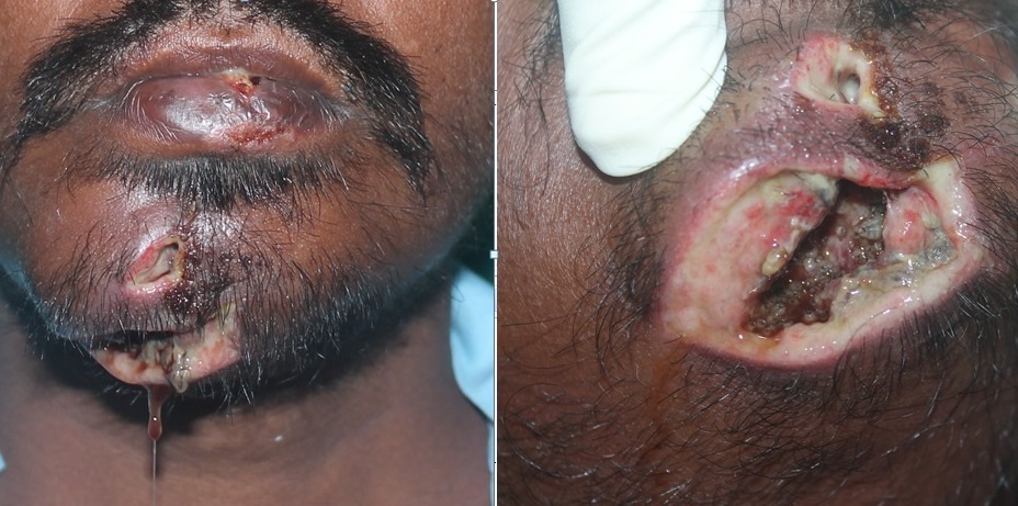
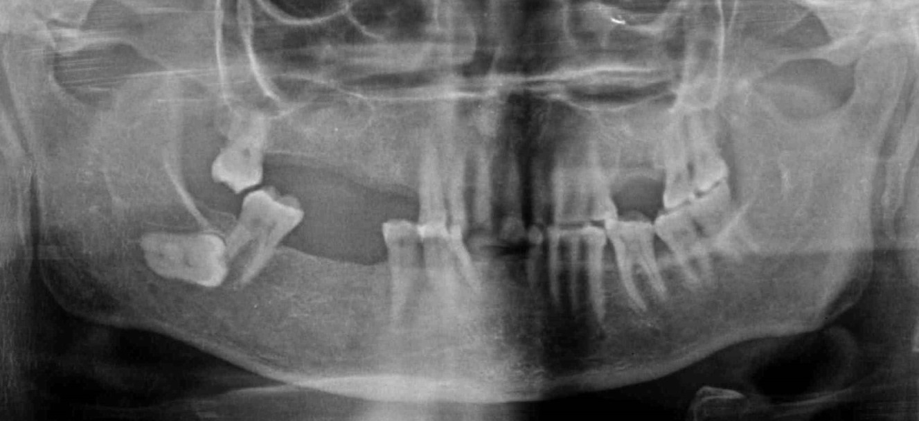
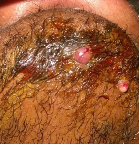
A 45-year-old male of low socioeconomic status, living in poor conditions reported To hospital with chief complaint of pain, swelling, discharge and crawling sensation at the chin region since three weeks. The patient was poor, chronic alcoholic, abandoned by family, living on street dishevelled and unkempt. History revealed trauma to face one month back, when he accidentally fell on the road. He got bruises and lacerations at chin region. He sought treatment at a nearby health centre but wound was not healing despite treatment. Also, patient was reluctant and unwilling for treatment there. Patient appeared undernourished and dehydrated. Extra oral examination revealed laceration at chin region with diffuse swelling, extending up to the submental region which was covered with necrotic slough and measuring approximately 3.0 x 2.5cm. No bleeding was present from the ulcers. There was watery discharge from the wound. No intraoral communication, intra-oral wound or bony exposure was present. There was no fracture mobility or sublingual hematoma evident. Moving worms, Maggots, were noticed burrowed deep inside the wound [Table/Fig-1]. Patient was febrile with 101oC temperature. Routine blood investigations were within normal limits. Based on the history and presence of maggots, provisional diagnosis of myiasis was made. Radiographic evaluation revealed no mandibular fracture [Table/Fig-2].
Indurated swelling and wound at chin region (left) and multiple maggots (larvae) within the ulcerated wound (right).
Orthopantamogram of the patient.
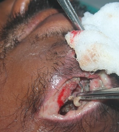
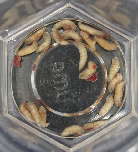
The patient was explained about the condition and informed consent was taken. Under local anesthesia, cotton bud impregnated with turpentine oil was applied at the wound site for approximately 15 minutes. After this application, maggots were manually removed with the help of tweezers and haemostat [Table/Fig-3] and sent for entomological examination. Around 20 maggots were removed [Table/Fig-4]. Further management included surgical debridement of dead necrotic tissue and irrigation done with saline and betadine. Larvae were removed consecutively for another three days until no more larvae were found. The patient was given Inj. Cefazolin 1g six hourly and Inj. Metronidazole 400mg eight hourly for five days postoperatively. Tetanus prophylaxis was also given to the patient. Regular cleaning, debridement and dressing were done to maintain wound hygiene and care. Adequate nutrition was maintained with protein rich fluid therapy. The lacerated wound at chin region was sutured on 5th day and the patient was discharged on the 7th day. After two weeks, the wound healed satisfactorily [Table/Fig-5].
Maggots were manually removed with the help of tweezers and haemostat.
Maggots (larvae) which were removed.
Wound healing after two weeks.
Discussion
‘Myiasis’ is a condition in which the body cavities, organs or tissues of live humans (or vertebrates) are invaded by the eggs or larvae of flies of order Diptera. The word ‘myiasis’ has been derived from Latin word ‘muia’ meaning ‘fly’ and ‘iasis’ meaning ‘disease’ [1]. Hope (1940) was first to introduce the term ‘myiasis’ and Zumpt (1965) defined it as ‘the infestation of live human and vertebrate animals with dipterous larvae which, at least for a certain period, feed on the host’s dead or living tissue, liquid body substances or ingested food [2]. The usual cause is oviposition of flies in open wound or decaying tissues followed by development of larvae. The larva of a fly (order Diptera) is also known as maggot. Any organ of the body accessible to the larvae may be involved. Myiasis may be ocular, nasal, aural, oral, oesophageal, intestinal or cutaneous. Laurence (1909) first described ‘oral myiasis’ as the condition wherein the soft tissues in the oral cavity are invaded by the eggs or parasitic larvae of flies [3].
Although myiasis is frequently seen in animals, it shows considerable incidence in human beings in rural, tropical and subtropical regions [1,4]. Around 86 different species of flies have been shown to cause myiasis in human beings. The fly may lay eggs or larvae onto foodstuffs causing chance infection following ingestion (known as accidental myiasis), or eggs or larvae may be laid directly on to necrotic tissue in wounds (semi-specific myiasis) or some species of flies may require living tissue and lay eggs directly onto undamaged skin (obligatory myiasis) [2].
Myiasis in maxillofacial region is more commonly associated with predisposing anatomical, physical and medical conditions which include mouth breathing habit, incompetence of lips, cerebral palsy, parkinsonism, epilepsy, extraction socket, neglected maxillofacial and mandibular trauma, patients undergoing mechanical ventilation, alcoholism and senility and certain local pathological conditions such as cancrum oris and oral malignancies. This condition is also found frequently in close association with state of mental deficiency, where maintenance of oral hygiene is very difficult due to poor dexterity [3,5].
Myiasis can cause infections of the skin, gut, bladder, nasal cavities, aural cavities, eyes and occasionally the oral cavity. Myiasis of maxillofacial region is an extremely rare entity, even in developing countries. Maxillofacial region get involved if there is neglected trauma to the region. There have been quite a few cases of maxillofacial myiasis reported in the literature [Table/Fig-6].
Cases of maxillofacial myiasis reported in the literature.
| Authors | Etiology / Predisposing Factor | Site of Presentation | Management |
|---|
| Abdo et al., [11] | Neurologic deficit and low socio-economic status | Upper lip | Topical use of gentian violet Oral therapy with Ivermectin (6mg) Surgical exploration.
|
| Schneider TR et al., [12] | Advanced periodontal diseases and senility | Upper lip and labial vestibule | Manual removal of larvae Subsequent management of periodontal disease.
|
| Ali FM et al., [13] | Mentally challenged | Maxillary anterior Vestibule | Removal of maggots by turpentine oil and simple tweezers. Ivermectin 6mg OD for 3 days, along with metronidazole 400mg for 5 days and analgesic (buprofen with paracetamol).
|
| Sharma et al., [14] | Mentally challenged | Palate, extending from incisors to first molar region on right side. | Wound was cleaned by 10% hydrogen peroxide Larvae were manually removed with the help of turpentine oil and serrated tweezers. Wound was irrigated and cleaned with 1% iodine solution Metronidazole 500mg eight hourly Anti-tetanus serum.
|
| Ramli et al., [15] | Cerebral palsy | Anterior palate | Cotton bud impregnated with turpentine.
|
| Pereira et al., [16] | Cerebral palsy | Anterior palate | Manual removal (tweezer).
|
| Reddy RMH et al., [17] | Learning disability Trauma
| Upper anterior vestibule and nasal floor | Turpentine swab Irrigation with hydrogen peroxide and betadine solution Broad spectrum antibiotics and analgesics.
|
| Sharma et al., [18] | Neurological deficit and low socioeconomic status with poor living conditions | Maxillary anterior palate.
| Pressure around the lesion and the use of forceps Cotton bud impregnated with turpentine oil Complete oral prophylaxis Suturing of buccal and lingual flaps Complete periodontal therapy Tablet doxycycline 100mg BD followed by OD for 7 days, tablet metronidazole 400mg TID for 5 days, tablet Ivermectin 3mg BD for 5 days, chlorhexidine 0.2% mouthwash rinse 3–4 times/day.
|
| Maheshwari VJ et al., [19] | | Maxillary anterior vestibular region | Copious irrigation with physiological saline and povidine iodine Delayed primary wound closure Antibiotic coverage with oral penicillin.
|
| Shikha S et al., [20] | Maxillofacial trauma one week ago leading to avulsion of teeth #11 and #21 Epileptic and mentally retarded since birth
| Maxillary anterior region especially in the gingiva.
| Cotton swab impregnated with turpentine Oral Ivermectin 6mg once daily for three days and oral amoxicillin 250mg three times daily for seven days.
|
| Sharma et al., [1] | Neurological deficit(hemiparesis) and low socioeconomic status Trauma to the face one month back leading to avulsion of the upper left anterior tooth.
| Orifice of the socket of 21. | Cotton bud impregnated with turpentine oil Manual removal with the help of tissue holding forceps Extraction of the periodontally involved teeth followed by curettage and placement of metronidazole pack. Oral therapy with Ivermectin 6mg once daily for two days.
|
| Antunes AA et al., (series of 10 cases) [21] | | Middle third of the face was the most frequently affected region (7 cases). | Surgical debridement whether or not associated with the use of oral Ivermectin. |
| Sankari LS et al., [22] | Epileptic attack 10 days back leading to fracture of the upper front teeth. | Maxillary anterior region involving the vestibular sulcus and labial mucosa extending from the right canine to the left canine | Turpentine oil Manual removal of maggots with tweezers Broad spectrum antibiotics amoxycillin with clavulanic acid and ibuprofen with paracetamol.
|
| Shinohara et al., [9] | Hypotonic cerebral palsy | Anterior maxillary gingiva and labial sulcus | Antibiotic therapy (1g cefalotin, i.v., every 6hour) Intravenous rehydration 6mg Ivermectin orally.
|
| Singh WR et al., [23] | Obstructive hydrocephalus | Maxillary labial sulcus and labial gingiva | Antibiotic (Inj. Ceftriaxone) on the first visit. Manual removal with tweezer Wound debridement.
|
The specific odour of the wound attract adult fertile female flies, they feed on wound exudates and lay their eggs in the injured and necrotic tissues, open wounds or discharging orifices. The conditions essential for egg laying and survival of the larvae are provided by these wounds [2]. These flies lay around 500 eggs at same time directly over these diseased and compromised tissues. The first instar larvae hatch after 12-24 hours and they enter the living tissues and feed for next 5–7 days. The larvae releases proteolytic enzymes and toxins which decompose the surrounding living and necrotic tissues and the larvae feed on it to obtain nutrition [1]. They create tunnels between soft tissue layers and separate mucoperiosteum from bone and burrow themselves deeper and deeper into the soft tissues [3]. Since these maggots are photophobic, they migrate deeper into the tissues to conceal and hide in a protective niche and continue their life cycle [5]. During this period, the larvae moult twice and the third instar (last stage) leaves the host to develop into pupa inside the ground. In the next 1–2 weeks, the adult fly emerges [6]. The larvae are tapering in shape, whitish grey in colour, their segments appear as transverse rows with a brown-black tip anteriorly [3].
The ideal and standard treatment plan includes removal of larvae manually using topical asphyxiating drugs, maintaining nutrition and administration of antibiotics to prevent any secondary infection. The simplest technique is mechanical removal of the maggots with tweezers, usually under local anesthesia. Because of the hooks that larvae uses to grip the tissue cavity, simple mechanical removal is not possible many times. So, topical asphyxiating agents are used which act as topical irritant and blocks the larva’s respiratory sinuses, forcing this aerobic organism to come up to the surface in search of oxygen and facilitating their removal with the help of forceps or tweezers [7]. This method is also known as Occlusion or Suffocation approach. The topical asphyxiating drugs include ether, chloroform, olive oil, turpentine oil, petroleum jelly and nail polish. Care must be taken to prevent tearing or laceration of the larva because any portion of remaining larval part left in the wound cavity will induce an undesirable inflammation or a bacterial infection. Also it becomes difficult to retrieve that left over lacerated portion of the larvae [1,8].
The administration of drugs to treat myiasis is incipient and a very few reports have been documented. Ivermectin is the only antibiotic documented till now which is very effective against maggots. This drug blocks nerve impulses at the nerve ending by releasing Gamma Amino Butyric Acid (GABA), causing palsy and death of larvae. Shinohara et al., first documented successful treatment of this condition with Ivermectin 6mg given orally, and repeated after 24 hours [9]. The main advantage of using Ivermectin is that it is not necessary to operate to remove the larvae [9]. More recently, a topical agent, Nitrofurazone, has been used with high success rate for the management of myiasis [10]. Also topical application of Polymyxin B has also been documented as an alternative for the occlusive treatment of myiasis [7].
In the present case, the patient suffered trauma and neglected the wound treatment for due course of time. Since he was poor and living a life of street beggar, he was not maintaining hygiene. The fly would have laid eggs in the open wound which led to the development of myiasis. The larvae were burrowed deep inside the wound. The case was managed with mechanical removal of the maggots, systemic antimicrobial administration and maintaining nutrition of the patient which gave good results; content and happy patient.
Conclusion
From the case presented, it can be concluded that the fly can deposit their eggs on untreated neglected open lacerated wounds of maxillofacial region. This condition often affects individuals with low socioeconomic level, poor hygiene habits and unhealthy patients with any psychiatric disorder, diabetic patients or immunocompromised state. Undoubtedly maintenance of good hygiene along with fly control measures is of utmost importance for prevention of myiasis in all maxillofacial lacerations and injuries. The treatment of this condition is mechanical removal of the maggots/larvae from the wound site and systemic management of the condition.
[1]. Sharma J, Mamatha GP, Acharya R, Primary oral myiasis: a case reportMed Oral Patol Oral Cir Bucal 2008 13(11):E714-16. [Google Scholar]
[2]. Novelli MR, Haddock A, Eveson JW, Orofacial myiasisBr J Oral Maxillofac Surg 1993 31:36-37. [Google Scholar]
[3]. Lata J, Kapila K, Aggarwal P, Oral Myiasis: a case reportInt J Oral Maxillofac Surg 1996 25:455-56. [Google Scholar]
[4]. Hope F, On insects and their larvae occasionally found in the human bodyTrans R Soc Entomol 1840 2:256-71. [Google Scholar]
[5]. Jain S, Gupta S, Jindal S, Singla A, Oral myiasis in a cerebral palsy patient: a case reportJ Clin Exp Dent 2010 2:e110-12. [Google Scholar]
[6]. Gomez RS, Perdigao PF, Pimenta FJGS, Leite ACR, Tanos de Lacerda JC, Neto AL, Oral myiasis by screwworm CochliomyiahominivoraxBr J Oral Maxillofac Surg 2003 41:115-16. [Google Scholar]
[7]. Krajewski A, Allen B, Hoss D, Patel C, Chandawarkar RY, Cutaneous myiasisJournal of Plastic, Reconstructive & Aesthetic Surgery 2009 62:e383-86. [Google Scholar]
[8]. Babu C, Rai BA, Nair MA, Kalola R, Bhut MK, Traumatic wound myiasis in maxillofacial region - A case reportInternational Journal of Dental Clinics 2010 2(2):53-57. [Google Scholar]
[9]. Shinohara EH, Martini MZ, Oliveira Neto HG, Takahashi A, Oral myiasis treated with ivermectin: case reportBraz Dent J 2004 15(1):79-81. [Google Scholar]
[10]. Lima Júnior SM, Asprino L, Prado ÂP, Moreira RWF, de Moraes M, Oral myiasis caused by Cochliomyiahominivorax treated nonsurgically with nitrofurazone: report of 2 casesOral Surg Oral Med Oral Pathol Oral RadiolEndod 2010 109(3):e70-73. [Google Scholar]
[11]. Abdo EN, Sette-Dias AC, Comunian CR, Dutra CEA, Aguiar EG, Oral myiasis: a case reportMed Oral Patol Oral Cir Bucal 2006 11:E130-31. [Google Scholar]
[12]. Schneider TR, Cherubini K, Yurgel LS, Salum F, Figueiredo MA, Oral myisias: a case reportJournal of Oral Sciences 2007 49(1):85-88. [Google Scholar]
[13]. Ali FM, Patil K, Kar S, Patil AA, Ahamed S, Oral myiasis affecting gingiva in a child patient: an uncommon case reportCase Reports in Dentistry 2016 2016:2197450 [Google Scholar]
[14]. Sharma N, Malhotra D, Manjunatha BS, Kaur J, Oral myiasis-a case reportAustin J Clin Case Rep 2014 1(8):1039 [Google Scholar]
[15]. Ramli R, Rahman A, Oral myiasis: case reportMalays J Med Sci 2002 9(2):4750 [Google Scholar]
[16]. Pereira T, Tamgadge AP, Chande MS, Bhalerao S, Tamgadge S, Oral MyiasisContemp Clin Dent 2010 1(4):275-76. [Google Scholar]
[17]. Reddy MR, Das N, Vivekananda MR, Oral myiasis in childrenContemp Clin Dent 2012 3(Supp.1):S19-22. [Google Scholar]
[18]. Sharma D, Kumar S, Parashar P, Naphade VV, Oral gingival myiasis: a rare case report and literature reviewContemp Clin Dent 2015 6(4):548-51. [Google Scholar]
[19]. Maheshwari VJ, Naidu SG, Oral myiasis caused by Chrysomyabezziana: a case reportPeople’s Journal of Scientific Research 2010 3(2):25-26. [Google Scholar]
[20]. Shikha S, Prasad Guru R, Ashutoshdutt P, Meenakshi S, Oral myiasis: a rare case report and literature reviewJ Dent (Tehran) 2015 12(6):456-59. [Google Scholar]
[21]. Antunes AA, Santos Tde S, Avelar RL, Martins Neto EC, Macedo Neres B, Laureano Filho JR, Oral and maxillofacial myiasis:a case series and literature reviewOral Surg Oral Med Oral Pathol Oral Radiol Endod 2011 112(6):e81-85. [Google Scholar]
[22]. Sankari LS, Ramakrishnan K, Oral myiasis caused by ChrysomyabezzianaJ Oral Maxillofac Pathol 2010 14(1):16-18. [Google Scholar]
[23]. Singh WR, Aheibam KK, Ningthoujam B, Devi NA, Worms in the oral cavity: a case of oral myiasisDHR International Journal of Medical Sciences 2013 4(2):46-51. [Google Scholar]