Triglyceride enters into the plasma and circulates as neutral core lipids of lipoproteins mainly chylomicrons and Very Low Density Lipoproteins (VLDL). Intact triglyceride cannot be entered into the tissues; instead it is hydrolyzed to glycerol and fatty acids by hepatic and lipoprotein lipase located on endothelium of the blood capillaries [1]. Therefore, the level of triglyceride in plasma is balanced by the rate of release and entrance into (Vr), degradation and clearance (Vc) from the plasma [2]:
Tyloxapol (Triton WR1339) is a prototype of these nonionic detergents that following intravenous or intra-peritoneal injection that causes the milky serum which last up to 48 hour [3]. Tyloxapol surround lipoproteins and inhibits the action of lipolytic enzymes [5]. Since the discovery of the detergent, it is used to study lipid metabolism. The detergent is applied to estimate VLDL secretion rate [5–10], to investigate the metabolic relationships of lipoproteins [11,12], and more recently to induce artificial hyperlipidemia [13,14]. The model of experimental lipemia in animals is used to study the pathogenesis of atherosclerosis its effects and mechanism of hypolipidemic drugs [13,14]. But it is difficult to define whether the decrease in plasma lipids is attributed to the treatment with hypolipidemic drugs or arise from the metabolism and clearance of tyloxapol from the blood stream. Several reports have addressed the short term changes of serum lipids and (apo) lipoproteins in tyloxapol injected animals [11,12]. However few studies have been conducted to investigate the kinetic of plasma lipids for a long period. The current research was performed to study the kinetic of plasma lipids and lipoproteins in tyloxapol injected rats over a period of two weeks.
Materials and Methods
Duration of the study was from March of 2013 and continued for ten months. Tyloxapol (Triton WR1339) was obtained from Sigma (USA), diethyl ether and ethanol were purchased from Merck. All other chemicals and solvents were of reagent quality and were obtained from local suppliers.
Animal Diet: Albino (Wistar) male rats were housed in a room with 12-h light/dark cycle under constant temperature (25oc) and humidity for 15 days. The rats weighing 200-220 g fed standard rodent laboratory food and were starved overnight (10 h) before blood sampling. All experimental procedures were performed in accordance with the Guide for the Care and Use of Laboratory Animals (GCULA) approved by National Research Council.
Tyloxapol injection: All experiments were commenced at 09.00 AM. Tyloxapol dissolved in isotonic saline 20% (V/V) with slight agitation and left standing overnight. 0.5ml of detergent (400 mg/kg) was injected into tail veins of rats under light ether anaesthesia. One hour after tyloxapol injection, the blood sample was taken and triglyceride was measured immediately. Fifteen male rats were injected with tyloxapol at any time, but about one third of rats showed elevated triglyceride level. Therefore, two thirds of the rats failed to increase plasma triglyceride and were omitted from the experiment as unsuccessful injections. The blood samples (0.5 ml) were taken on EDTA contained tubes from both tail vein cut and puncture in the early phase (1-6 h), but by vein puncture on the second and third phases (2-9 days). The blood serum or plasma was separated by 10 min centrifugation at 3000 rpm and stored at 4°C before analysis or at –70°C for longer preservation. All measurements were done on the plasma except of electrophoresis that was performed on the fresh serum. However, no differences were found in lipid levels in serum and plasma.
Measurement of plasma lipids: Plasma triglyceride and total cholesterol were measured by enzymatic methods CHOD-PAP and GPO-PAP respectively (Pars-Azmon Inc., Tehran). We observed that the serum of tyloxapol injected rats had some detergent to slightly inhibit the lipase used in the kit for enzymatic measurement of triglyceride. The existence of detergent in the assay medium underestimates the measurement. Thus, we preferred to measure the triglyceride manually (instead by auto-analyser) in water bath with regular shaking to overcome the probable inhibition. All plasma samples taken after 1 hour injection of detergent had high triglyceride levels and must be diluted appropriately by isotonic saline before the measurement; otherwise the assay will have a serious negative error. For measurement of total phospholipids, 250 μl of plasma was extracted by chloroform: methanol by method of Folch as described previously [15]. The extract was dried by flow of gaseous nitrogen and the total extract was hydrolyzed by heating with sulfuric acid and hydrogen peroxide [16]. The final volume was adjusted to 1 ml by distilled water and the inorganic phosphate (25 μl) was measured as molybdenum blue (Zist-Shimi Inc., Tehran). Serum lipoproteins were fractionated by electrophoresis on agarose gel (Sebia, Moulineaux, France). Four μl of serum samples were loaded on the gel by slit applicator. The lipoproteins were fractionated at 75 V for 25 minutes and finally stained with amidoblack.
Statistical Analysis
The results are presented as the means ± SEM of three inter-assays performed at least in five different rats. The significant differences between samples and corresponding control were accessed by student’s t-test.
Results
Short term kinetic of plasma lipids following injection of tyloxapol
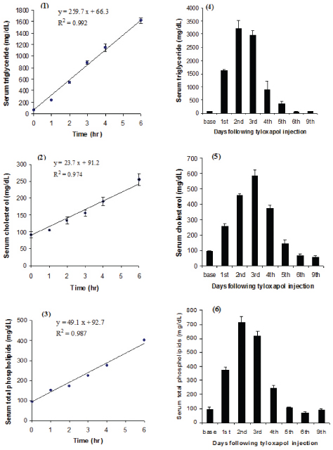
Time course of plasma lipids have been shown in [Table/Fig-1-3] following injection of tyloxapol. The basal fasting concentrations of triglyceride, cholesterol and total phospholipids were 66.3 ± 10.4, 91.2 ± 8.5 and 92.7 ± 6.4 mg/dl respectively and increased linearly during 6 hour. The mean rate of triglyceride secretion was 259.7 ± 8.1 mg/h.dl in this phase.
Time course of lipids accumulation in plasma of tyloxapol injected rats. Male rats starved for 12 hour were injected with tyloxapol and the blood samples were taken before the injection and at the time intervals 1, 2, 3, 4 and 6 hour thereafter [Table/Fig-1-3]. The intercept was set as the concentration of basal state to obtain the equation of each line. The overall changes of plasma lipids in the first (at 6 h), second (1-2 days) and third phase (3-9 days) after tyloxapol treatment [Table/Fig-4-6]. The concentrations of plasma lipids plotted in mg/dl on ordinate and the time after tyloxapol injection is shown in hours [Table/Fig-1-3] and days [Table/Fig-4-6] on abscissa.
Long term kinetic of plasma lipids following injection of tyloxapol
The overall changes of plasma lipids have been presented in [Table/Fig-4-6] in the time intervals 1, 2, 3, 4, 5, 6 and 9 days after tyloxapol injection. Triglyceride continued to accumulate in plasma during the second phase (1-2 days) but at a lower rate. The rate of triglyceride secretion was determined in the second day by measuring the difference of triglyceride levels in two successive hours. The rate of triglyceride secretion was 105 mg/h.dl in this phase and triglyceride reached to maximum level of 3200 mg/dl after 24 h. Cholesterol level increased in plasma slightly later than triglyceride and rose up to maximum level of approximately 586 mg/dl in 3rd day. [Table/Fig-6] showed that the total phospholipids increased in the second day to maximum level of 714.5 ± 40.4 mg/dl. In the last phase (3-6 days), the level of plasma lipids decreased slowly and returned to the basal.
The mean rate of triglyceride secretion into the plasma
Liver is the major source of plasma triglyceride in fasted state. Since it is proposed that tyloxapol inhibits completely the degradation and clearance of triglyceride from the plasma, the rate of its accumulation in plasma is a good estimate of the rate of secretion. On the other hand, cholesterol and phospholipids are not a direct goal for tyloxapol, therefore this method underestimates their secretion rate. The rate of triglyceride secretion (Vr) is equal to the slope of triglyceride mass changes versus the time. The Vr for triglyceride in early 6 h (first phase) is calculated from the slope of the curve-1 to be equal to 259.7 ± 8.1 mg/h.dl. The mean rate of triglyceride secretion in starved male rat in vivo using tyloxapol method are gathered from different references [3–11,17–24] and depicted in [Table/Fig-7]. It was needed to change the units of secretion presented by some references into mg/h.dl for simplicity of the comparison. The average rate of triglyceride secretion was calculated as 250.6 ± 37.0 mg/h.dl but it was reduced to 241.6 ± 11.4 when the aberrant values (references of 7,8) were omitted as outliers.
The rate of triglyceride secretion into the plasma after intravenous injection of tyloxapol. The secretion rate of triglyceride is expressed as units of mg/h.dl or mg/h.100 g body weight invivo studies, but as mg/h.g liver or μg/h.mg cell protein invitro studies (isolated hepatocytes or perfused rat liver). The different units could be converted to each other by assuming the plasma volume as 4.1% (2) and liver weight as 4% of body weight (10) and each gram of liver is equivalent to 156 mg cell protein (18). (a) The plasma was dialyzed for 72 h against phosphate-buffered saline, pH 7.4 to remove tyloxapol before assay of lipids. (b) The triglyceride was measured manually in water bath at 37°C with regular shaking to overcome the probable inhibitory effect of tyloxapol. (c) The mass of triglyceride was measured in serum-VLDL instead of whole serum. (d) The rats were anaesthetized with urethane for the duration of 2.5 h experiment and the VLDL-lipids were separated by using ultra-centrifugation and chromatography, but the extent of loses of lipids has not been determined.
| Strain | Weight (g) | Starved(h) | Triton dose(mg/body) | Sampling period (h) | Vrmg/h.dl | References |
|---|
| Albino Wistar | 190-220 | 48 | 100 | 3 | 271 | 3 |
| Sprague-Dawley | 220-250 | 10 | 60 | 2 | 261 | 11(a) |
| Albino Wistar | 200-220 | 10 | 100 | 6 | 260 | 23(b) |
| Albino Wistar | 240-270 | 24 | 100 | 3 | 197 | 10(c) |
| Albino Wistar | 300-325 | 0 | 124 | 2 | 85 | 8(c,d) |
| Sprague-Dawley | 140-200 | 2 | 100 | 2 | 573 | 7 |
| Holzman | 110-170 | 16 | 140 | 6 | 220 | 24 |
| Sprague-Dawley | 190-200 | 48 | 200 | 3 | 220 | 6 |
| Hebrew Univ. | 200-220 | 12 | 150 | 2 | 201 | 9 |
| Sprague-Dawley | 300-325 | 4 | 187 | 3.5 | 303 | 22 |
| Evans | 250-275 | 4 | 75 | 3 | 166 | 21 |
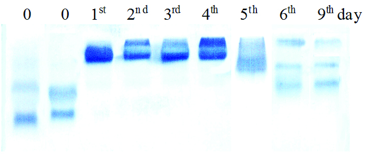
A typical electrophoregram of serum lipoproteins before and after tyloxapol injection
The result of electrophoresis of serum lipoproteins following tyloxapol injection are presented in [Table/Fig-8]. The first and second lanes are from two starved rats before tyloxapol injection. The electrophoregram of serum lipoproteins in starved normal rat shows three distinctive fractions LDL, VLDL and HDL migrating toward the anode. The percent of HDL was higher than LDL fraction in rat relative to fasted normal human [17,18]. The other lanes are from 1, 2, 3, 4, 5, 6 and 9 days after tyloxapol injection. The electrophoregram pattern of serum lipoproteins was the same in 2, 4 and 6 hours after tyloxapol injection. Therefore, we showed the electrophoregram after 2 hour injection as the sample in the first day of injection. [Table/Fig-8] indicates that after tyloxapol injection HDL disappears from the serum and the levels of VLDL and LDL increase. Tyloxapol alter not only the amount of the fractions but also reduced the electrophoretic mobility of all fractions. Tyloxapol as a nonionic detergent binds to lipoproteins and by increasing the weight reduces their migration [19]. The resulting pattern was similar to type III Fredrickson’s hyperlipoproteinaemia and continues up to 5 days that normal pattern begin to appear again.
An agarose gel electrophoresis of serum lipoproteins before and after tyloxapol injection. The cathode is at the top and the anode is at the bottom. Three distinctive bands indicate LDL, VLDL and HDL from top to bottom respectively. Four μL of each serum sample was put by a template on the top of the gel and separated by applying 75 volts for 25 minute. The specimen in the first and second lanes is before tyloxapol injection; the other lanes are from 1, 2, 3, 4, 5, 6 and 9 days after the injection.
Discussion
Short term kinetic of plasma lipids following tyloxapol injection
The results of the current study showed three distinctive phases in the kinetic of plasma lipids following intravenous injection of tyloxapol. The first phase begins from the administration of tyloxapol and extends at least 6 hr. Triglyceride accumulated linearly and sharply at the mean rate of 259.7 ± 8.1 mg/h.dl in this phase [Table/Fig-1]. Otway and Robinson [3] showed this phase continues for 3 h, but others indicated for a longer period [4,12]. This phase is convenient and used to measure the secretion rate of VLDL associated triglyceride from the liver [6–10] and to study the effects and mechanism of hypolipidemic drugs that probably acts on VLDL secretion [13,14]. The rate of triglyceride secretion in rats reported by some references was gathered and presented in [Table/Fig-7]. We calculated the average rate of triglyceride secretion in starved male rat as 250.6 ± 37.0 mg/h.dl or 102.8 ± 15.2 mg/h.kg body, which was comparable to the result of the current study. This value is also comparable to the mean rate of triglyceride secretion in starved mice which collected from several references and reported to be as 99.1 ± 7.3 mg/h.kg body [4]. This value is about twice of the secretion rate reported on the isolated rat hepatocytes [18] and perfused rat liver [8], on which some of the secreted triglyceride is exposed to hydrolysis and uptake by the liver cells [20]. Recently this phase has been used to study the effects of hypolipidemic drugs in artificial hyperlipidemia [13,14]. Since there is a great variation on hyperlipidemic values, we suggest that the researchers measure the concentration of triglyceride manually over the time instead of one time and compare the slope of the lines in the control and case groups [7,21,22].
Long term kinetic of plasma lipids following tyloxapol injection
The second phase refers to second and third days of detergent treatment. Triglyceride continued to accumulate in plasma in this phase but with a lower rate (105 mg/h.dl) and reached to maximum level of 3200 mg/dl after 24 hour. Millar et al., have shown that triglyceride accumulates linearity in plasma during this phase, but the secretion rate reduces gradually [4]. They concluded that the reduction in the rate of triglyceride accumulation is not due to the lack of inhibition of its hydrolysis but more likely is due to a decrease in the hepatic production rate. But Brindley et al., demonstrated that tyloxapol had not influenced the rate of triglyceride synthesis in rat hepatoctye [25].
In the last phase (3-5 days) the plasma lipids fell down toward the normal ranges. The biological half-life of tyloxapol is reported to be 23 hour in vivo [26]. By assuming the plasma volume of about 8.2 ml [3,10], single doses of 100 mg detergent per 200 g body results a plasma concentration approximate to 10 mg/dl. Orbans et al., used a single low dose of tyloxapol (40 mg/body) and observed that the levels of serum lipids elevated to maximum level at 6 h but fell down after 24 hour [27]. Conversely, Levin and Saltzman induced sustained hyperlipidemia by three injection of detergent per week [28]. The kinetics of plasma lipids observed in the current study is confirmed by the pathological changes of the liver reported by Trout and Viles [29]. They examined the pathological changes of the liver tissue after treatment with tyloxapol for a long time by an electron microscope. They observed that the liver uptakes tyloxapol and accumulates in hepatocyte lysosomes as early as one hour post-injection. The detergent induced autophagic vacuoles, the size and numbers of vacuoles increased during 12 hour and continued for three days. The autophagic lysosomes cleared into the bile canaliculi by mechanism resembling exocytose. The size and number of tyloxapol-filled lysosomes begin to decrease after 3-5 days and normal morphology reappeared following 15-30 days.
The changes of serum lipoproteins following tyloxapol injection
The results of electrophoresis of serum lipoproteins reveal several important points in accordance with the changes of plasma lipids. The electrophoretic pattern of serum liopoproteins of starved rat relative to human showed that the percent of HDL is more than LDL fraction [18]. In addition, the HDL fraction disappeared and VLDL fraction increased 2 h after tyloxapol injection. This pattern is similar to type III Fredrickson’s hyperlipoproteinaemia. Korolenko et al., determined the composition of lipoprotein fractions before and one day after tyloxapol injection by X-ray diffraction analysis [12]. They found that the levels of VLDL increased to more than ten times but the levels of LDL and HDL did not change significantly. Yamamoto et al., performed an in vitro study with various concentration of tyloxapol on gel of electrophoresis using whole plasma or isolated HDL [19]. They found that the detergent induces progressive structural changes in HDL in concentration dependent manner. Tyloxapol at concentration of 10 mg/ml causes dissociation of apoproteins A and C from HDL fraction without loss of lipids [4,11,19], increases the size of the particles and gradually decreases the electrophoretic mobility. They showed that apoAI deficient HDL remains at the place of application on the level of 10 mg/ml of tyloxapol, the concentration that is available by IV dose of 100 mg per 200 g body of rat.
The mechanisms of hyperlipidemic action of tyloxapol
Different mechanisms have been proposed whereby the detergent causes hyperlipidemia [4]. Some evidences indicated that tyloxapol forms a surface coat around lipoproteins rending them inaccessible as substrate. Some researchers suggested that the detergent acts directly on the lipolytic enzymes, hepatic and lipoprotein lipase [5] and lecithin- cholesterol acyltransferase [26], rather than physical modification of the substrate. Two groups of researchers investigated the kinetic of plasma apo (lipo) proteins after administration of tyloxapol at the initial minutes up to 20 hours [4,11]. They observed that HDL apoproteins AI and C begin to decrease in initial minutes and disappear completely after 2 hours. They suggested that the intact HDL may be essential for rapid catabolism of VLDL [11]. The absence of HDL cofactors follows tyloxapol injection (AI cofactor of LCAT and CII activator of LPL) may cause the massive increase in plasma triglyceride. The results of the kinetic of plasma lipids in conjunction with the lipoprotein electrophoresis are significant. Although in the electrophoregram the increment of VLDL and disappearance of HDL fraction occurs simultaneously following injection of detergent, the reduction of VLDL (at the 3rd day) anticipates the reappearance of HDL fraction (after 5 days). Therefore, the latter hypothesis was discarded and it could be concluded that, the changes of VLDL are probably independent of HDL fraction following treatment of detergent. Zeniya and Reuben observed that the detergent suppressed biliary cholesterol and phospholipids secretion rates but the bile flow and biliary lysosomal discharge did not alter [30]. This mechanism may also participate partly in the elevation of plasma lipids.
Simultaneous increment of triglyceride, cholesterol and phospholipids
The current findings showed that tyloxapol causes to increase the levels of total plasma cholesterol and phospholipids as well as triglyceride [Table/Fig-1-3]. The results confirm the previous studies [13,26,30–32]. Several mechanisms have been involved by which tyloxapol causes to elevate the levels of total cholesterol and phospholipids. The simplest explanation is that the components of VLDL increase coordinately. Plasma VLDL phospholipids are subject to hydrolysis by lipoprotein and hepatic lipases, as it is demonstrated that they possess phospholipase-A1 activity [11]. Cholesterol and phospholipids are also hydrolyzed by LCAT, which is inhibited partly by tyloxapol [26]. Argen et al., have shown that in tyloxapol injected rats the composition of phospholipids changes similar to the nascent VLDL [32]. Tyloxapol also displaces apoproteins from HDL which inhibit the transfer of phospholipids between lipoproteins [4,11,19].
Conclusion
A single intravenous injection of tyloxapol at dose of 400mg/kg body weight shows three distinctive phases, sharp linear increment, slow linear increment and slow decrement of plasma lipids toward the basal levels.