It is widely accepted that the oral and general health can influence quality of life. According to the World Oral Health Report of 2003, oral diseases impede activities in school and work causing many productive hours to be lost each year all over the world. Despite worldwide improvements in the oral health, dental carious is still a major oral health problem in most industrialized countries, affecting 60%-90% of school children and the vast majority of adults [1]. The current concept regarding cariogenesis is that a caries lesion, either clinically invisible or detectable, is the accumulation of numerous episodes of demineralization and remineralization, rather than a unidirectional demineralization process. The periods during which there is return to the resting pH is when remineralization occurs. Thus, it can be stated that remineralization is the process by which partly-dissolved crystals are induced to grow by accretion of calcium and phosphate ions from solution. Remineralization is an important natural repair process that counteracts cariogenic challenge [2].
Fluoride is recognized as a remineralizing agent, interacting with oral fluids on the interface of enamel and subsurface regions of teeth, and combining with calcium and phosphate ions to form fluorapatite. The anticaries benefits of fluoride depend on the use of an effective concentration and frequency of application [3].
Casein Phosphopeptide-Amorphous Calcium Phosphate (CPP-ACP) was introduced as a remineralizing agent in the year 1998 [4]. It comprises of nanocomplexes of milk protein CPP with ACP. It has been claimed that it promotes remineralisation of the carious lesions by maintaining a supersaturated state of essential minerals, at the same time it also hinders colonization of dental surfaces by cariogenic bacteria [5].
Functionalized Tricalcium Phosphate (fTCP) has tricalcium phosphate particles which have been ball milled with sodium lauryl sulphate. It is known that the fluoride ion has the ability to remineralize enamel subsurface lesions.
All the three remineralizing agents mentioned above differ in their composition and mechanism of action, yet each one has a promising ability to remineralize the enamel. Thus, the present study was undertaken to compare and evaluate the remineralization potential of Fluoride Varnish, Casein Phosphopeptide - Amorphous Calcium Phosphate (CPP-ACP) Paste and functionalized Tricalcium Phosphate (fTCP) Paste using confocal microscope.
Materials and Methods
The present in-vitro study was conducted in Department of Pedodontics and Preventive Dentistry, A.E.C.S. Maaruti College of Dental Sciences and Research Centre, Bangalore, India, for a period of 40 days.
Sample selection and preparation- Sixty permanent central incisors extracted for periodontal reasons were included in this study as they have sufficient cervico-incisal length to accommodate two windows of 3X3mm one below the other. All soft debris and calculus were removed from the teeth with a periodontal scaler. The teeth were then stored in 10% formalin solution. The samples included were free of hypoplasia, caries, discolouration, developmental defects and enamel fractures. This was done as any pre-existing alteration of surface morphology of the tooth can directly influence the caries progression [6].
Preparation of enamel window- Two square piece of plaster adhesive tape measuring 3mm length x 3mm width (mesio-distal) were placed on the labial incisal and cervical one third surfaces of each of the 60 permanent maxillary central incisors and the remaining portion of the teeth were painted with acid resistant nail varnish. After drying of varnish, the adhesive tape was removed.
Preparation of samples for demineralization- Artificial carious lesions were created on the exposed enamel by suspending all teeth in a demineralizing solution which was a mixture of calcium chloride [2.0mmol/L], tri sodium phosphate [2.0mmol/L] in acetate buffer [75mmol/L] solution at 4.6 pH for four days [7]. Techniques involving the direct use of acid to produce caries like lesions have lately replaced the ones involving the microbial production of acid. These methods have improved our understanding of the mechanism of de and remineralization processes. Further they provide information about the effects of caries preventive agents on the de/remineralization dynamics at the surface and in the subsurface of the teeth [8–10]. After four days the teeth were removed from the demineralizing solution and were randomly divided into three groups of 20 each.
Experimental groups
Group-I Fluoride Varnish (Fluorprotector; Ivaclar Vivadent).
Group-II CPP-ACP (GC Tooth Mousse, Recaldent; GC Corp; Japan).
Group-III functionalized Tricalcium Phosphate (CLINPRO Tooth crème, 3M).
Preparation of samples for remineralization- Before proceeding with remineralization one demineralized window in all the specimens was covered with nail varnish to serve as a control. The specimens in Group I were painted with Fluoride Varnish once whereas CPP-ACP Paste and fTCP Paste were applied in paste form for 2 minutes; twice a day for 20 and 40 days. These specimens were then placed in artificial saliva throughout the duration of study to stimulate oral condition. The solution was kept at room temperature.
Post-treatment analysis- Following the treatment regimen the specimens were mounted in self-cure acrylic resin. The samples were sectioned longitudinally perpendicular to the varnished area so that each section included the varnish-covered baseline lesion area and the uncovered, post-treatment lesion area with a hard tissue microtome (Leica SP 1600) to obtain specimens that were 150-200 microns thick. The sections were stained with freshly prepared 0.1mM Rhodamine B solution for 1 hr. Rhodamine B from the solution incorporates in demineralized tooth structure and does not penetrate sound tooth structure [11].
The stained samples were washed thoroughly with phosphate buffer solution until there was no dye leaching out of the sample. All samples were mounted on frosted glass slides with 80% glycerol mountant and the edges of the cover slip were coated with transparent nail enamel for further analysis through the confocal laser scanning microscope, Leica TCS SL inverted microscope [7].
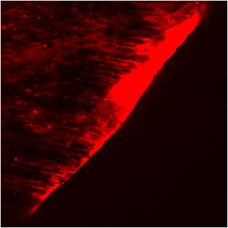
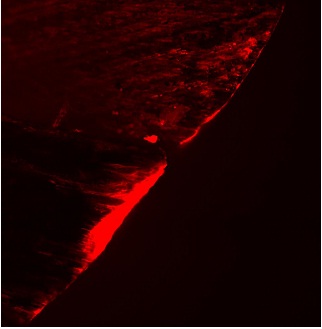
The images with CLSM were captured from the labial surface that is one each from either side of the midpoint measured from the occluso-cervical length of the tooth at [5X] magnification and argon laser was used at 488 nm wavelength for excitation and emission range of 498-514 nm wave length [Table/Fig-1,2]. The captured images were calibrated for linear depth of fluorescence and also the average/total fluorescence of the lesion using the Leica TCS SL in-built software, Germany [7]. These values were noted and tabulated. These values were compared by statistical analysis using Fisher’s t-test, One Way ANOVA test and multiple comparisons with Post-hoc Bonferroni test.
Photomicrograph of demineralized area under confocal microscope.
Photomicrograph of remineralized area under confocal microscope.
Results
Group I (Fluoride Varnish) showed no significant difference in the remineralization at both the time intervals. Group II (CPP-ACP Paste) and Group III (functionalized Tricalcium Phosphate) showed a significant difference in the remineralization at the two time intervals (p<0.01*) showing that the remineralization potential of CPP-ACP paste and fTCP increased significantly at 40 days [Table/Fig-3].
Evaluation of mean depth of remineralization of the study groups by Fisher’s exact test at 20 days & 40 days.
| Study groups | 20 days | 40 days | Fisher’s exact test | p-value |
|---|
| Depth of demineralized area (before) (μ) | Depth of remineralized area (μ) | Depth of demineralized area after remineralization (μ) | Depth of demineralized area (before) (μ) | Depth of remineralized area (μ) | Depth of demineralized area after remineralization (μ) |
|---|
| Group I | 151.80 ± 10.12 | 115.60 ± 7.01 | 36.20 ± 4.54 | 151.33 ± 11.85 | 115.97 ± 8.48 | 35.30 ± 4.67 | 0.12 | p>0.05 |
| Group II | 160.06 ± 6.09 | 107.56 ± 4.95 | 52.50 ± 4.80 | 149.13 ± 8.30 | 112.04 ± 7.08 | 37.05 ± 2.54 | 1.65 | p<0.01* |
| Group III | 156.90 ± 3.58 | 94.19 ± 9.43 | 62.71 ± 3.52 | 145.43 ± 7.61 | 97.81 ± 7.37 | 47.62 ± 2.91 | 0.96 | p<0.01* |
| Depth (μ) before application of remineralizing agents. (ANOVA) | F value | 2.670 | Depth (μ) after application of remineralizing agents. (ANOVA) | F value | 28.977 |
| p-value | 0.083 | p-value | <0.001* |
* significant
[Table/Fig-3] shows ANOVA results for Analysis of depth (μ) before application of remineralizing agents showed that higher mean depth was recorded in Group II followed by Group III and Group I respectively. No significant difference was observed between the groups (p>0.05) with respect to the mean depth. Analysis of depth (μ) after application of remineralizing agents showed that higher mean depth was recorded in Group I followed by Group II and Group III respectively. The difference in mean depth among the groups was found to be statistically significant (p<0.001). In order to find out among which pair of groups there existed a significant difference, we carried out multiple comparisons using Bonferroni test [Table/Fig-4] which depicted that Fluoride Varnish showed a significant increase in the remineralization potential as compared to CPP-ACP paste and functionalized Tricalcium Phosphate at both time intervals (p<0.05*) CPP-ACP paste showed a significantly higher remineralization potential than functionalized Tricalcium Phosphate when the two were compared (p<0.05*).
Comparison of mean depth of remineralization between the study groups using Bonferroni’s test at 20 days and 40 days.
| (I)group | (J) group | 20 days | 40 days |
|---|
| Mean difference (I-J) | p -value | Mean difference (I-J) | p –value |
|---|
| Group I | Group II | 8.039 | 0.021* | 3.93 | 0.01* |
| Group III | 21.415 | <0.001* | 18.16 | 0.0001* |
| Group II | Group I | -8.039 | 0.021* | 3.93 | 0.01* |
| Group III | 13.376 | 0.001* | 14.23 | 0.0001* |
| Group III | Group I | -21.415 | <0.001* | 18.16 | 0.0001* |
| Group II | -13.376 | 0.001* | 14.23 | 0.0001* |
* significant
Discussion
Fluoride is the cornerstone of the noninvasive management of non-cavitated carious lesions [12]. It helps in repair or healing of the lesion mostly by deposition of the mineral on the existing damaged crystals (crystal growth) or nucleation and de novo crystal formation.
Fluoride varnishes were first developed around the late 1960s and early 70s. The idea was that by lengthening the time the fluoride is in contact with the teeth the fluoride uptake should be increased. Zero et al., stated that the primary anti-caries activity of fluoride occurs topically [13]. The inhibition of caries by fluorides was ascribed to the reduced solubility of enamel due to the incorporation of fluoride ions into the crystal lattice of enamel in the form of fluorapatite.
CPP are peptides that are derived from the milk protein casein that are complexed with calcium and phosphate. CPP contain a cluster of phosphoseryl residues that stabilize nanoclusters of ACP in metastable solution. The CPP binds to spontaneously forming ACP nanoclusters and prevents their growth to the critical size required for nucleation and precipitation. It has been proposed that the mechanism of anticariogenicity for CPP-ACP is that it substantially increases the level of calcium phosphate in plaque, which decreases enamel demineralization and enhances remineralization. CPP will bind to surfaces such as plaque, bacteria, soft tissue and dentin (owing to its sticky nature), providing a reservoir of bioavailable calcium and phosphate in the saliva and on the surface of the tooth.
Recently, a new prospective calcium system was prepared by reacting beta-TriCalcium Phosphate (β-TCP) with Sodium Lauryl Sulfate (SLS) using a mechanochemical ball milling process to form a functionalized calcium phosphate. During this process, Tri-Calcium Phosphate is functionalised resulting in an organic calcium phosphate hybrid being formed. This prospective calcium system interacts with demineralized enamel to help boost remineralization benefits of fluoride. It delivers more bio-available fluoride and calcium to the teeth. The present study evaluated and compared the remineralization potential of fTCP with Fluoride Varnish and CPP-ACP Paste.
In the present study, fluoride varnish group showed no significant difference in the remineralization depth at 20 and 40 days. Research to date has shown no difference in efficacy with multiple applications of varnish within a short period [14].
When compared to CPP-ACP paste group and fTCP group at both time intervals there was a significant increase in remineralization in the fluoride varnish group which is in accordance with the study by Lata et al., which concluded that; CPP-ACP cream is effective in remineralizing early enamel caries at surface level but to a lesser extent than Fluoride Varnish [15]. The CaF2 layer functions as a pH-controlled fluoride reservoir and is the most important supplier of free fluoride ions during the cariogenic attack [16]. Prabhakar et al., concluded that sodium fluoride showed relatively greatest remineralizing and dentinal tubule occlusion property when compared with GC MI Paste Plus and Clinpro Tooth Crème [8]. Jayarajan J et al., showed that the added benefit of fluoride (NaF 0.2%), CPP-ACPF (Tooth Mousse-Plus) showed marginally more amount of remineralization than CPP-ACP (Tooth Mousse) [17].
In the present study a significant increase in remineralization potential was seen at the end of 40 days in CPP-ACP paste group as compared to 20 days. Hegde MN and Moany A showed that 10% CPP-ACP paste is capable of remineralizing subsurface enamel lesions. The samples treated for 35 days showed significantly higher remineralization compared to those treated for 7, 14, 21 and 28 days. This result is in accordance with the present study which states that the remineralization was dose-dependent [18].
However, when CPP-ACP group was compared to the Fluoride Varnish group, the Fluoride Varnish was significantly better. The presence of plaque, bacteria and saliva, observed in the in situ condition significantly enhances the remineralization process which was not possible in an in-vitro condition. This could be the possible reason why the Fluoride Varnish group was better than the CPP-ACP group. Similar to our study, Shirahatti et al., concluded that fluoridated dentifrice provides substantial protective effect against lesion formation and lesion depth progression however, CPP-ACP paste did not have any additional ability in reducing lesions depth progression and effect was similar to non-fluoridated dentifrice group [19].
When CPP-ACP group was compared to fTCP group, CPP-ACP group was significantly better. The results of present study are similar to the study by Balakrishnan et al., which concluded that CPP-ACP showed better remineralizing potential than the fTCP and Calcium Sodium Phosphosilicate (CSP) groups. The enhanced remineralization potential by the CPP-ACP containing products can be attributed to the high level of bio-available calcium and phosphate ions released by it [20]. The fTCP is poorly soluble with the large particle size which explains the poor release from the product and the inability to significantly increase salivary calcium and inorganic phosphate levels [12].
Balakrishnan et al., also concluded that the micro hardness of enamel treated with fTCP + NaF was lower than that of CPP-ACP group after 30 days remineralization [20]. Similarly the current study too showed a significant increase in the remineralization potential of CPP-ACP group as compared to fTCP group after 40 days.
The fTCP group had a low level of total (acid soluble) calcium phosphate. This may be related to the known instability of fluoride ions in oral care formulations containing poorly soluble calcium-based abrasives [12]. The fTCP product contains sodium fluoride hence, the fluoride ion would be susceptible to a reduction in bioavailability in the presence of added calcium phosphate without a stabilizer. This material described as fTCP is poorly soluble and together with the large particle size and low amount added would explain the poor release from the product and the inability to significantly increase salivary calcium and inorganic phosphate levels. Hence, it is not surprising that the product failed to show enhanced remineralization over other materials in this study. The results of present study are in accordance with study conducted by Shen et al., which concluded that fTCP was not significantly different to 1000 ppm F whereas CPP-ACP and CPP-ACP Plus were superior to 5000ppm F with Tooth Mousse Plus producing the highest level of enamel lesion remineralization [12]. Contrary to this Karlinsey et al., concluded that the combination of a new functionalized calcium phosphate technology plus 1000ppm F, produced significantly greater remineralization relative to both the 1000ppm F test dentifrice and MI Paste Plus, and was statistically equivalent to Theramed SOS (1450ppm) [21]. Also Amaechi BT et al., observed that combining fluoride with tricalcium phosphate could provide more anti-caries benefits compared to using fluoride alone [22].
The uniqueness of the present study is that it evaluates and compares the remineralization potential of Fluoride Varnish, CPP-ACP Paste and fTCP Paste. The fTCP Paste used in the study is a relatively newer remineralizing agent so its comparative studies are scarce. Also the confocal laser scanning microscope used overcomes the limitations of conventional wide-field microscopy. CLSM images are of notable higher quality than those obtained with the conventional optical microscope because the generated images contain volumetric and texture details impossible to obtain with the conventional ones. From the observations of the present study, it can be inferred that among the groups tested, fluoride varnish is the most effective remineralizing agent followed by CPP-ACP Paste and fTCP.
Limitation
It is understandable that remineralization in vitro may be largely different when compared with dynamic biological system in oral cavity in vivo. Thus, straightforward assumptions to clinical conditions must be bestowed with caution due to glaring constraints of in vitro studies. There is immense need for improved and groundbreaking remineralization methods to halt the caries process, especially in individuals with a high cariogenicity.
Conclusion
Based on the data from the present study it can be concluded that all the three remineralizing agents successfully showed the remineralization of artificial carious lesions at both 20 and 40 days time intervals. However, fluoride varnish group showed the highest remineralization potential followed by CPP-ACP paste group and lastly by fTCP group at both 20 and 40 days. Both CPP-ACP paste group and fTCP group showed a significant increase in the remineralization potential after 40 days as compared to 20 days unlike fluoride varnish group where no significant difference was observed.
Nonetheless, it must be restated that our results were obtained in an in vitro ideal environment, independent of any anti-microbial effect. Saliva, plaque and many other confounding factors may affect the release of remineralizing agents through such systems. We therefore, need further in vivo studies to vindicate our findings and descry the true clinical efficacy of these newer remineralizing agents.
* significant
* significant