Congenital Ichthyosis - Collodion Baby Case Report
Priyanka Srivastava1, Anuj Srivastava2, Prachi Srivastava3, Anupama Vithal Kumar Betigeri4, Minakshi Verma5
1 Assistant Professor, Department of Physiology, PGIMER and Dr. RML Hospital, New Delhi, India.
2 Consultant Paediatrician and Consultant Gynaecologist Respectively, Shiv Radhika Memorial Hospital, Gorakhpur, U.P, India.
3 Consultant Paediatrician and Consultant Gynaecologist Respectively, Shiv Radhika Memorial Hospital, Gorakhpur, U.P, India.
4 Assistant Professor, Department of Physiology, PGIMER and Dr. RML Hospital, New Delhi, India.
5 Assistant Professor, Department of Anatomy, Lady Harding Medical College, New Delhi, India.
NAME, ADDRESS, E-MAIL ID OF THE CORRESPONDING AUTHOR: Dr. Priyanka Srivastava, E-2, Flat No. 340, Vasant Kunj, New Delhi-110070, India.
E-mail: drpriyanka25@gmail.com
Collodion baby, Lamellar ichthyosis, Harlequin ichthyosis, Non bullous congenital ichthyosiform erythroderma
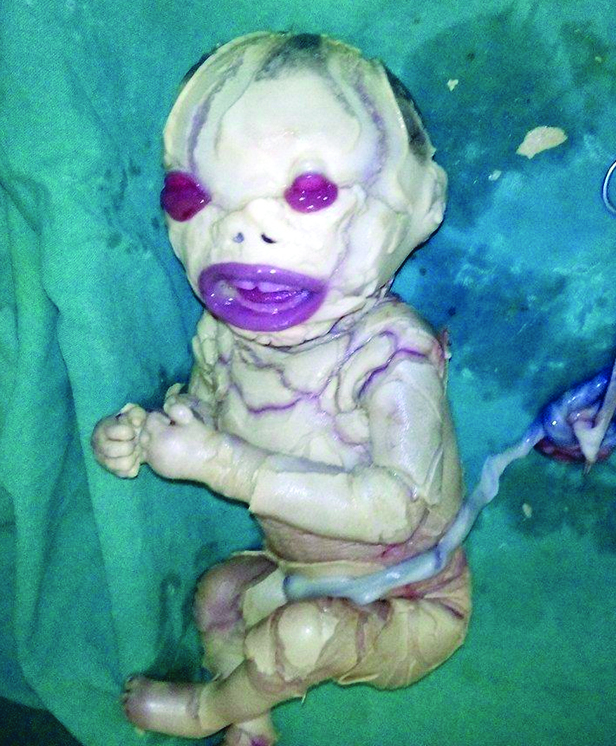
A term female neonate was delivered by LSCS to a primigravida mother with the parents being non-consanguineous. The infant had a birth weight of 2.85 kg (appropriate for gestational age), with length and head circumference being 51cm and 32cm, respectively. On examination the vitals of the neonate was normal and the Apgar score was normal 6/9/9 at 1, 5, and 10 minutes respectively. A detailed physical examination showed broad thick scales all over the body with predominance over the head, chest, abdomen and arms. On head and neck examination there is presence of abnormal parchment like membrane covering the head and the hairs are sparse. There was associated ectropion and eclabium too. Due to excess of scales around the mouth gave the typical fish-like appearance. The newborn had generalized erythema and oedema over the body [Table/Fig-1]. No other associated obvious anomalies were seen. Thorough systemic examination was carried out which revealed no abnormalities. The family history was taken and pedigree analysis was done. There was no history of dermatological-related disorder or any other genetic disease. There was no history of consanguinity. During pregnancy there is no history of maternal complications or any drug exposure. The neonate was admitted in NICU and managed with adequate humidification in an incubator. The dermatological opinion was taken and proper monitoring of body temperature and adequate fluid and electrolyte replacement was done. Tube feeding was done at age of 1 day. As advised by the dermatologist topical emollients were regularly applied. Artificial tears were applied to prevent drying of eyes. The infant was closely monitored for any sepsis like condition in the form of off color, shock or delayed perfusion. The baby was then referred to a tertiary care hospital for further management. However, the neonate was later lost to follow-up and so we were unable to ascertain whether the neonate has any kind of genetic mutation. As we know that TGM1 gene mutation is the commonest cause of Collodion baby and it could not be ascertain whether the infant had Lamellar icthyosis Autosomal Recessive (AR) type or Non Bullous Congenital Icthyosiform Erythroderma (NBCIE). The written informed consent was taken from parents for case reporting and image publication.
Figure showing generalized erythema and oedema over the body.
Discussion
Hallopeau and Watelet were the first who gave the term Collodion baby (CB) [1]. The skin of the newborn is replaced by a cornified substance, which gives the body a parchment like appearance or a varnished appearance [2]. This condition is inherited primarily as autosomal recessive ichthyosis either LI or NBCIE. CB is an extremely rare dermatological condition with an estimated incidence of 1 in 50,000 to 100,000 birth [3]. A new form of the disease has been notified as “self-healing collodion syndrome’ in these cases newborn completely recovers within few months after birth [4].
Collodion baby is described as a congenital condition characterized by presence of parchment –or- cellophane like membrane encompassing the whole body. This is secondary to disorder of cornification. These are usually prematurely born and diagnosed at the time of birth only. Due to presence of tight membrane these babies develop many complications like ectropion, eclabium, restricted extremities and digits movements due to pseudocontractures, absence of eyebrows, sparse hairs on head, deformed nose and ears due to hypoplasia of nasal and ear cartilage. The babies has poor sucking, distal limb ischemia and oedema of extremities.
A scoring system has been developed for neonate with collodion membrane based on various parameters to know the outcome [5]. In one of the previous case report CB association to drug ingestion like infliximab has been seen [6] and the biopsy reveals thickened stratum corneum [5].
It is seen that the collodion membrane sheds off in next 2-4 weeks after birth revealing the underlying skin disorder. In long term course, approximately 75% of collodion baby cases will develop an AR Congenital ichthyosis (lamellar ichthyosis or congenital ichthyosiform erythroderma) [7].
The exact cause of the CB syndrome is not known but in most of the cases autosomal recessive inheritance pattern is seen [8] and they are very rare and may be associated with consanguinity. In just 10% of these cases the membrane sheds off and underlying skin is normal for rest of the life [1]. In rest 15% cases association with various entities is seen like ichthyosis vulgaris, trichothidystrophy, metabolic and endocrinal disorders which involve keratinization disorders [7]. Lamellar ichthyosis is an AR type of congenital ichthyosis characterized by hyperkeratosis. Incidence of LI is 1 per 140,000- 300,000 population. Commonly the LI cases is caused by various genetic mutations including TGM1(commonest), NIPAL4, ALOX12B, ALOXE3, ABCA12, CYP4F22, PNPLA1 (OMIM 615024), (17) and CERS3 (OMIM 615023). TGM1 is located on chromosome 14q11.2 and has 15 exons (Genbank NM- 000359.2) [9].
NBCIE is also an AR disorder characterized by mild clinical features and mutations in ALOX12B and TGM1 genes is seen which leads to abnormal TGM1. Management requires combined effort of dermatologist, neonatologist and in some cases ophthalmologist and ENT specialist. The aim of treatment is to eliminate fish like scales and reduce the excessive irritation. The parents should be counselled and regular follow-up of the patient is needed [10].
In CB there is high risk of dehydration and electrolyte imbalances due to high insensible loss from the skin, leading to hypernatraemic dehydration. So it requires the patient to be placed under high humified incubators and regular monitoring of the body temperature and proper nutrition is required [11,12].
An increase risk of infections both cutaneous and systemic like candida and bacterial, fissures, ischemia and oedema of limbs due to membrane compression [1,13]. The first line of management is moisturizers and topical keratolytic agents, they enhances skin barrier and facilitate desquamation. Sodium chloride, urea, vitamin E acetate, glycerol and petroleum jelly are various agents available as moisturizers and lubricants. These infants are at increased risk of intoxication by absorption of topical products, like salicylates or keratolytics due to impaired skin. In severe cases with marked hyperkeratosis keratolytical agents like lactic acid, glycolic acid, salicyclic acid, N- acetyl- cystine, glycol can be used. Ectropion is managed by application of artificial tears and eye lubricants. In severe cases surgical correction is done. Administration of retinoids have keratolytic effect and help in elimination of scales and prevent hyperkeratosis of skin. Isotretinoin and retinoids both have proven to be effective in these cases. External auditory canal must be regularly cleaned by an ENT person so that accumulation of scales is prevented and so hearing loss can be prevented [14].
Collodion syndrome is a rare disease so it is essential to have a protocol for the treatment of these patients, the instructions to follow in treatment and proper management of the complications that can arise. In long-term course the etiological cause of the disease should be established so that appropriate measures can be given to the patient.
The treatment is mainly supportive like use of incubators and IV fluids and tube feeding and use of emollients. In our experience, we can suggest that reassurance to the parents is needed that the newborn can be survived and condition will get better. Special care to the skin is needed and collodion membrane should not be peeled off, it will shed after 1-2 week and infection should be prevented.
[1]. van Gysel D, Lijnen RLP, Moekti SS, De Laat PCJ, Oranje AP, Collodion baby: a follow-up study of 17 casesJournal of the European Academy of Dermatology and Venereology 2002 16(5):472-75. [Google Scholar]
[2]. Cortina WJ, Cruz MJ, Villalobos OA, Espinoza MA, Ictiosis congenital (feto arlequín)Bol Med Hosp Infant Mex 1975 32:699-702. [Google Scholar]
[3]. Dyer JA, Sparker M, William M, Care of newborn with ichthyosisDermatol Ther 2013 26(1):1-15. [Google Scholar]
[4]. Shwayder T, Akland T, Neonatal skin barrier: structure, function and disordersDermatol Therapy 2005 18:87-103. [Google Scholar]
[5]. DiGiovanna Robinson-Bostom L, Ichthyosis: etiology, diagnosis, and managementAm J Clin Dermatol 2003 4(2):81-95. [Google Scholar]
[6]. Offiah M, Brodell RT, Campbell LR, Wyatt JP, Collodion-like membrane in a newborn exposed to infliximabJ Am Acad Dermatol 2014 71(1):e22-23. [Google Scholar]
[7]. Dermatology at the Millennium. By Delwyn Dyall-Smith, Robin Marks, Page 586, Published by Informa Health Care, 1999, ISBN [Google Scholar]
[8]. Moslehi R, Signore C, Tamura D, Mill JL, DiGiovanna JJ, Tucker MA, Adverse effects of trichothiodystrophy DNA repair and transcription gene disorder on human fetal developmentClin Genet 2010 77:365-73. [Google Scholar]
[9]. Fachal L, Rodriguez-Pazos L, Ginarte M, Charaterization of TGM1 c.984+1G>A mutation identified in a homozygous carrier of Lamellar ichthyosisInt J Dermatol 2012 51(4):427-30. [Google Scholar]
[10]. Taïeb A, Labrèze C, Collodion baby: what’s newJ Eur Acad Dermatol Venereol 2002 16(5):436-37. [Google Scholar]
[11]. Schmuth M, Martin V, Janecke AR, Inherited ichthyosis/generalized Mendelian disorders of cornificationEur J Hum Genet 2013 21(2):123-33. [Google Scholar]
[12]. Rubio-Gomez GA, Development of a disease severity score for newborns with collodion membraneJ Am Acad Dermatol 2014 70(3):506-11. [Google Scholar]
[13]. Roberts JB, Adelson D, Case report: prolonged collodion membrane causing constrictive bands of the digits and treatmentDermatology Online Journal 2010 16(1):15 [Google Scholar]
[14]. Harvey HB, Shaw MG, Morrell DS, Perinatal management of harlequin ichthyosis: a case report and literature reviewJ Perinatol 2010 30(1):66-72. [Google Scholar]