Expression of Cytokeratin-19 and Thyroperoxidase in Relation to Morphological Features in Non-Neoplastic and Neoplastic Lesions of Thyroid
Hemanathan Guhanandam1, Revathishree Rajamani2, Naseen Noorunnisa3, Manimaran Durairaj4
1 Post Graduate Student, Department of Pathology, Shri Sathya Sai Medical College and Research Institute, Ammapettai, Kancheepuram, Tamil Nadu, India.
2 Assistant Professor, Department of Pathology, Shri Sathya Sai Medical College and Research Institute, Ammapettai, Kancheepuram, Tamil Nadu, India.
3 Professor, Department of Pathology, Shri Sathya Sai Medical College and Research Institute, Ammapettai, Kancheepuram, Tamil Nadu, India.
4 Professor, Department of Pathology, Shri Sathya Sai Medical College and Research Institute, Ammapettai, Kancheepuram, Tamil Nadu, India.
NAME, ADDRESS, E-MAIL ID OF THE CORRESPONDING AUTHOR: Dr. Hemanathan Guhanandam, Shri Sathya Sai Medical College and Research Institute, Ammapettai-603108, Kancheepuram District, Tamil Nadu, India.
E-mail: skillers_04@yahoo.co.in; mail2rrevathi@gmail.com
Introduction
Thyroperoxidase (TPO) is a protein involved in thyroid hormone synthesis. TPO gene suppression and mutation were involved in thyroid tumours. CK-19 plays important role in the structural integrity of epithelial cells. Reduced TPO expression with increased CK-19 immunoreactivity has been implicated as a marker for differentiating non neoplastic and neoplastic thyroid lesions.
Aim
To study the histopathological features of thyroid lesions and to evaluate the diagnostic role of thyroperoxidase and CK-19 in non-neoplastic and neoplastic thyroid lesions.
Materials and Methods
Prospective observational study of 65 thyroid specimens was studied for detailed histopathological examination and Expression of Immunohistochemical Markers Cytokeratin-19 (CK-19) and Thyroperoxidase.
Results
TPO IHC marker was expressed by non-neoplastic and benign lesions of thyroid but not in malignancy. CK-19 was expressed 100% in papillary carcinoma of thyroid and its variants, focal and weak staining noted in goitre and hyperplastic areas.
Conclusion
Most of the non-neoplastic and neoplastic lesions were diagnosed based on histopathological features. When the histopathological diagnosis are equivocal, immunohistochemical markers aids in diagnosing malignancy. Diffuse and strong TPO expression indicates non-neoplastic thyroid lesions whereas diffused and strong CK-19 expression indicates thyroid malignancy.
Multinodular goitre, Papillary carcinoma, Thyroid disorders
Introduction
Thyroid disorders are the most common endocrine disorder worldwide. About 42 million peoples in India are suffering from thyroid disease [1]. The spectrum of thyroid disorders classified as euthyroid, hypothyroid and hyperthyroid depending upon their thyroid function test [2]. In western countries 50% of the people in the community have microscopic nodule, 15% have palpable goiter, 10% demonstrate abnormal TSH and 3.5% have papillary carcinoma of thyroid [3]. Among thyroid disorders Multinodular Goitre is the commonest presentation. Among thyroid cancer, papillary carcinoma of thyroid is the commonest tumour [4]. Thyroid lesions can be diagnosed using histopathological criteria which allow us to differentiate between benign and malignant lesion [5]. But in some cases suspicious areas were noted in few foci. In such cases Immunohistochemistry (IHC) is used to differentiate between benign and malignant lesion. Thyroperoxidase (TPO)is a membrane bound protein needed for the thyroid synthesis. TPO immunoreactivity was reported in non neoplastic and benign thyroid lesions. Lack of TPO expression indicates malignancy [6]. Cytokeratin-19 (CK-19) has diffuse expression in papillary thyroid carcinoma and its variants and also has focal and weak staining in non neoplastic thyroid lesions. Panel of two markers, CK-19 and TPO increases the diagnostic accuracy in thyroid lesions than a single marker [7].
Aim
In this study, the histopathological features of thyroid lesions and the diagnostic role of thyroperoxidase and CK-19 in non-neoplastic and neoplastic thyroid lesions were studied.
Materials and Methods
This prospective observational study comprising of 65 cases of thyroidectomy, carried out in the Department of Pathology at our Institute during the period of August 2013 to June 2015. The approval from our Institutional Medical Ethics Committee was obtained IEC NO: 2013/148. The specimens were fixed in 10% neutral buffered formalin, processed routinely, paraffin embedded and stained with haematoxylin and eosin. Types of specimen received were lobectomy, hemithyroidectomy, subtotal and total thyroidectomy. All the specimens were subjected to gross and histopathological examination and IHC expression of TPO and CK-19 was done. An Immunohistochemical Score (IHS) was calculated for CK-19 and TPO for each section. This IHS took into account the percentage of cells (0-100%) as well as the staining intensity category (0-3). The extent of positivity was estimated and classified on a five point scale as follows grade 0- no staining; grade 1-10 % to ≤ 30%; grade 2 >30 % to ≤ 60%; grade 3 >60% to ≤ 90%; grade 4-100%. Positivity staining intensity was graded as follows -weak 1; moderate 2; strong 3. Final immunohistochemical score for the extent and staining intensity is as follows: 0- negative; 1-4 weakly positive (+); 5-8 moderate positive (++); 9-12, strongly positive (+++). Results were tabulated
Statistical Analysis
Statistical analysis was carried out using SPSS version 19.0 (IBM SPSS, US) software with Regression Modules installed. Chi-square test was done to find out the significance. One-way ANOVA Test was done to correlate histopathology with CK-19 and TPO IHC expression.
Results
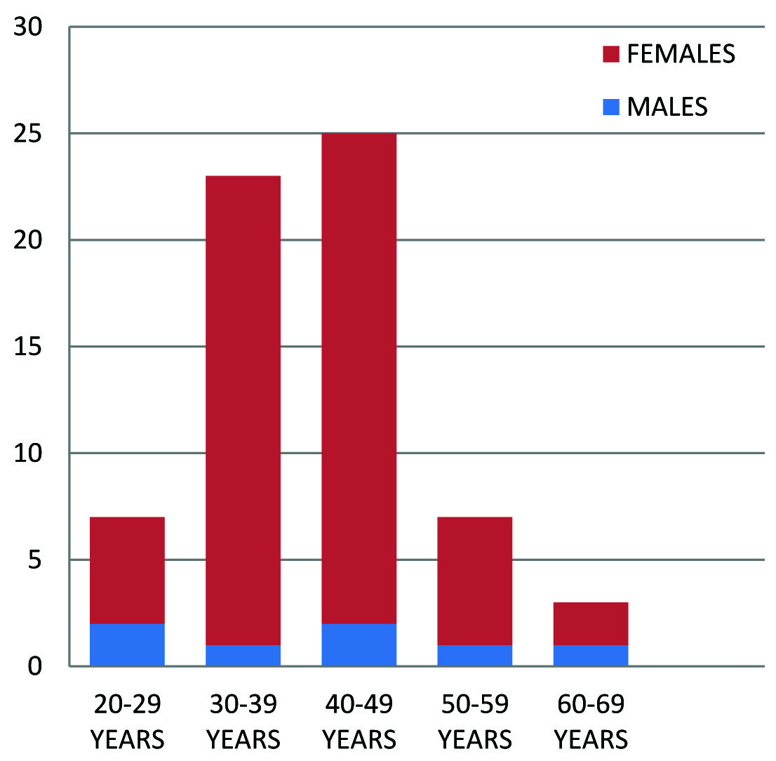
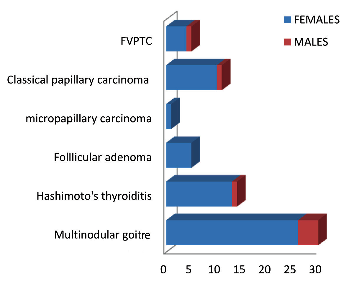
Among 65 cases 7 (11%) cases were males and 58 (89%) cases were females. Common age group affected was between 30-49 years. Among 16 malignant cases, 14 cases were in age group between 30-49 years of age and only 2 cases were above 60 years of age [Table/ Fig-1]. Among 65 cases of thyroid lesions received in our department, 29 cases were Multinodular goitre, 15 cases were Hashimoto’s thyroiditis, 5 cases were follicular adenoma, 10 cases were classical papillary carcinoma, 1 case was micro Papillary carcinoma and 5 cases were Follicular variant of Papillary carcinoma of thyroid [Table/Fig-2]. Out of 44 non neoplastic lesions, 40 cases showed 61-90% TPO positivity, 3 cases showed 31-60% positivity, 6 case showed 11-30% positivity. All cases of Follicular adenoma showed 11-30% positivity. All malignant cases showed less than 10% positivity [Table/Fig-3]. CK-19 expression was found to be more than 90 % in all malignant cases (Papillary carcinoma and its variants). A total of 19 cases of Multinodular goitre showed 11-30% positivity; 9 cases of goitre showed 31-60 % positivity [Table/Fig-4,5]. Histopathological correlation with TPO and CK-19 using one-way ANOVA was found significant [Table/Fig-6].
Age incidence of thyroid disorders.
Distribution of thyroid disorders.
Immunohistochemical expression of TPO
| <10% | 11-30% | 31-60% | 61-90% | >90% | |
|---|
| Multinodular goiter | | | 2 | 27 | | 29 |
| Hashimoto’s thyroiditis | | 1 | 1 | 13 | | 15 |
| Follicular adenoma | | 5 | | | | 5 |
| Papillary carcinoma of thyroid and FVPTC | 16 | | | | | 16 |
| Total | 16 | 6 | 3 | 40 | 0 | 65 |
Immunohistochemical expression of CK-19.
| Cases | <10% | 11-30% | 31-60% | 61-90% | >90% | |
|---|
| Multinodular goiter | | 19 | 9 | 1 | | 29 |
| Hashimoto’s thyroiditis | | | 13 | 2 | | 15 |
| Follicular adenoma | | | | 5 | | 5 |
| Papillary carcinoma of thyroid and FVPTC | | | | | 16 | 16 |
| Total | 0 | 19 | 21 | 8 | 16 | 65 |
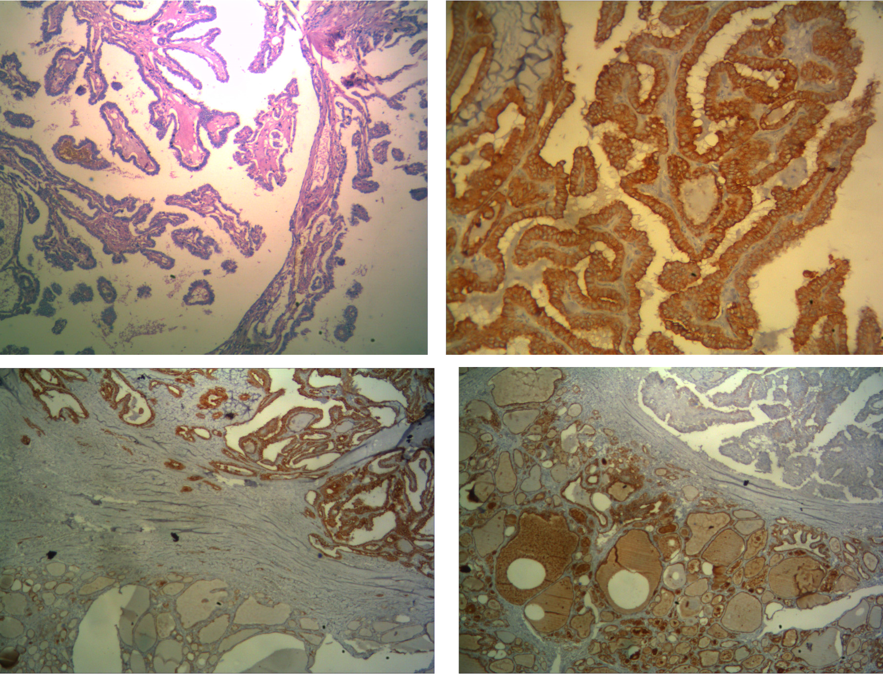
(a) H&E stain-Papillary carcinoma. (b) CK-19 stain 3+ in Papillary carcinoma. (c) 4x- CK-19 expression in papillary carcinoma with mild staining in nodular goitre. (d) 4X- TPO expression in goitre area with loss of expression in Papillary carcinoma area.
One-way Anova for HPE Correlation with TPO and CK-19.
| | Sum of Squares | DF | Mean Square | F | Sig. |
|---|
| TPO score | Between Groups | 820.339 | 4 | 205.085 | 134.607 | .000 |
| Within Groups | 91.415 | 60 | 1.524 | | |
| Total | 911.754 | 64 | | | |
| CK-19 score | Between Groups | 1232.248 | 4 | 308.062 | 159.387 | .000 |
| Within Groups | 115.968 | 60 | 1.933 | | |
| Total | 1348.215 | 64 | | | |
Discussion
Distinction between benign and malignant tumour is vital for the management and prognosis. When the histopathological features are equivocal, immunohistochemistry helps in arriving at final diagnosis. In our study, females are affected more than males. In a study by Burn and Taylor et al., female to male ratio was 2.5:1 [8]. A similar female preponderance was observed by Vicker et al., with female to male 2.1:1 [9]. In our study, non-neoplastic lesions were more common. Among non-neoplastic lesions, multinodular goitre was more common. In neoplasm, papillary carcinoma thyroid was more common. Thyroid carcinoma was more common in females than in males. TPO expression was found to be 90-100% in non-neoplastic and benign lesions and its expression was lost completely in malignancy. This is in concordance with the study done by De Micco et al., where they demonstrated anti TPO antibody MoAb 47 expressed in all normal and benign thyroid tissue but only in 3% of malignant lesion [6]. Various authors have implicated TPO expression in differentiating benign and malignant thyroid tumours [Table/Fig-7]. In our study CK-19 immunoreactivity was found to be diffuse and strong in malignancy and pale and focal staining in benign conditions. This is in concordance with the study done by various authors [Table/Fig-8].
Comparative analysis of TPO expression in Thyroid lesions.
| Study | Non-neoplastic lesions | Benign lesions | Malignant lesions |
|---|
| De micco et al., [6] | 100% | 100% | 3% |
| Zeming liu et al., [7] | 100% | 86.2% | 12% |
| Lima et al., [10] | 100% | 78.8% | 20% |
| S Savin et al., [ 11] | Not included | 68% | 21% |
| Present study | 100% | 100% | 0% |
Comparative analysis of CK-19 expression in Thyroid lesions.
| Study | Non-neoplastic lesions | Benign lesions | Malignant lesions |
|---|
| Michel R Nasr et al., [12] | 50% | 68% | 100% |
| Bose D et al., [13] | 50% | 75% | 100% |
| Cheung et al., [14] | 20% | 17% | 80% |
| Beesley et al., [15] | 25% | 25% | 100% |
| Won young et al., [16] | 50% | 50% | 90% |
| Present study | 100% | 100% | 100% |
In our study CK-19 expression lost in goitre with papillary areas and its positivity indicates malignancy transformation. This is correlated with the study done by Erkilic et al., where they have observed CK-19 expression differentiates papillary carcinoma thyroid and MNG showing papillary areas [17]. De Matos et al., demonstrated CK-19 and Galectin 3, HBME 1 these three immune markers were accurate in diagnosing benign and malignant thyroid lesions [18]. Majority of studies have reported that when CK-19 combined with panel of markers differentiates benign and malignant thyroid lesions [19–21]. Our study recorded CK-19 staining was weak and diffuse in most of the benign conditions. This indicates that CK-19 cannot be used as a single marker to establish the malignancy. Negative staining for CK-19 is strong evidence against papillary carcinoma thyroid. This is important in distinguishing papillary hyperplasia from papillary carcinoma. Bose et al., demonstrated weak and focal staining of CK-19 in MNG and follicular adenoma, strong and intense positivity in papillary carcinoma of thyroid [13]. The results from our study implicates that a combination of positive and negative marker is essential in distinguishing benign and malignant conditions. In our study, TPO expression indicates benign condition and loss of its expression implicates malignancy transformation and strong membranous CK-19 indicates malignancy. This is in concordance with the study done by Zeming Liu et al., where they demonstrated gain of CK-19 expression and loss of TPO expression were both considered significant in the progression of thyroid carcinoma [7].
Limitation
There are few limitations in the present study. CK-19 and TPO expression was observed in papillary carcinoma and its variants. Its expression was not studied in other thyroid tumours like Medullary carcinoma and Anaplastic carcinoma which were not received during the study period. CK-19 and TPO expression in cytology and its correlation with histopathology was not done. If done it would have provided the additional evidence for the diagnostic utility of these markers in thyroid lesions.
Conclusion
Although no single immunohistochemical marker is completely sensitive in differentiating benign from thyroid malignancy, the combination of TPO and CK-19 attains higher diagnostic yield. Loss of TPO expression in suspicious papillary areas indicates malignancy. Although CK-19 expression is demonstrated in both benign and malignant thyroid conditions, negative stain is the good evidence against papillary carcinoma thyroid.
[1]. Unnikrishnan AG, Menon UV, Thyroid disorders in India: An epidemiological perspectiveIndian J Endocr Metab 2011 15:78-81. [Google Scholar]
[2]. Pedersen IB, Laurberg P, Knudsen N, An increased incidence of overt hypothyroidism after iodine fortification of salt in Denmark: a prospective population studyJ Clin Endocrinol Metab 2007 92:3122-27. [Google Scholar]
[3]. Wang C, Crapo LM, The epidemiology of thyroid disease and implications for screeningEndocrinol Metab Clin North Am 1997 26(1):189-218. [Google Scholar]
[4]. LiVolsi VA, Papillary thyroid carcinoma: an updateMod Pathol 2011 24(2):S1-S9. [Google Scholar]
[5]. Vriens MR, Schreinemakers JM, Suh I, Guerrero MA, Clark OH, Diagnostic markers and prognostic factors in thyroid cancerFuture Oncology 2009 5(8):1283-93. [Google Scholar]
[6]. De Micco C, Vassko V, Henry JF, The value of thyroid peroxidase immunohistochemistry for preoperative fine-needle aspiration diagnosis of the follicular variant of papillary thyroidcancerSurgery 1999 126:1200-04. [Google Scholar]
[7]. Zeming Liu, Pan Yu, Significance of CK-19, TPO, and HBME-1 expression for diagnosis of papillary thyroid carcinomaInt J Clin Exp Med 2015 8(3):4369-74. [Google Scholar]
[8]. Burn JI, Taylor SF, Natural history of thyroid carcinoma-A study of 152 patientsBrit Med J 1962 :1218-23. [Google Scholar]
[9]. Vicker P, Thyroid cancer – A 10 year study from general hospitalInc J Surg 1981 43:817-20. [Google Scholar]
[10]. Lima MA, Gontijo VA, Santos MC, Schmitt FCL, Thyroid peroxidase expression in diseased human throid glands endocrine pathologyEndocrine Pathology 1999 10(3):223-28. [Google Scholar]
[11]. Savin S, Cvejic D, Thyroid peroxidise and galectin 3 immunostaining in differentiated thyroid carcinoma with clinic pathological correlationHuman Pathology 2008 39:1656-63. [Google Scholar]
[12]. Nasr MR, Mukhopadhyay S, Immunohistochemical markers in the diagnosis of papillary thyroid carcinoma: utility of HBME1 combined with CK-19 immunostainingModern Pathology 2006 19:1631-37. [Google Scholar]
[13]. Bose D, Das RN, Chatterjee U, Banerjee U, Cytokeratin 19 immunoreactivity in the diagnosis of papillary thyroid carcinomaIndian J Med Paediatr Oncol 2012 33:107-11. [Google Scholar]
[14]. Cheung CC, Ezzat S, Freeman JL, Immunohistochemical diagnosis of papillary thyroid carcinomaMod Pathol 2001 14(4):338-42. [Google Scholar]
[15]. Beesley MF, McLaren KM, Cytokeratin 19 and galectin 3 immunohistochemistry in the differential diagnosis of solitary thyroid nodulesHistopathology 2002 41(3):236-43. [Google Scholar]
[16]. Park WY, Muk S, Diagnostic value of decreased expression of CD 56 protein in papillary carcinoma of the thyroidBasic and Applied Pathology 2009 2:63-68. [Google Scholar]
[17]. Erkiliç S, Aydin A, Koçer NE, Diagnostic utility of cytokeratin 19 expression in multinodular goiter with papillary areas and papillary carcinoma of thyroidEndocr Pathol 2002 13(3):207-11. [Google Scholar]
[18]. De Matos PS, Ferreira AP, Usefulness of HBME-1, cytokeratin 19 and galectin-3 immunostaining in the diagnosis of thyroid malignancyHistopathology 2005 47(4):391-401. [Google Scholar]
[19]. Nga ME, Lim GS, Soh CH, HBME-1 and CK-19 are highly discriminatory in the cytological diagnosis of papillary thyroid carcinomaDiagn Cytopathol 2008 36:550-56. [Google Scholar]
[20]. Saleh HA, Jin B, Barnwell J, Utility of immunohistochemical markers in differentiating benign from malignant follicular-derived thyroid nodulesDiag Pathol 2010 5:9-11. [Google Scholar]
[21]. Wu G, Wang J, Combined staining for immunohistochemical markers in the diagnosis of papillary thyroid carcinoma: Improvement in the sensitivity or specificityJournal of International Medical Research 2013 41(4):975-83. [Google Scholar]