Unusual Origin of a Double Upper Subscapular Nerve from the Suprascapular Nerve and the Posterior Division of the Upper Trunk of the Brachial Plexus: A Case Report
George Paraskevas1, Konstantinos Koutsouflianiotis2, Kalliopi Iliou3, Theodosios Bitsis4, Panagiotis Kitsoulis5
1 Associate Professor, Department of Anatomy, Faculty of Medicine, Aristotle University of Thessaloniki, Greece.
2 Postgraduate Medical Student, Department of Anatomy, Faculty of Medicine, Aristotle University of Thessaloniki, Greece.
3 Postgraduate Medical Student, Department of Anatomy, Faculty of Medicine, Aristotle University of Thessaloniki, Greece.
4 Postgraduate Medical Student, Department of Anatomy, Faculty of Medicine, Aristotle University of Thessaloniki, Greece.
5 Assistant Professor, Department of Anatomy-Histology-Embryology, University of Ioannina, Ioannina, Greece.
NAME, ADDRESS, E-MAIL ID OF THE CORRESPONDING AUTHOR: Dr. George K. Paraskevas, Associate Professor, Department of Anatomy, Faculty of Medicine, Aristotle University of Thessaloniki, Greece-54124.
E-mail: g_paraskevas@yahoo.gr
A double upper subscapular nerve on the right side was detected in a male cadaver, with the proximal one arising from the suprascapular nerve and the distal one from the posterior division of the upper trunk of the brachial plexus. Both of them penetrated and supplied the uppermost portion of the right subscapularis muscle. That anatomic variation was associated with a median nerve formed by two lateral roots. The origin and pattern of the upper subscapular nerve displays high variability, however the presented combination of the variable origin of a double upper subscapular nerve has rarely been described in the literature. The knowledge of such an anatomic variation is essential for the surgeon operating in the region especially in instances of brachial plexus’ repair after any traumatic injury. Moreover, the awareness of the precise origin and topography of these nerves is important for the physician attempting to block these nerves or utilizing these nerves as grafts for neurotization of adjacent damaged nerves of the brachial plexus.
Abnormal origin, Anatomic variation, Duplication
Case Report
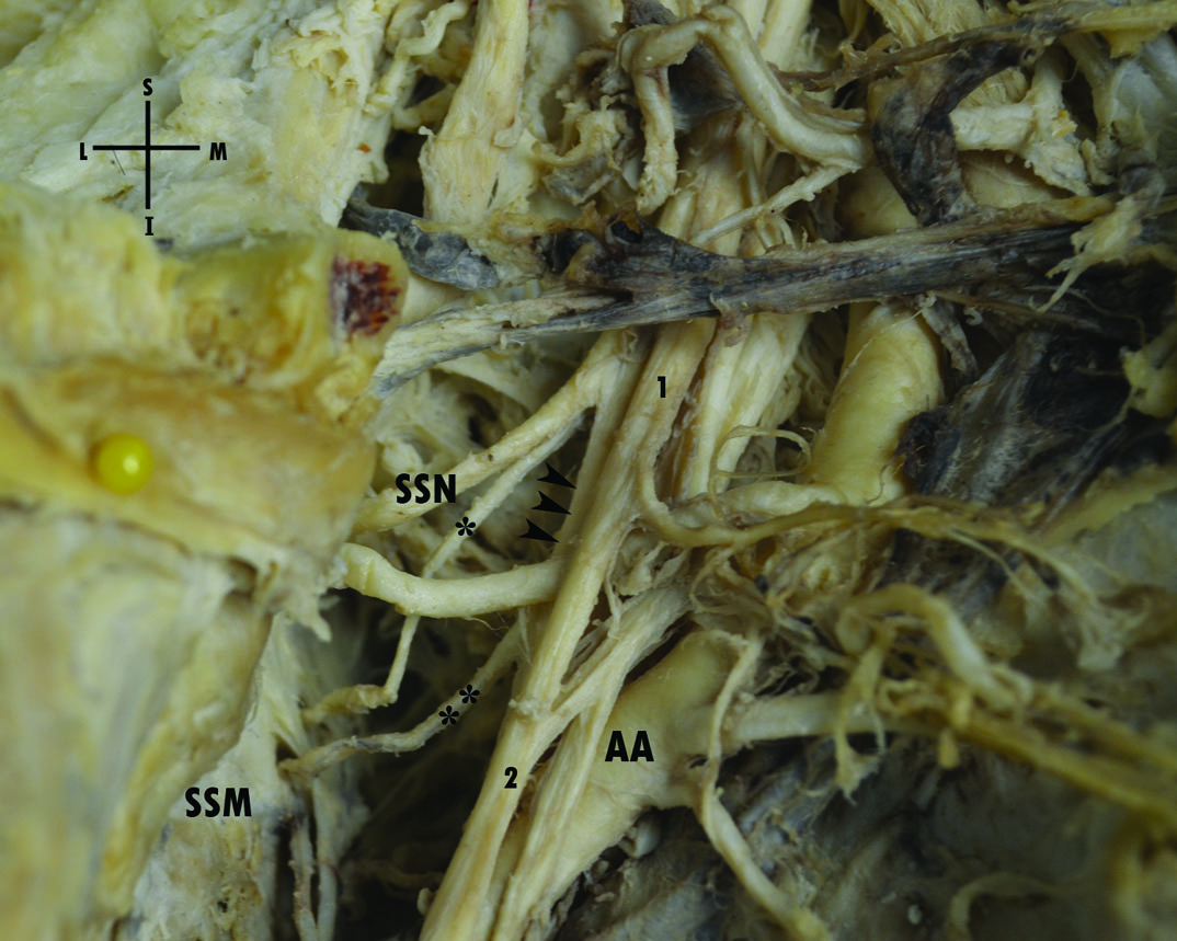
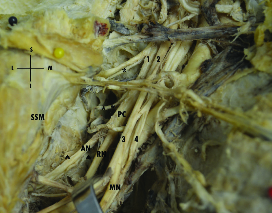
During the routine dissection in our Laboratory of Anatomy we encountered a double Upper Subscapular Nerve (USN) in a 78-year-old formalin-fixed male cadaver used for educational and research purposes. The cause of death was unrelated to the present case report. In specific, after meticulous dissection of the right cervical and axillary region by means of the classical method of anatomical dissection we detected a nerve trunk emerging from the suprascapular nerve just after its abnormal origin from the posterior division of the upper trunk of the brachial plexus. That nerve trunk was observed entering into the uppermost portion of the ipsilateral subscapularis muscle without providing additional branches. Moreover, the presence of a supernumerary nerve trunk, the so-called accessory USN was noticed. It was arising from the termination of the posterior division of the upper trunk of the brachial plexus, which after an oblique course pierced the upper part of the right subscapularis muscle, just lateral to the entry point of the main USN [Table/Fig-1a]. The posterior cord of the brachial plexus was directed normally downwards and outwards providing the thoracodorsal and lower subscapular nerve and terminating into the radial and axillary nerve. Moreover, the above mentioned anatomic variant was associated with a median nerve formed by two lateral roots [Table/Fig-1b]. No other anomalies of the adjacent nerves or evidence of previous surgical interventions on the region were present. The origin, course and supply area of the unilateral double USN were repeatedly documented as photographs during the course of the dissection.
The proximal upper subscapular nerve (*) is demonstrated arising from the initial part of the right SupraScapular Nerve (SSN). The distal upper subscapular nerve (**) is seen as well originating from the termination of the posterior division of the upper trunk of the brachial plexus (arrowheads) and penetrating the right SubScapularis Muscle (SSM) just lateral to the entry point of the proximal homonymous nerve. (1:anterior division of the upper trunk of the brachial plexus, 2:lateral cord of the brachial plexus, AA: axillary artery, S: superior, I: inferior, L: lateral, M: medial).

The right proximal upper subscapular nerve (*) and the ipsilateral distal upper subscapular nerve (**) are demonstrated, as well as the posterior cord of the brachial plexus (PC) with the lower subscapular nerve (arrowheads) along with the Radial (RN) and Axillary Nerve (AN). (1: posterior division of the upper trunk of the brachial plexus, 2: anterior division of the upper trunk of the brachial plexus, 3: lateral cord of the brachial plexus, 4: additional lateral root of the median nerve, SSM: subscapularis muscle, MN: median nerve, S: superior, I: inferior, L: lateral, M: medial).

Discussion
The USN is a small nerve that is originating from the posterior cord of the brachial plexus from cervical 5 and 6 fibers (C5 and C6) and pierce the uppermost portion of the subscapularis muscle [1]. The USN is distributed to the proximal and middle portion of the subscapularis muscle, whereas the lower subscapular nerve innervates the axillary border of the subscapularis muscle and the teres major muscle [2]. According to the various studies done the USN is noted as a single nerve trunk in the studies done by Kerr AT (53.50%), Tubbs RS et al., (90.3%), Saleh D et al., (45.46%) and Kato K (’16%); as a double nerve in 40.76% [2], 8% [3], 48.48% [4] and 60% [5]. USN is also reported as three independent nerve trunks in 5.73% [2], 1.6% [3], 6.06% [4] and also with a high incidence of 80%, as reported by Kim JH et al., [6]. It has been mentioned that in 30% of the cases, the USN arises as three to five small branches [5]. Kim et al., noted that in 20% of their material the USN was composed of four branches [6]. The USN is described as originating in 97% or 100% from the posterior cord of the brachial plexus [3,4]. However, Kerr observed that nerve arising from the posterior division of the upper trunk of brachial plexus in 56.68% and only in 28.02% from the posterior cord [2]. On the contrary, Chaudhary et al., noted the USN originating from the posterior division of the upper trunk only in 8.33% [7].
Our reported case is unique since a double USN was detected with the proximal one originating from the initial portion of the suprascapular nerve that was abnormally arising from the posterior division of the upper trunk of the brachial plexus, whereas the distal USN the so-called accessory USN was arising from the termination of the posterior division of the upper trunk of the brachial plexus. Kerr analytically displayed the various morphological patterns of USN; in specific, he noted the single USN originating from the posterior division of the upper trunk in 25.48% and the single USN emerging from the suprascapular nerve in 0.64%. Although, Kerr documented precisely two distinct USNs, both of them arising from the posterior division of the upper trunk (32.81%), or a case (0.64%) where the one USN was originated from the suprascapular nerve and the other one from the cord formed by the union of the posterior division of the upper and intermediate trunk [2], the same author did not detect a case similar to the one displayed in the current study.
It has been noticed that in some hemiplegic patients or patients with cervical cord injury a remarkable spasticity of subscapular muscle is observed resulting in a painful shoulder with consequent limitation of range of motion of shoulder joint [8,9]. As alternative therapy the subscapular nerve blocks to the motor points of the subscapular muscle using phenol has been suggested. The needle is introduced horizontally under the medial edge of the scapula. Afterwards, the needle must be driven upwards to form a 6.630 angle with the horizontal plane in order to block the USN [9]. Alternatively, weak electric stimulation may be delivered through the needle and at the point where the strongest subscapular muscle contraction is observed, the phenol solution is injected [8]. USN should be potentially a donor nerve for grafting or neurotization to the adjacent nerves, such as suprascapular or thoracodorsal nerve. It has been found that the mean USN’s length is 5cm, whereas it’s mean diameter 2.3mm [3]. A similar research has been conducted for the lower subscapular nerve, that in a remarkable incidence (57.5%) originated from the axillary nerve and it was concluded that this nerve constitutes an ideal donor nerve for the musculocutaneous and axillary nerves in instances of its selective lesions [10–12].
Conclusion
A deeper awareness of such a combination of variable origin of a double USN is important in avoiding complications during brachial plexus repair in case of its lesions. Furthermore, the knowledge of such neural variation is important especially for the anaesthetists during their attempt to block the USN, as well as for the neurosurgeon utilizing the USN as a potential nerve graft or for neurotization of the adjacent nerves in cases where they have been damaged.
[1]. Williams PL, Gray’s Anatomy 1995 38th editionEdinburghChurchill Livingstone [Google Scholar]
[2]. Kerr AT, The brachial plexus of nerves in man, the variations in its formation and branchesAm J Anat 1918 23:285-95. [Google Scholar]
[3]. Tubbs RS, Loukas M, Shahid K, Judge T, Pinyard J, Shoja MM, Anatomy and quantitation of the subscapular nervesClin Anat 2007 20:656-59. [Google Scholar]
[4]. Saleh D, Callear J, McConnel P, Kay S, The anatomy of the subscapular nerves- a new nomenclaturePlast Reconstr Surg 2012 130(25):480 [Google Scholar]
[5]. Kato K, Innervation of the scapular muscles and its morphological significance in manAnat Anz 1989 168:155-68. [Google Scholar]
[6]. Kim JH, Woo JS, Hur MS, Lee KS, Anatomical studies of the upper and lower subscapular nervesKorean J Phys Anthropol 2014 27(4):219-23. [Google Scholar]
[7]. Chaudhary P, Singla R, Kalsey G, Arora K, Branching pattern of the posterior cord of the brachial plexus: a cadaveric studyJ Clin Diag Res 2011 5(4):787-90. [Google Scholar]
[8]. Uchikawa K, Toikawa H, Liu M, Subscapularis motor point block for spastic shoulders in patients with cervical cord injurySpinal Cord 2009 47:249-51. [Google Scholar]
[9]. Menezes CC, Cricenti SV, Mendes FMP, Souza EM, Maia-Filho EM, Anatomical and clinical study of the subscapular nervesJ Morphol Sci 2010 27(2):74-76. [Google Scholar]
[10]. Bhosale SM, Havaldar PP, Study of variations in the branching pattern of lower subscapular nerveJ Clin Diag Res 2014 8(11):AC05-07. [Google Scholar]
[11]. Tubbs RS, Khoury CA, Salter EG, Acakpo-Satchivi L, Wellons JC, Blount JP, Quantitation of the lower subscapular nerve for potential use in neurotization proceduresJ Neurosurg 2006 105:881-83. [Google Scholar]
[12]. Tubbs RS, Tyler-Kabara EC, Aikens AC, Martin JP, Weed LL, Salter EG, Surgical anatomy of the axillary nerve within the quadrangular spaceJ Neurosurg 2005 102:912-14. [Google Scholar]