A Novel Technique To Correct Multiplanar Maxillary Hypoplasia
Sibu Sajjan Simon1, Arun Paul Charlu2, Rabin Kurudamannil Chacko3, Saurabh Kumar4
1 Associate Professor, Department of Dental Surgery, Unit 1, Christian Medical College and Hospital, Vellore, Tamil Nadu, India.
2 Associate Professor, Department of Dental Surgery, Unit 1, Christian Medical College and Hospital, Vellore, Tamil Nadu, India.
3 Professor, Department of Dental Surgery, Unit 1, Christian Medical College and Hospital, Vellore, Tamil Nadu, India.
4 Assistant Professor, Department of Dental Surgery, Unit 1, Christian Medical College and Hospital, Vellore, Tamil Nadu, India.
NAME, ADDRESS, E-MAIL ID OF THE CORRESPONDING AUTHOR: Dr. Sibu Sajjan Simon, Department of Dental Surgery, Unit 1, Christian Medical College and Hospital, Vellore-632001, Tamil Nadu, India. E-mail : simonprostho@gmail.com
Dental malocclusion and facial deformity are frequent observations in patients with clefts of the orofacial region. These patients have a low self perception secondary to their aesthetic appearance. Cleft palate patients are further affected in their speech and oral function with direct impediment to their quality of life. Early identification and treatment in cleft lip and palate patients may directly enhance their overall well being and productivity with sustainable prognosis when managed by skilled and evidence informed operators. We present a successful case management of a patient with a cleft palate and dentofacial deformity with a past surgical history, treated with an anterior maxillary advancement osteotomy, stabilized with an interpositional non vascular iliac bone graft. The posterior open bite was corrected using overlay full coverage crowns. Both these techniques are rarely reported in the literature. The procedure positively improved the quality of life in our patient with regards to her aesthetics, speech and function. This treatment approach could be considered in similar cases to achieve predictable outcomes.
Anterior maxillary advancement osteotomy, Cleft palate, Illiac crest graft, Overlay crowns, Posterior open bite
Case Report
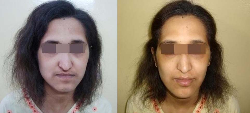
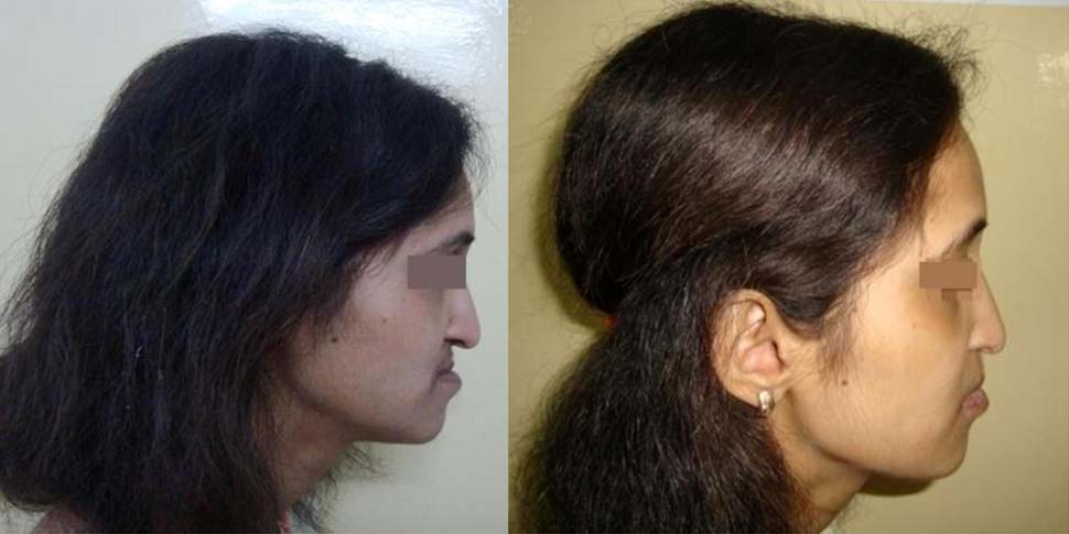
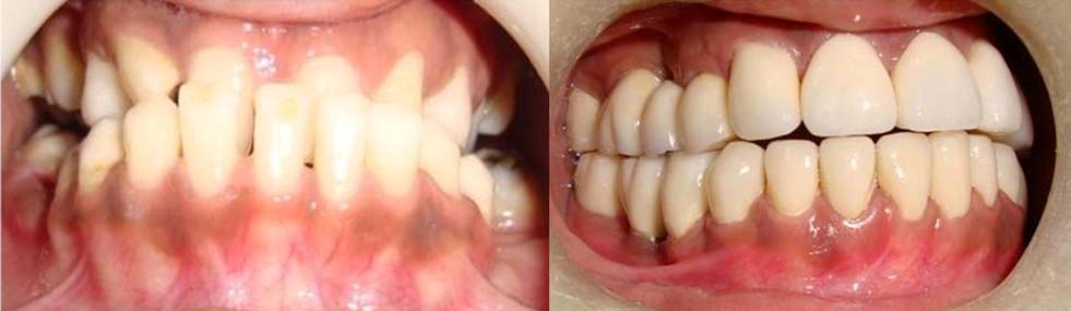
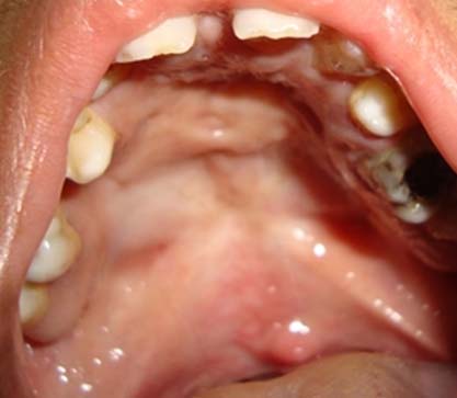
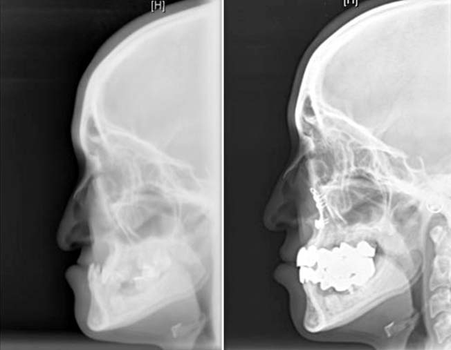
A 17-year-old female patient with a history of cleft palate presented to CMCH, Vellore, India with concern regarding her aesthetics, masticatory function and speech. She alleged unsatisfactory outcomes with four previous surgical corrections the last one being one year before her presentation to our outpatient unit. The patient seemed to be quite withdrawn socially and appeared insecure with a direct effect to her quality of life. Extraoral examination revealed a skeletal class III jaw relationship with forward positioning, overclosure and reduced lower facial height [Table/Fig-1,2]. The concave facial profile and the hooked nasal tip projected a “witch like” appearance. The nasolabial groove was quite pronounced. Intraoral examination presented a reverse overjet with deep bite in the anterior region and retraction of the mandible could only get the maxillary and mandibular anteriors in an edge to edge position with an exaggeration of the posterior open bite and the patient was observed to habitually occlude into a position of convenience to obtain a functional occlusion [Table/Fig-3]. The palatal surface was free of any fistulas or openings [Table/Fig-4]. Preoperative cephalometric measurements helped to correlate the soft tissue findings [Table/Fig-5].
Pre and Postoperative front view.
PRE And Postoperative profile view.
PRE and Postoperative Intraoral view.
preoperative intraoral view.
preoperative and postoperative cephalometric view
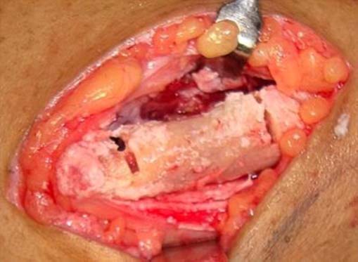
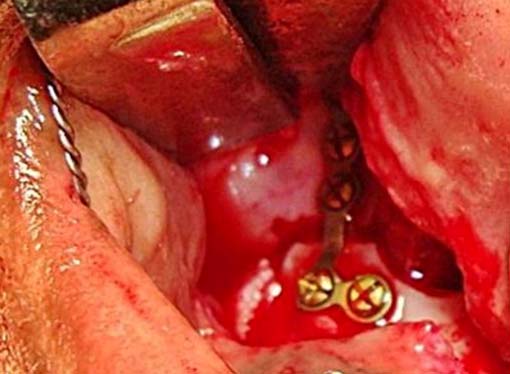
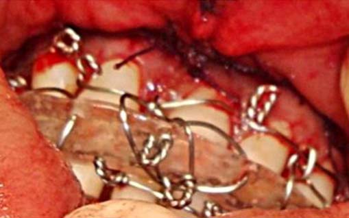
A treatment plan was formulated to include an Anterior Maxillary Advancement Osteotomy (AMAO) with the insertion of a non vascular iliac bone graft (NVIBG) to stabilise the anterior maxilla through the premolar region [Table/Fig-6]. The anterior nasal spine was torqued forward and positioned in a preplanned overjet relation. The segment that was mobilized 7mm anteriorly was stabilized bilaterally with titanium “L” plates with the NVIBG at the posterior maxilla [Table/Fig-7]. A prefabricated surgical splint with an occlusal ramp helped to stabilize the splint with the opposing arch and the maxillary and mandibular arches were approximated using Intermaxillary Fixation (IMF) [Table/Fig-8]. The patient had an uneventful recovery and was reviewed at regular intervals for the first six weeks, following which the IMF was removed. The wired splint was disengaged and cemented to the maxillary teeth and instructions for oral hygiene were given. At a six month review, the patient was without complaint and radiographs supported favourable bone healing pattern at the site of the graft. The maxillary splint was debonded and the dentition was prepared for a full arch rehabilitation with zirconia full coverage crowns with an emphasis to correct the posterior open bite [Table/Fig-3].
Non Vascular Iliac Bone Graft (NVIBG).
NVIBG secured with l plate.
Splint with intermaxillary fixation.
Post-treatment the cephalometric measurements showed dramatic improvement [Table/Fig-5]. The ANB was reduced by 8° indicating a significant reduction in the anteroposterior relation of the maxilla to the mandible. The lower facial height measured from the inferior orbital rim to Pogonion had increased by 7mm. The Frankfort Mandibular plane angle had increased by 10° [Table/Fig-9]. Consequent to the treatment, the patient’s facial appearance improved considerably with an increase in the vertical facial height, correction of the antero-posterior jaw discrepancy, reduced prominence of the nasolabial groove and a functional occlusion [Table/Fig-1,2]. The patient’s satisfaction with regard to her function, aesthetic outlook and speech was complete. On a scale of 1 to 10, the patient gave an overall 10 points for the proposed treatment outcome. She was placed on regular review. At a five year telephonic interview the patient seemed satisfied with the overall outcome and improved quality of life.
PRE and postoperative cephalometric measurements.
| Points of reference | Preoperative | Posoperative |
|---|
| SNA | 69.40 | 730 |
| SNB | 820 | 78.10 |
| ANB | 130 | 50 |
Discussion
Cleft palate is the second most common birth defect in the world with a multifacotrial aetiology with substantial physical and social implications [1]. India has a current crude birth rate of 20.22 births/1,000 population. Current studies of orofacial deformities in India reveals reported statistics with the number of infants born every year with cleft lip and cleft palate being 28,600, which means that on an average 78 affected infants are born every day, or three infants with clefts born every hour [2]. Outcomes with regards to aesthetics are a major concern in patients affected with Cleft Lip and Palate (CLP) especially in socially stigmatising societies. Early identification and treatment can address aesthetic and functional outcome with a direct impact to the quality of life. There is a general consensus that 6-12 months is the ideal time for palatoplasty for early rehabilitation [3]. Awareness of the impediments caused by cleft palate and access to skilled operators plays an important role. Since the 1970s, CLP deformities have been conventionally corrected by orthognathic surgery, and since the late 1990s, anterior maxillary distraction osteogenesis (AMD) has been practiced to address maxillary hypoplasia in patients with CLP. Significant correlations have been observed between surgical movement and postoperative relapse in both the horizontal and vertical planes in studies identifying factors associated with relapse following treatment with Lefort 1 osteotomy in CLP patients [4]. The risk of shortening the soft palate and inducing a velopharyngeal incompetence maybe associated with the sagittal advancement of the entire maxilla in CLP patients. Observations of hard and soft tissue changes in CLP patients treated with either AMD and advancement LeFort I osteotomy (ALO) have revealed a significant and predictable change in patients treated with AMD and a higher probability of relapse in ALO group [5,6]. The use of AMAO is an alternative method to conventional Le Fort I osteotomy and rigid external distraction systems for CLP patients with severe velopharyngeal incompetence, as it does not affect the velopharyngeal sphincter with results being enhanced in patients who have a class I molar relationship, negative or zero overjet, or impacted or malaligned anterior teeth. Treatment outcomes can be achieved in these patients by creating space for the eruption of impacted anterior teeth and their alignment by increasing the arch length and maintaining a class I molar relationship. AMAO with the proposed technique maybe used in indicated patients to address the anterior crossbite with a concave profile.
AMAO with a sandwich graft provides a three dimensional stabilization along with the fixation plates, and the splint that is reinforced by the intact posterior maxillary segment which provides a buttress. Repositioning of the maxilla with internal fixation and without interpositional graft may affect the stability of the repositioned segment [7]. The technique described in this report has the advantage of differential tipping of the Anterior Nasal Spine (ANS). A retruded ANS is often seen in patients with cleft palate deformity and is the main cause for the hook shaped appearance of the nasal tip. The forward tipping in AMAO not only corrects the nasal tip but also helps to diminish the lateral nasal insufficiency manifested by the deep nasolabial groove.
Posterior open bite poses extreme challenges for correction especially in CLP patients. Extrusion of posterior teeth with intermaxillary elastics in conjunction with conventional multiloop edgewise archwire mechanics has shown to cause extrusion of the posterior teeth in the correction of posterior open bite [8]. Various options exists in terms of external restorations to address the posterior open bite situations ranging from direct and indirect composite restorations to partial and full coverage crowns and can be chosen based on the severity of enamel and dentin wear, the number of caries, and the size of existing restorations [9,10]. Overlay full coverage restorations could be an alternative approach to manage posterior open bite in CLP patients as compared to orthodontic management that mainly focuses on orthodontic extrusion and uprighting with intraoral mini implants which would require frequent visits and adjustments. Quality of life in regards to aesthetics, speech and function are important parameters in patient satisfaction towards treatment outcomes. Surgical maxillary advancement in CLP patients has reports of high levels of satisfaction when the aesthetic and functional needs are addressed [11].
Conclusion
Rehabilitation in cleft lip and palate patients is a highly complex task. A multidisciplinary evidence based approach is essential for successful outcomes. Rehabilitation of patients presenting with retrognathic maxilla secondary to cleft palate with compromises in speech, function and aesthetics can be effectively treated using anterior maxillary advancement osteotomy with illiac crest graft sandwich and overlay crowns to correct multiplanar maxillary hypoplasia.
[1]. Levi B, Brugman S, Wong V, Wan D, Palatogenesis (Engineering, pathways and pathologies)Organogenesis 2011 7(4):242-54. [Google Scholar]
[2]. Mossey P, Little J, Addressing the challenges of cleft lip and palate research in IndiaIndian J of Plast Surg 2009 42(Suppl):S9-18. [Google Scholar]
[3]. Agrawal K, Cleft palate repair and variationsIndian J of Plast Surg 2009 42(Suppl):S102-09. [Google Scholar]
[4]. Hirano A, Suzuki H, Factors related to relapse after Le Fort I maxillary advancement osteotomy in patients with cleft lip and palateCleft Palate Craniofac J 2001 38(1):1-10. [Google Scholar]
[5]. Kumari P, Roy SK, Roy ID, Kumar P, Datana S, Rahman S, Stability of Cleft maxilla in Le Fort I Maxillary advancementAnn of Maxillofac Surg 2013 3(2):139-43. [Google Scholar]
[6]. Markose E, Paulose J, Paul ET, Soft Tissue Changes in Cleft Lip and Palate Patients: Anterior Maxillary Distraction versus Conventional Le-Fort I OsteotomyJ Maxillofac Oral Surg 2013 12(4):429-35. [Google Scholar]
[7]. Santos SE, Moreira RW, de Moraes M, Asprino L, Araujo MM, Skeletal stability after inferior maxillary repositioning without interpositional graftInt J Oral Maxillofac Surg 2012 41(4):477-81. [Google Scholar]
[8]. Küçükkelş N, Acar A, Demirkaya AA, Evrenol B, Enacar A, Cephalometric evaluation of open bite treatment with NiTi archwire and anterior elasticsAm J Orthod Dentofacial Orthop 1999 116:555-62. [Google Scholar]
[9]. Nam J, Raigrodski AJ, Heindl H, Utilization of multiple restorative materials in full-mouth rehabilitation: A clinical reportJ Aesthet Restor Dent 2008 20(4):251-63. [Google Scholar]
[10]. Ammannato R, Ferraris F, Marchesi G, The "index technique" in worn dentition: a new and conservative approachInt J Aesthet Dent 2015 10(1):68-99. [Google Scholar]
[11]. Andersen K, Norholt SE, Kuseler A, Jensen J, Thomas K, A retrospective study of cleft lip and palate patients satisfaction after maxillary distraction or traditional advancement of the maxillaJ Oral Maxillofac Res 2012 3(2):e3 [Google Scholar]