Hip Pain and Gait Disturbance Associated with Idiopathic Hypoparathyroidism
Rana Kaynar1, Murat Uludag2, Nilay Yalcin3, Mehmet Hamza Turkcanoglu4, Mahir Cengiz5
1 Department of Physical Medicine and Rehabilitation, Cerrahpasa Medical Faculty, Istanbul University, Istanbul.
2 Department of Physical Medicine and Rehabilitation, Cerrahpasa Medical Faculty, Istanbul University, Istanbul.
3 Department of Physical Medicine and Rehabilitation, Cerrahpasa Medical Faculty, Istanbul University, Istanbul.
4 Department of Radiology, Istanbul Education and Research Hospital, Istanbul.
5 Department of Internal Medicine, Cerrahpasa Medical Faculty, Istanbul University, Istanbul.
NAME, ADDRESS, E-MAIL ID OF THE CORRESPONDING AUTHOR: Dr. Murat Uludag, Tahtakale Mah. Begonya Sok. Bizimevler 1 Sitesi A6 D9, Avcilar-Istanbul/Türkiye.
E-mail: muludag1@yahoo.com
Idiopathic Hypoparathyroidism (IHP) is a disease characterized by diminishes in production of parathyroid hormone due to dysfunction of the parathyroid glands [1]. IHP is characterized by soft-tissue ectopic calcifications and skeletal abnormalities. Subcutaneous calcifications around the hips and shoulders may be seen in the course of this disease [2]. Muscle cramps involving the back and legs, paresthesias, and convulsions are common complaints. The clinical manifestations of this condition are varied and nonspecific. Consequently, IHP is usually diagnosed many years after its onset [3]. Back and hip pain and stiffness are is commonly overlooked in these patients [1].
We hereby report a case of a 40-year-old man who presented with bilateral hip pain that had hindered his normal walking for 6 months. He also had a 30-year history of epilepsy and cataract surgery 10 years earlier. His family history was non-contributory. He had no history of surgical excision or damage to the parathyroid glands, radiation, or neoplastic/granulomatosis infiltration.
He had no muscle weakness or cranial nerve involvement and no difficulty with speech, apathy, or mental slowness. The FADIR (flexion, abduction, and internal rotation) and FABERE (flexion, abduction, external rotation, and extension) tests were positive and painful at 15° of flexion due to hip contractures.
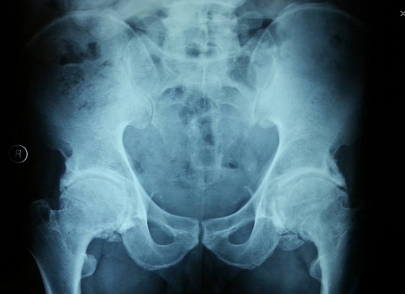
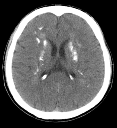
Laboratory results demonstrated the typical findings of hypoparathyroidism: parathormone value 2 (normal level = 15–65) pg/mL, serum calcium 4.6 (normal level= 8.4–10.6) mg/dL, and phosphate 7.2 (normal level = 2.3–4.7) mg/dL. Pelvis radiography revealed bilateral ossification extending along the acetabulum [Table/Fig-1]. Computed tomography of the brain showed calcifications in the pallidum and caudate nuclei bilaterally [Table/Fig-2]. The electroencephalogram was normal.
Pelvis radiography shows bilateral ossification around the acetabulum. Note the preservation of the joint space.
Head computed tomography shows bilateral symmetrical calcifications in the pallidum and caudate nuclei.
No other cause of hypoparathyroidism was found. Tests for polyendocrinopathy were normal. A diagnosis of idiopathic primary hypoparathyroidism was made. Calcium, vitamin D supplementation, paracetamol 500 mg tid, diclofenac sodium 50 mg bid, and physical therapy with range of motion and strengthening exercises were started and continued until the pain limit was reached. These therapies reduced the severity of his complaints within 1 week.
IHP causes hypocalcaemia by hindering calcium reabsorption in the renal tubules and bone, and to a insufficiency in the production of 1,25-dihydroxyvitamin D. The most frequent osseous findings of IHP are localized or generalized sclerosis. Extraosseous calcification is typically seen in the basal ganglia, periarticular tissue, blood vessels, and ocular lens. The development of calcification may be associated not only parathyroid hormone deficiency but also to the duration of hypocalcaemia and hyperphosphataemia [3]. The imaging findings of IHP closely resemble those of diseases such as diffuse idiopathic skeletal hyperostosis and ankylosing spondylitis [1–5].
Our patient’s main complaint was pain and gait difficulty and the diagnosis of IHP was delayed until the age of 40 years. Delaying diagnosis and treatment resulted in cataracts, speech difficulty, pain, limited range of motion of the hip and gait disturbance.
Disclosures: Financial disclosure statements have been obtained, and no conflicts of interest have been reported by the authors or by any individuals in control of the content of this article.
[1]. Kajitani TR, Silva RV, Bonfá E, Pereira RM, Hypoparathyroidism mimicking ankylosing spondylitis and myopathy: a case reportClinics (Sao Paulo) 2011 66(7):1287-90. [Google Scholar]
[2]. Korkmaz C, Yaşar S, Binboğa A, Hypoparathyroidism simulating ankylosing spondylitisJoint Bone Spine 2005 72(1):89-91. [Google Scholar]
[3]. Unverdi S, Oztürk MA, Inal S, Selek H, Göker B, Haznedaroglu C, Idiopathic hypoparathyroidism mimicking diffuse idiopathic skeletal hyperostosisJ Clin Rheumatol 2009 15(7):361-62. [Google Scholar]
[4]. Iwai S, Kabata T, Maeda T, Kajino Y, Ogawa K, Kuroda K, Hyperostosis around the bilateral acetabulum associated with hypoparathyroidismMod Rheumatol 2012 22(5):766-68. [Google Scholar]
[5]. Zabihiyeganeh M, Jahed SA, Akbari H, Longstanding hypoparathyroidism in a fifty-two-year-old woman misdiagnosed as spondyloarthropathyIran Red Crescent Med J 2014 16(12):e22489 [Google Scholar]