Cervical fibroid, Postmenopausal women, Vaginal hysterectomy
Leiomyomas of the uterus are extremely common neoplasms with incidence of 4 to 11%. Lipoleiomyoma is an extremely rare benign tumour of uterus arising mainly in the uterine corpus even though only few cases of cervical lipoleiomyomas reported in the literature [1]. We present this case because of its rarity and unusual clinical presentation.
A 39-year-old multiparous woman presented with complaints of mass descending per vagina for three months and stress incontinence for one week. Gynecological examination revealed a fleshy mass of size approximately 8x5cm seen arising from the posterior lip of cervix which bled on touch. All the standard serological and hematological parameters were within normal range. The patient underwent vaginal hysterectomy with pelvic floor repair.
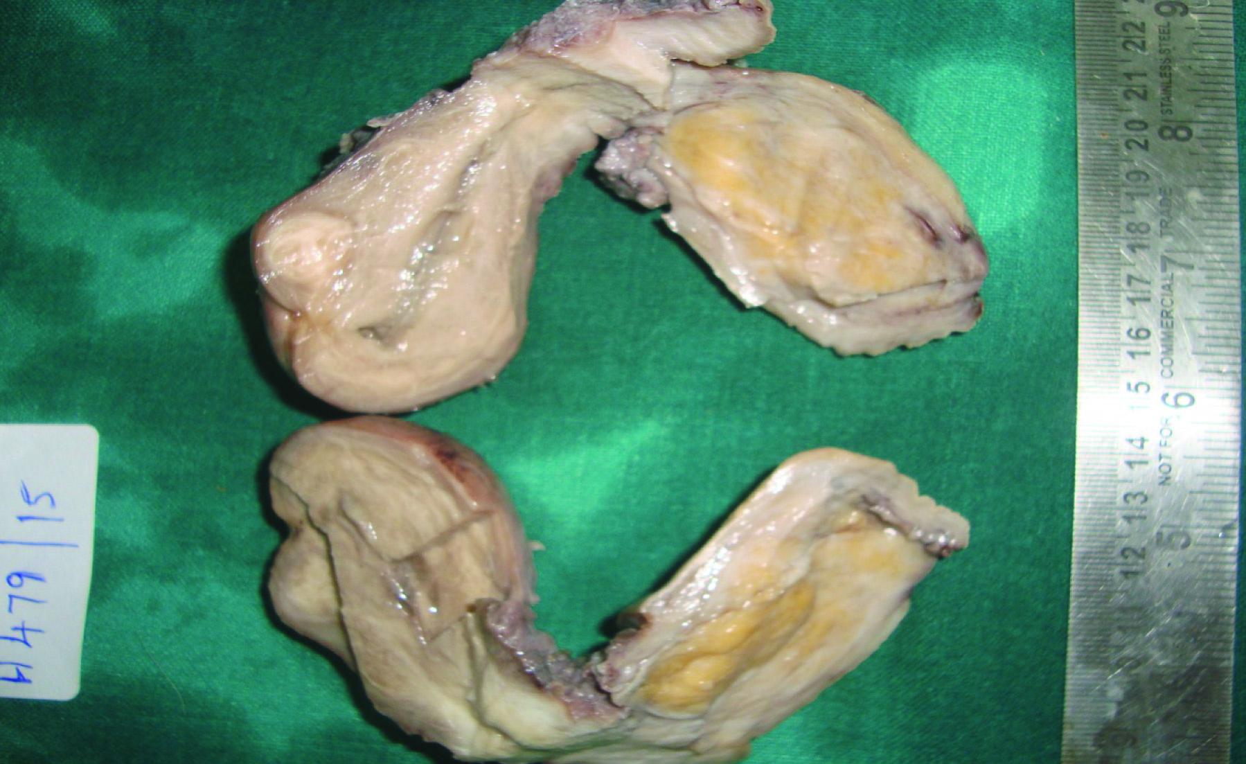
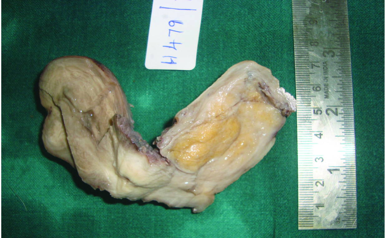
On gross examination of the specimen [Table/Fig-1,2] the uterus with cervix measured 12x5x4cm with elongated cervix. Cut section revealed a grey yellowish mass measuring 8x5cm occupying the posterior lip of the cervix and was soft in consistency. Another intramural fibroid measuring 1cm in diameter was also identified and was firm with whorled appearance.
Gross photo of cervical leiomyoma.
Gross picture showing lipoleiomyoma involving posterior lip of cervix.
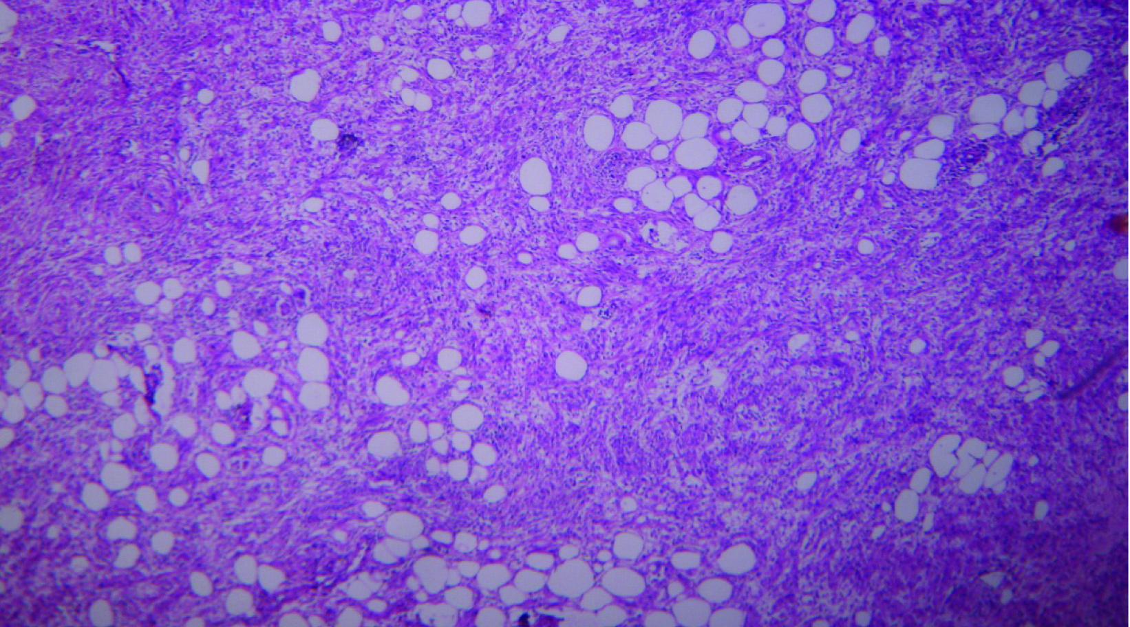
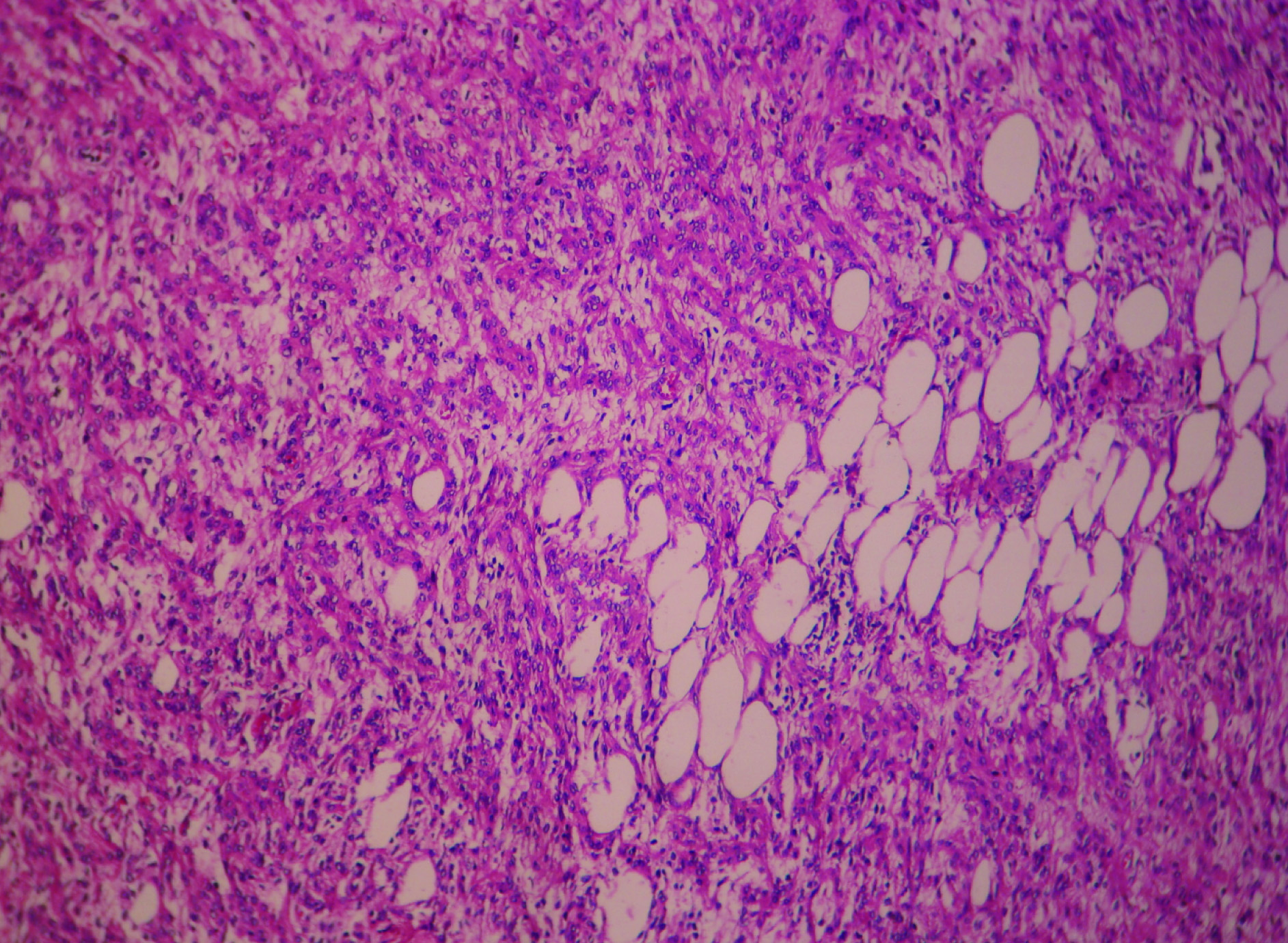
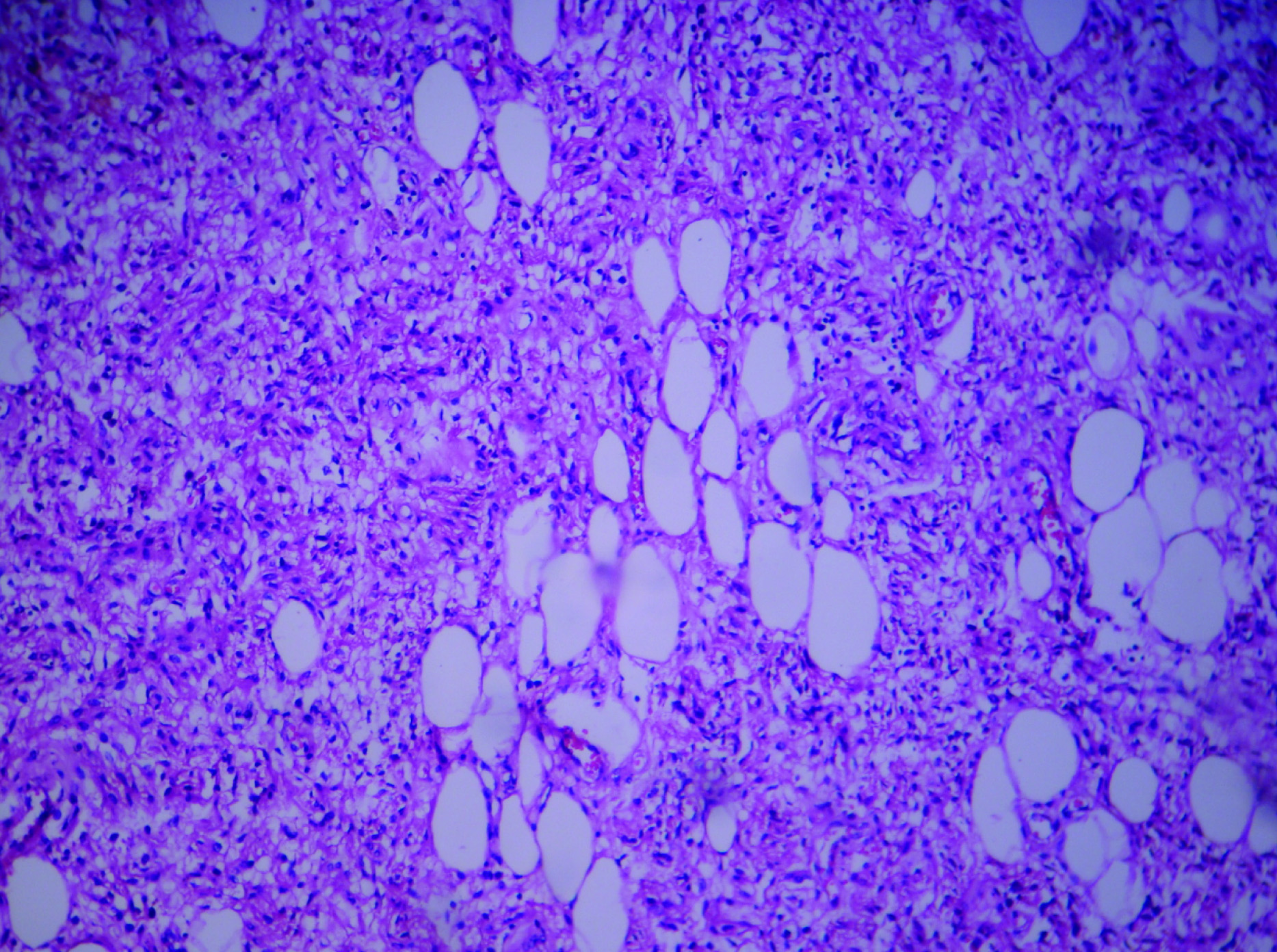
Histopathological examination of cervical growth showed mixture of spindle shaped smooth muscle cells arranged in whorled pattern with mature adipocytes [Table/Fig-3,4,5 and 6]. There was no nuclear atypia, mitosis and necrosis. Thick walled blood vessels and capillaries were also present. Based on the above findings, a diagnosis of benign lipoleiomyoma of cervix was made. Section from uterus showed leiomyoma and endometrium was in secretory phase.
A 10x magnification spindle cells with mature adipocytes.
A 40x adipocytes and spindle cells.

A 40x mature adipocytes and spindle cells
Lipoleiomyomas of uterus are rare and their histologic spectrum includes lipomas, lipoleiomyomas and fibromyolipomas [2]. They commonly occur in postmenopausal women in the age group of 50-60 years and most common location is in the uterine corpus. Cervical fibriods constitutes 1-2% of total fibroids. The presence of adipocytes in the myometrium has been interpreted as lipomatous degeneration, fatty metamorphosis or metaplasia [3]. Their commonest location is in the uterine corpus and is rarely found in the cervix as in our case. The symptoms vary according to their location and includes dysuria, urgency, ureteral obstruction and dyspareunia [1]. Occasionally a cervical fibroid may become pedunculated and prolapse through the external os [4].
The pathogenesis of these tumours has been attributed to various causes including fat cell metaplasia of smooth muscle cells or connective tissue, lipocytic differentiation of primitive connective tissue cell, perivascular fat cells and degeneration of connective tissue.
The differential diagnosis of lipomatous tumour in pelvic region includes benign cystic teratoma of ovary, uterine lipomas, spindle cell lipoma, angiolipoma, angiomyolipoma & well differentiated liposarcoma [3]. Treatment of lipoleiomyoma is similar to that of leiomyoma.
Cervical lipoleiomyoma are rare than uterine lipoleiomyomas. They frequently occur in perimenopausal or postmenopausal women. Although radiological investigation aids in diagnosis, histopathological examination establishes the diagnosis.
[1]. Kalyankar V, Kalyankar B, Rare case of cervical lipoleiomyomaJournal of Evolution of Medical and Dental Sciences 2014 3:5529-33. [Google Scholar]
[2]. Houser LM, Carrasco CH, Sheehan CR, Lipomatous tumour of the uterus: Radiographic and ultrasonic appearenceBr J Radiol 1979 52:992-93. [Google Scholar]
[3]. Bolat F, Kayaseluck F, Canpolat T, Erkanli S, Tuncer I, Histogenisis of lipomatous component in uterine lipoleiomyomasTurkish Journal of Pathology 2007 23(2):82-86. [Google Scholar]
[4]. Sharma S, Ahluwalia C, Mandal AK, A Rare Incidental Case of Lipoleiomyoma CervixAsian Pacific Journal of Health Science 2015 2(1):186-90. [Google Scholar]