Edentulous patients show changes in residual ridge relationships caused by resorption of residual alveolar ridges after extractions. The changes are characterized by an upward rotation of mandible, with a resulting decrease in occlusal vertical dimension and increase in mandibular prognathism [1].
Craniovertical posture relations were studied by Solow and Tallgren in a series of radiographic cephalometric studies of young adults. The result indicated that the posture of the head and cervical column is related to the morphologic characteristics of the facial skeleton [2].
The centre of the head and the articulation of the head with the spine are not in the same long axis, it means that muscular effort must resist gravity to attain head posture. However, the maintenance of natural head posture is influenced by various known and unknown afferent stimuli [3].
Longitudinal studies of denture wearers have shown that changes in hyoid position are coordinated both with the changes in head and the cervical posture [4]. In a previous cephalometric investigation of the individuals, who were provided with complete maxillary and mandibular denture, the changes in hyoid position and craniovertical posture were studied after immediate placement of the dentures and during one year of denture use. In this study, analysis of individual changes indicated that a pronounced decrease in mandibular inclination because of residual ridge resorption was associated with a retroclination of cervical column and a decrease in craniocervical angulation. These changes were regarded as adaptive postural changes resulting from the rapid initial alteration in mandibular position [4].
According to Vig et al., respiratory adaptations such as changes in the bore of the airway may play an important role in controlling head posture [3]. Whereas, Fish has suggested that, the tongue in forming part of the anterior border of the pharynx could influence the bore of the airway by continual adjustment of muscle tension in response to intrapharyngeal pressure accompanying inspiration and expiration [5]. Fish, also stated that the progressive loss of oral bulk through tooth loss and bone resorption in the edentulous patient is accompanied by commensurate shortening and broadening of the tongue [6]. Thus, it is possible that changes in tongue position could indirectly initiate changes in head posture.
From the previous studies it is convincible, that replacement of the lost oral bulk by means of complete dentures can produce changes in tongue shape and position, which in turn could bring about changes in head posture [6]. Since, there was no study/literature available regarding the effect of complete dentures on head posture in different age groups at various time intervals among Indian individuals; this clinical study was planned.
The aim of the present clinical study was to examine the effect of complete dentures on the head posture/craniovertical angle, with the following objectives:
Materials and Methods
A pilot invivo study was conducted in the Bapuji Dental College Hospital, Davangere, India, for a period of 5 months in year 2014. Ethical clearance was obtained by the concerned authority and subjects were also made aware of the study then their consent was procured. Twenty completely edentulous patients/subjects, who required complete denture fabrication, were selected. Sample size was decided with the consent of biostatistician to make statistical analysis possible for this pilot study. All the patients were randomly selected irrespective of their sex following these criteria i.e. they were of Indian origin, and being completely edentulous for at least 6 months or with no previous denture experience, between the ages of 45 to 75 years, good neuromuscular control and without any facial asymmetry. Simple laboratory armamentariums were used in the study [Table/Fig-1&2].
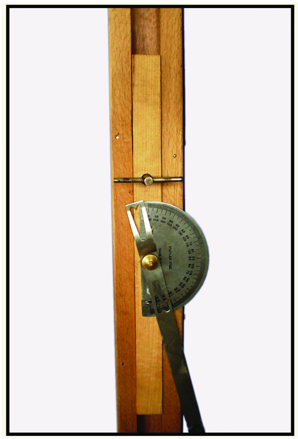
Showing 40 cm ruler and protractor assembly attached to vertical right-angled column.
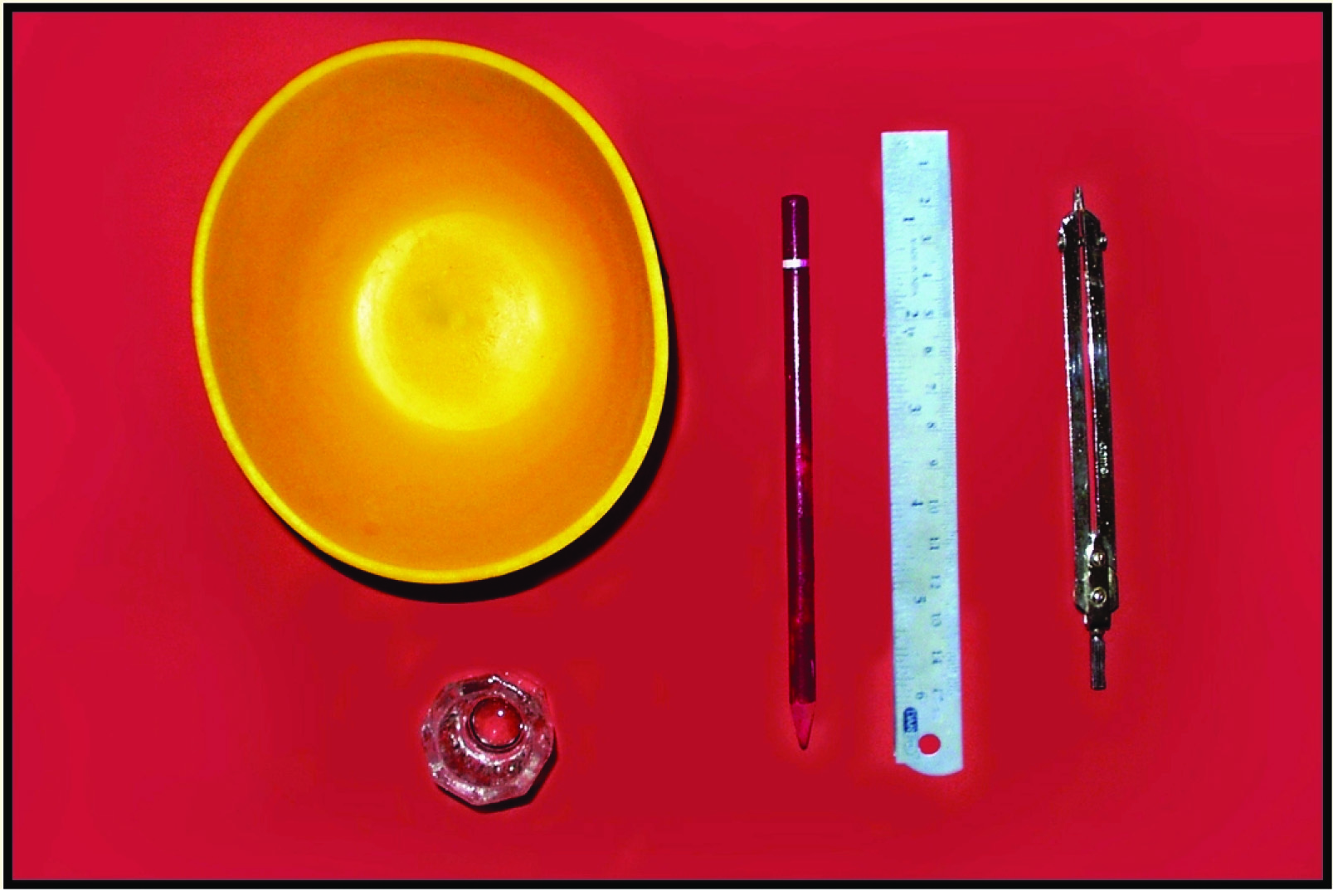
Showing armamentarium used in the study.
The patients selected for the study, were divided into two groups (Group ‘A’ and ‘B’), each group consisting of 10 patients. First group (Group ‘A’) consisting of patients who were aged between 45-60 years and the second group (Group ‘B’) patients were aged between 61-75 years. To divide the patients into two groups, physical wellbeing, at or below 60 years of age and above the same was considered and hypothysed that, age may also play some role in changing the head-posture. For all the study group patients, 20 maxillary and mandibular complete dentures were fabricated by conventional method. While construction of the dentures, all the necessary precautions were taken to minimize the errors in mechanical and laboratory steps. Special attention was given to record jaw relation accurately. Then the craniovertical angle measurement was planned.
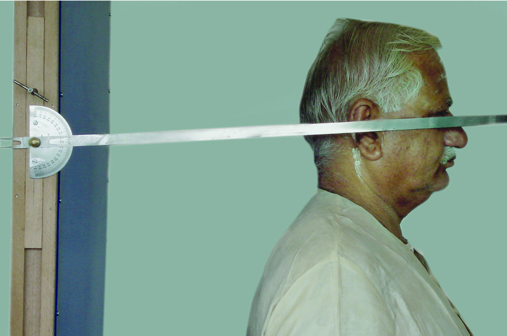
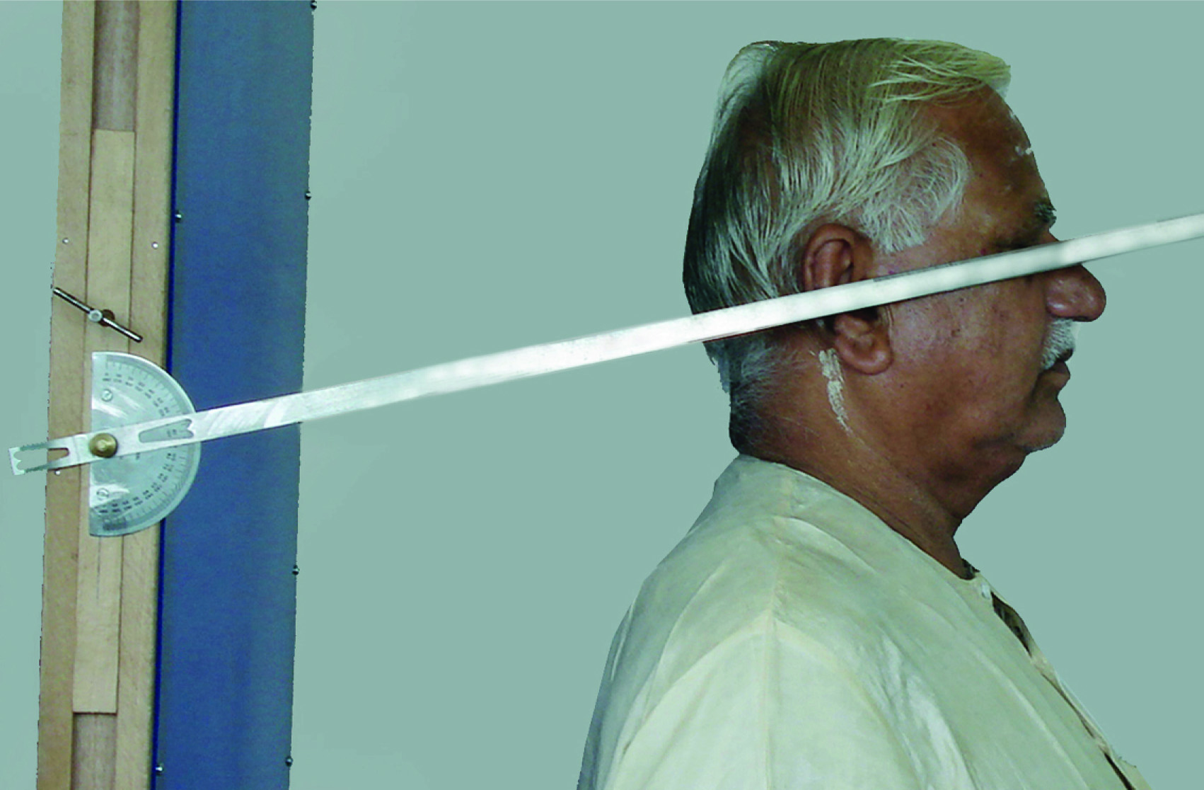
To measure the angle, before the placement of the complete denture, two dots were placed. First dot was placed 1-2 cm anterior from the upper border of the tragus of the ear, then with the help of a scale and divider another dot was placed approximately 6-7 cm apart over the upper border of zygomatic arch of the patients. Patients were asked to stand with their back against a vertical right-angled column. From this position each patient was asked to assume “Intention” position, which is achieved by asking the patient to take small step forward. Next the patients were asked to tilt the head forward and backward with decreasing amplitude until a position of comfortable head balance was attained. A 40-cm ruler was placed over the skin to coincide the marked dots. Measurements of craniovertical angle were obtained by recording the angle formed by ruler on the protractor [Table/Fig-1&3]. Readings were taken at different time intervals i.e. before denture placement [Table/Fig-3], immediately after denture placement [Table/Fig-4], then 30 minutes, 24 hours and 30 days after the denture placement.
Showing cranioverticle angle measurement made on the patient before complete denture placement.
Showing cranioverticle angle measurement made on the patient immediately after complete denture placement.
Statistical Analysis
All the readings obtained were tabulated and statistically analysed with the help of Student paired t-test and Student unpaired t-test. The obtained results were expressed as mean±SD and range values. Changes in the craniovertical angle/head posture compared to base line value were tested by Student paired t-test (for intra group) and Student unpaired t-test (for inter group). In the statistical analysis for significance, probability (p) was considered significant if p-value was < 0.05 however; if p-value was > 0.05, changes were considered as not significant. The obtained values were tabulated and statistically analysed.
Results
In this study total of 20 completely edentulous patients of Indian origin were selected and divided into two age groups, Group ‘A’ (45-60 years) and Group ‘B’ (61-75 years). Each group was consisting of 10 patients. For all the patients conventional complete dentures were fabricated. Before denture placement measurement of craniovertical angle was recorded and obtained value was considered as a base line. Thereafter, craniovertical angle was measured at various time intervals.
The descriptive information on craniovertical angle (in degree) was assessed at various time intervals in two study groups [Table/Fig-5].
Showing descriptive information on cranio-vertical angle (in degrees) assessed in two study groups at different time intervals
| Age Groups | Before denture placement | Immediately after denture placement | After 30 minutes of denture placement | After 24 h of denture placement | After 30 d of denture placement |
|---|
| A(45-60) years | 77.1 ±2.5(73-80) | 74.2 ±2.4(70-79) | 73.7 ±4.1(69-83) | 73.7 ±2.5(70-77) | 74.8 ±3.3(69-79) |
| B(61-75) years | 81.6 ±7.5(73-92) | 76.3±5.9(70-85) | 76.0 ±6.7(68-87) | 77.1 ±7.4(67-89) | 80.1 ±7.2(72-92) |
Values are expressed as Mean±SD (Range).
In the subjects of Group ‘A’, craniovertical angle were ranged between 73° to 80° with an average of 77.1° (SD±2.5), before denture placement which was considered as a base line value. In Group ‘B’ base line value was little higher i.e. ranged between 730 to 92° with an average of 81.6° (SD±7.5). Immediately after denture placement all the subjects showed noticeable change with extension in the craniovertical angle with average of 74.2° (SD±2.4) in Group ‘A’ and 76.3° (SD±5.9) in Group ‘B’. After 30 minutes of denture placement craniovertical angle showed significant change in comparison with its base line with an average of 73.7° (SD±4.1) in Group ‘A’ and 76.0° (SD±6.7) in Group ‘B’. Twenty four hours after complete denture placement craniovertical angle showed minimum or no change with an average of 73.7° (SD±2.5) in Group ‘A’ and 77.1° (SD±7.4) in Group ‘B’. After 30 days, all the subjects showed flexion in craiovertical angle with an average of 74.8° (SD±3.3) in Group A and 80.1° (SD±7.2) in Group B. However, in both the groups values obtained after the denture placement at different time intervals, were higher than their base line.
In the statistical analysis for significance [Table/Fig-6], t-value was measured with the student t-test (paired) for intra group values. In the Group ‘A’, changes in craniovertical angle were significant with increasing values from 3.02 to 7.52 up to 24 hours then after 30 days of the denture placement that decreased to 4.44 but which still remained significant. In Group ‘B’ the t-values showed significant variable changes with the range of 5.36 to 9.64 till 24 hours of the denture placement, however after 30 days changes were not found significant, with the t-value of 1.63 and p-value was 0.14.
Showing changes in craniovertical angle (in degrees) in two study groups at different time intervals.
| Time of assessment | Group A | Group B |
|---|
| Mean± SD | Difference from the base line | Significancet p | Mean± SD | Difference from the base line | Significancet p |
|---|
| Before denture placement (Base Line) | 77.1 ±2.5 | – | – | 81.6 ±7.5 | – | – |
| Immediately after denture placement | 74.2 ±2.4 | 2.9 ±3.0 | 3.02 < 0.05 | 76.3 ±5.9 | 5.3 ±3.1 | 5.36 < 0.05 |
| 30 min after denture placement | 73.7 ±4.1 | 3.4 ±4.7 | 2.29 <0.05 | 76.0 ±6.7 | 5.6±1.8 | 9.64 < 0.05 |
| 24 h after denture placement | 73.7 ±2.5 | 3.4 ±1.4 | 7.52 <0.05 | 77.1 ±7.4 | 4.5 ±3.9 | 3.65 <0.05 |
| 30 d after denture placement | 74.8 ±3.3 | 2.3 ±1.6 | 4.44 <0.05 | 80.1 ±7.2 | 1.5 ±2.9 | 1.63 0.14 NS |
Test – student t-test (paired) p, < 0.05 - significant, p > 0.05 - Not significant.
In the comparative statistical analysis [Table/Fig-7], t-value was measured with student unpaired t-test for intergroup values. In Group ‘A’ change in craniovertical angle before to immediately after placement of denture showed the mean value of 2.9° (SD ± 3.0) whereas Group ‘B’ showed little higher values i.e. 5.3° (SD±3.1). In the comparative analysis t-value was measured to 1.74 and p-value was obtained to 0.10. These values showed no significant changes between the two groups.
Showing the comparative changes in craniovertical angle (in degree) between the two study groups at different time intervals.
| Difference | Group AMean changes ±SD | Group BMean changes ±SD | A Vs B |
|---|
| t-value p-value |
|---|
| Before to Immediate after DP | 2.9 ±3.0 | 5.3 ±3.1 | 1.74 0.10, NS |
| Before to 30 min after DP | 3.4 ±4.7 | 5.6 ±1.8 | 1.38 0.20, NS |
| Before to 24 h after DP | 3.4 ±1.4 | 4.5 ±3.9 | 0.84 0.42, NS |
| Before to 30 d after DP | 2.3 ±1.6 | 1.5 ±2.9 | 0.76 0.46, NS |
Test – Unpaired t- test, P > 0.05, Not Significant, DP: Denture placement.
The change in craniovertical angle before denture placement to 30 min after denture placement showed the mean of 3.4° (SD±4.7) in Group ‘A’ and bit higher 5.6° (SD±1.8) in Group B. The t-value was 1.38 and p-value was 0.20, which denotes that changes were not significant. Likewise after 24 hours and after 30 days the changes were not significant.
The comparative statistical analysis showed that, after placement of complete denture, the pattern of changes in craniovertical angle were same and not significant between two age groups, however values were little higher in the Group ‘B’ subjects.
Discussion
Head posture/craniovertical angle is defined as the angulation relative to the gravity-defined vertical [3]. In the study done by Solow and Tallgren, natural head positions have been observed in different time interval, ranging between extension and flexion [7]. These postural features showed statistical association with both craniofacial and dentoalveolar morphology.
Extension is the positive deviation of the head to vertical from the initial base line (i.e. head lifts upwards); while negative deviation represented degree of head flexion (i.e. downward head tilt).
In another cephalometric study conducted by Solow and Tallgren, dentoalveolar morphology was also examined for association with craniovertical posture [2]. Dentoalveolar height and occlusion plane inclination showed a set of positive correlation with the craniovertical and sella nasion to vertical angulations. These findings seem to indicate that dentoalveolar compensations for the differences in vertical jaw relationship may be casually related to head posture.
Placement of complete dentures may bring change in position and function of the mandible and tongue. These functional relationships are dependent on the head and cervical posture. The changes in mandibular and tongue position in denture wearer are related to reposition of the residual alveolar ridge and the resultant positional changes of the dentures on the underlying tissue [4].
In the previous published literature, different methods were described to attain “natural head position” [8–12]. Some authors considered that most relaxed head position which was obtained without any outer influence, was the natural head position [2,13–14]. Usumez and Orhan described the natural head position is a dynamic and suggested that most common attained position should be selected from the narrow range of positions around the static natural head position [15].
Complete dentures essentially required to improve the facial esthetics of edentulous patients. However, very limited information is available in the various scientific literatures regarding the effect of complete dentures on the natural position of the head. Various methods have been used in the previous studies to measure the changes in head posture i.e. by Cephalographs [4], by Inclinometer [16], by Fluid level method [17]. Protractor and Plumb line device [3] and with custom made Protractor and ruler assembly [11].
In the present study Protractor and ruler assembly method was used to measure the change in craniovertical angle/head posture. The result of this study indicated that the majority of the patients (90%) showed significant change in head posture (extension) immediately after placement of the complete dentures. Only two patients in Group ‘A’ showed flexion. This immediate change also occurred in both the patient age group (Group ‘A’ and ‘B’) whereas in Group ‘B’ craniovertical angle was recorded little higher (t-value 5.3) than Group ‘A’ (t-value 3.02). [Table/Fig-6]. These findings are in agreement with the findings of the study done by Theron W et al., and Usumez A et al., [11,16].
After 30 minutes of denture placement 90% of the patients showed highest extension of craniovertical angle, with the t-value of 3.4(SD±4.7) in Group ‘A’ and t-value of 5.6 (SD±1.8) in Group ‘B’ [Table/Fig-6]. On the observation after 24 hours of complete denture placement the patients sustain the extended craniovertical angle, although some of the patient showed flexion as compare to their previous readings.
After 30 days of complete denture placement, readings of craniovertical angle showed flexion of the head which is higher than their base line value. However, they still remain higher marginally. This statistical analysis indicated that in Group ‘A the changes in head posture were significant but in Group ‘B’ changes were not significant.
The reasons for the differences between individuals in this study are not clear but several factors might play a role. Differences in tongue size and form exist from one individual to another. A larger tongue might be forced more posteriorly by the dentures than a smaller one with a subsequently more reduced airway bore. Further, the tongue might spread out more and be in a more anterior position after tooth extraction in some patients, producing a different response to the insertion of complete dentures than in others. The mandibular dentures vary in size from patient to patient. Those with larger and thicker lingual flanges might force the tongue more posteriorly thereby reducing the airway bore more than thinner dentures. A more extended head posture would result.
Solow and Tallgren found a relationship between craniofacial morphology and head posture that is relevant to the present investigation [7]. The natural extended or flexed head posture of an edentulous patient, resulting from cranial morphology that might influence the change in degree of head posture after insertion of complete dentures.
Daly, implicated occlusal height as one of the criteria in his study [18]. He found that an increased occlusal height in dentate orthodontic patients led to a more extended head posture. Increasing the vertical dimension from the edentulous state through the placement of complete dentures might affect head posture by the same mechanism.
Changes in lip and tongue pressures that accompany edentulousness or the insertion of dentures could have an effect on head posture. This conclusion is supported by the findings of Archer and Vig, who demonstrated that resting lip and tongue pressures are altered in skeletal class I and class II subjects with changes in head posture from flexion to extension [19].
Whatever the causative factor, dentists should be aware that changes in head posture are likely to occur after placement of complete dentures in patients who have been completely edentulous for some time or have not used dentures for at least 6 months. This change will usually be a mild extension of the head.
Limitations
Although this study has come to its aims, there were some quantitative limitations. This study was conducted only on small size of population therefore to generalize the results for the larger groups; the study should have involved more participants at different time intervals.
Conclusion
Within the limitations of this pilot study in which 20 edentulous patients without previous denture experience were examined, and following conclusions are drawn i.e. head posture was significantly altered by the placement of complete dentures in all the edentulous patients, although after 30 minutes and 24 hours, the extension of head posture remains constant with slight variation in craniovertical angle.
Age of the complete denture patients also showed some significance, as the observed changes in craniovertical angle/head posture in Group ‘A’ patients after 30 days of denture placement were significant and all the values remained slightly higher than their base line values. However, in Group ‘B’ changes observed were neither significant nor remained stable.
Values are expressed as Mean±SD (Range).
Test – student t-test (paired) p, < 0.05 - significant, p > 0.05 - Not significant.
Test – Unpaired t- test, P > 0.05, Not Significant, DP: Denture placement.