Lymphocytes are often termed to be isomorphic, having a monotonous light microscopic appearance. Morphological aspects of lymphocytes in tissue sections thereby are not routinely taken notice of, as their morphology seems to vary only in case of lymphoid malignancies, haematological malignancies apart from certain viral infections. However, in chronic inflammatory conditions including dental periapical infections, there is an antigenic stimulus which leads to proliferation of lymphocytes [1]. Atypical lymphocyte refers to unusual structure of lymphocytes that are a part of the cell mediated immune system of the body. Also, referred to as reactive lymphocytes, atypical lymphocytes have large than normal size sometimes with a diameter of more than 30 microns. The large size of the lymphocytes is attributed to antigenic stimulation. The nucleus feature of the reactive lymphocytes are also unusual; it can be circulated, ovoid, folded or may have a dent or a cleft. Cytoplasm is also found to be abundant and can be basophilic with vacuoles. [2]. These cells routinely raise suspicion regarding the nature of the pathosis. The other rare feature which is recently coming under light is “Cellular Cannibalism” which is defined as a large cell enclosing a slightly smaller one within its cytoplasm [3]. Previously, this feature was noted only in cases of malignant tumours [3]. Recently, Sarode et al., had described this phenomenon in oral squamous cell carcinoma as well as in benign lesions such as central and peripheral giant cell granulomas [3,4]. Cannibalism is said to influence biologic behaviour in these lesions [4,5]. Our tissue sections revealed cannibalistic cells at many places.
Thus, the objectives of the present study were to determine the proportion of atypical lymphocytes & cellular canabalism in periapical granulomas and cysts.
Materials and Methods
This observational study was conducted in the Department of Oral Medicine and Radiology and Oral Pathology and Microbiology VYWS Dental College and Hospital, Amravati, Maharashtra.
Cases (slides) showing marked plasma lymphocytic infiltrates and clear-cut cannibalistic cells and whose clinical Performa were available were chosen for evaluation.
Poorly stained slides or slides with fade nuclear stain were excluded from the study.
Ethical clearance was obtained from the institutional ethical and review board.
Haematoxylin and Eosin (H&E) stained 30 slides of chronic periapical granulomas and 20 slides of cysts reported in the year 2014-15 and the clinical Performa’s of the patients were retrieved from the files of the Department of Oral Medicine and Radiology and Oral Pathology and Microbiology. These slides were evaluated by 3 experts from the specialization of Oral Pathology and Microbiology to determine the presence of atypical lymphocytes and cellular cannibalism under high power magnification (400X).
Quantification of atypical lymphocyte was done by selecting 4 hot spots showing maximum density of plasma lymphocytic infiltrates. Quantification of cannibalistic cells, however, was done by thorough screening of the histopathological sections. Parameter that was used to identify cannibalistic cells was that these cells showed cytoplasmic extensions (pseudopodia) around the cell which is to be internalized or engulfed. The engulfed cell however, shows shrunken size and other features suggesting degeneration [4–6]
Inter examiner reliability was done before commencing the study which was found to be 0.8. As this was a proportional based study, percentage analysis was carried out.
Results
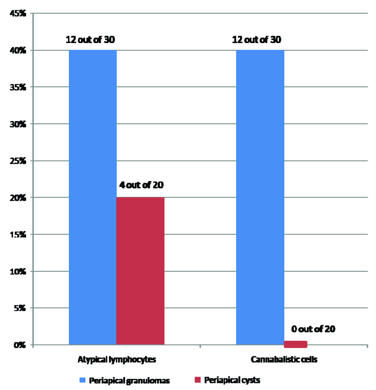
Out of the 30 slides of chronic periapical granulomas, about 12 slides (40%) revealed presence of atypical lymphocytes [Table/Fig-1,2]. Out of these 12 slides, 7 slides showed marked populations of atypical lymphocytes i.e. atypical cells >5 per high power field (HPF) and the remaining showed a relatively less number of atypical cells i.e. about 2 per 10 HPF. In case of slides of chronic periapical cysts, however, only 4 out of the 20 slides (20%) examined histopathologically showed presence of atypical lymphocytes. Also, number of atypical lymphocytes in cysts did not exceed 3-4 per 10 HPF. Again, notably, only those tissue sections which contained marked plasma cell infiltrations showed this kind of cell population. An interesting feature of cellular cannibalism was noted in tissues with atypical cells [Table/Fig-3]. Cannibalistic cells were present in 12 out of the 30 slides of chronic periapical granulomas (40%). None of the cysts, however, revealed cannibalistic cells (0%). Also, the total number of cannibalistic cells was found to be directly proportional to the number of atypical lymphocytes and the amount of plasma cell infiltration [Table/Fig-4].
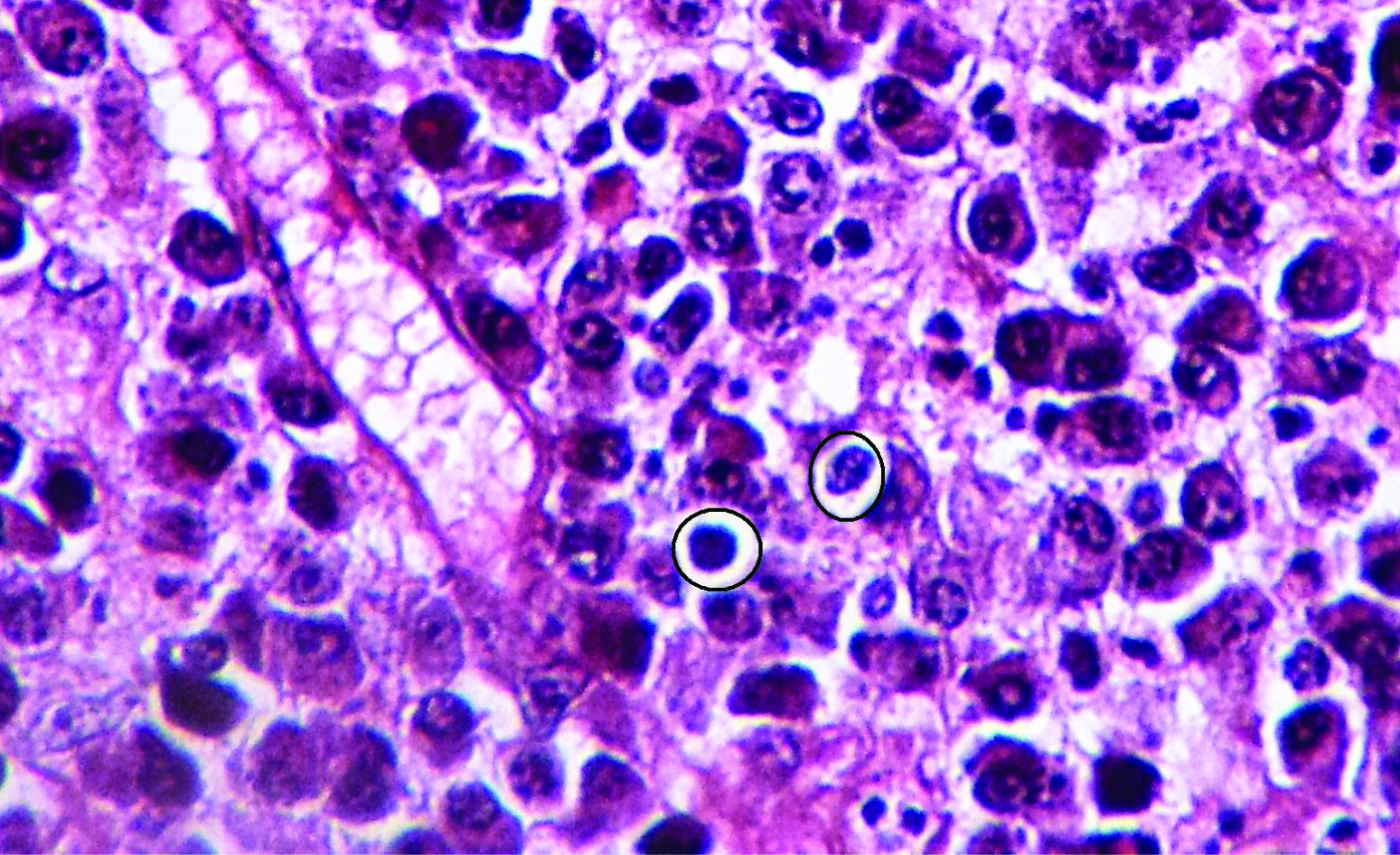
Revealing atypical bilobed cells with two nuclei and nucleoli closely mimicking a typical “Owl eye” appearance.

Revealing atypical cells in the mixed inflammatory cell infiltrates.
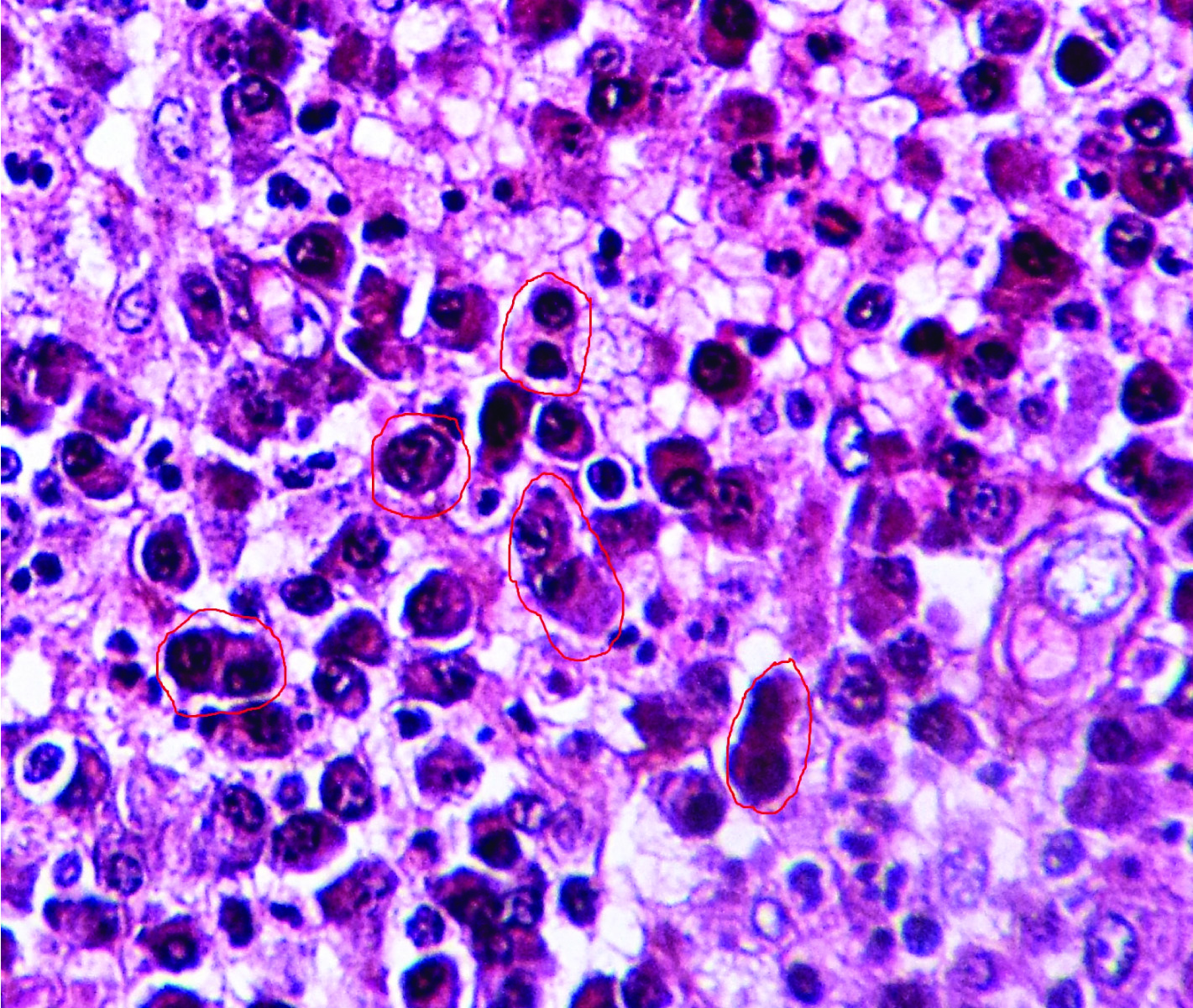
Revealing characteristic Cellular Cannibalism
Revealing the relative percentages of atypical lymphocytes and cannibalistic cells observed in chronic periapical lesions studied
Discussion
The present study was a descriptive, observational study conducted in the Department of Oral Medicine and Radiology and Oral Pathology and Microbiology with objectives of this study being to determine the proportion of atypical lymphocytes in chronic periapical granulomas and cysts; to determine the proportionate cellular cannibalism in these periapical lesions. However, it was observed that out of the 30 slides of chronic periapical granulomas examined, those 12 slides in which atypical cells and cellular cannibalism was evident, the size of the granuloma was found to be relatively larger as was evident radiographically and which it had acquired in a short span of time (less than a week) as was observed on going through their case histories while in the rest of the cases, the size of the pathosis was comparatively smaller and did not show much change in dimensions from the time it had occurred, thus, indicating aggressive behaviour of the lesions having atypical cells and cellular cannibalism.
Plump fibroblasts as well as budding capillaries were also observed in these sections confirming their biologically aggressive behaviour. No such correlation was seen in cases of periapical cysts. The reason for the presence of such atypical cells in the tissue sections could be understood by a deeper understanding of the pathogenesis of these chronic periapical pathoses. Immunological reactions which are provoked by bacterial antigens from the necrotic pulp of the teeth are the basis for the pathogenesis of these lesions. Thus, immune component plays a crucial part in the pathogenesis of these lesions. Both humoral and cell-mediated immune responses are said to play a role in the formation of these lesions [6]. It has been hypothesized that antigen-activated B cells differentiate into centroblasts (rapidly proliferating large cells). Centroblasts then differentiate into centrocytes (non- dividing, smaller, notched cell). Antigen-selected centrocytes eventually differentiate into memory B cells or plasma cells. The antigens that cause B-cell activation and differentiation are defined as T-cell independent (TI), and T-cell dependent (TD). TI antigens, which are able to elicit responses, include bacterial cell wall components such as polysaccharides. These cells are specifically responsive to bacteria such as those of Haemophilus family of the beta subgroup, Streptococcus family and Neisseria meningitidis [7,8]. The continuous antigenic stimulus is responsible to evoke the TI challenges which are long lasting. These responses cause continuous production of plasmablast and they are seen in tissue sections. These plasmablasts produce natural antibodies which cause clearance of pathogens.
This role is in addition to the early response described by Klein et al., for B cells, where rapid proliferation and differentiation creates a ‘wave’ of plasmablasts in the first few days following infection [7]. As also, most of the commensal bacteria from the mucosa are able to elicit these reactions. This is also confirmed from the studies which state that there is a seeding of these proliferating B cells and Plasma cells from the germinal centers in bone marrow [7]. The above mentioned findings were also confirmed in the study done by Yang et al., that bacterial lipopolysaccharide, which basically is a phosphatidylcholine compound, is highly antigenic [8]. This antigen is known to stimulate the migration of the B lymphocytes from different sites with such cells being recognized as immigrant cells. This antigenic compound causes proliferation of native as well as immigrant B cells and their differentiation into antibody producing plasma cells. It is proven in this study too that when antigen provoked immigrant lymphocytes differentiate into antibody producing plasma cells, they divide at least once into two identical daughter cells that is by the process of mitosis [8]. Such incompletely dividing plasma cells were seen in the tissue sections of our slides of periapical lesions which suggests strong antigenic stimulus.
This also explains the strong association of atypical cells and predominance of plasma cells in tissue sections. Thus, from above discussion, it is known that lymphocytes have to pass through various functional milestones after antigenic stimulus, thereby providing an array of morphologies through various stages. Thereby, it can be concluded that these lymphocytes can appear unfamiliar and atypical in tissue sections due to their incomplete division or larger size during proliferation as mentioned above or due to their large size etc. These features are typically pronounced when there is strong and complex bacterial antigenic stimulus. Another feature associated with the presence of these cells was the correlation of on an average, larger size, of the chronic periapical granulomas. Thus, the question arises that whether presence of atypical cells from the tissue sections denotes aggressive tendency and should be treated with an altogether different line of treatment.
Bacteria associated with such kind of antigenic stimulation should be isolated so that combination of antibiotics could be administered to the patients accordingly. As also, another feature noted was the presence of cellular cannibalism in the sections of periapical lesions. One of the major reasons which was put forth by Sarode et al., for the presence of cannibalistic cells was the lack of nutritional supply in these lesions and thereby for their survival, tumour cells were assumed to engulf other tumour cells and also, the leucocytes as well [4]. This hypothesis can even be applied to chronic periapical granulomas, as it is known that during growth of these lesions, there is a reduction in the vascularity eventually, leading to the death of central placed cells, a process known as avascular necrosis.
Thus, cellular cannibalism might probably be the natural survival attempt by these cells. Since cyst did not suffer from such lack of blood supply, cannibalism was not seen in the sections from cystic lesions. Studies have denoted that cellular cannibalism generally denotes aggressive tendency of both benign and malignant tumours, however, no such study till date has been carried-out on periapical pathologies and to the best of our knowledge, this has been the first of the studies to report such kind of features in periapical pathologies in the English medical literature. The chronic periapical granulomas, thus reflect a complex interaction of both the immunologic and non-immunologic mechanisms and are the most common pathologies routinely encountered [9]. Thus, further studies are required in this direction so that these histopathological features can direct us regarding the biologic behaviour of these lesions and thereby lead to proper treatment protocols.
Limitations
However, there were certain limitations in the study that it was a short term study not many cases were studied; as also molecular marker studies are needed which will be of help to further broaden our knowledge on this aspect. Thus, present study can be used as a pilot study for further studies.
Conclusion
Chronic periapical pathologies are the most routinely encountered lesions in the dental practice. However, not much importance is given on their diagnostic and prognostic aspects. In the present study, we have quoted our observations on the unique cellular composition that was seen in histopathological sections. These H/P findings on correlating with the clinical and radiological parameters indicated larger size and a faster growth pattern of periapical lesions showing atypical cells and cellular cannibalism. Thus, the question arises that whether presence of atypical cells from the tissue sections denotes an aggressive clinical behaviour and should be given a due consideration in deciding the treatment protocols for such cases to provide an optimum patient care. Hence, more studies with large sample size are recommended in this direction.