Introduction
Indian ocean islands and India have experienced massive severe Chikungunya outbreak from 2005 up till now and then Chikungunya became epidemic in India. The mutations that occurred in E1 gene were responsible for increased infectivity, virulence and host adaptability. It is important to find out the genotype and its probable evolvement and novel mutations in the E1 gene reported during 2006-2009 from the current isolates, which may affect the local protein structure.
Aim
To perform Molecular diagnosis and Molecular Characterisation of Chikungunya virus isolates.
Materials and Methods
A total of 33 samples were included in the study. RNA was isolated from 33 serum samples and Real time PCR was carried out. Further, Nested PCR and E1 partial gene sequencing was performed. Phylogenetic analysis, mutational analysis and protein modelling studies were carried out.
Results
Out of 33 samples tested, 31 were found positive for CHIK RNA. Phylogenetic analysis showed that isolates belongs to ECSA genotype and E1K211E, E1M269V and E1D284E mutations were observed from all the isolates.
Conclusion
The isolates may have evolved from ECSA Reunion island strains and identified unique mutations in E1 gene were maintained. These mutations have not affected local protein structure.
E1 gene, Phylogenetic analysis, Mutational analysis
Introduction
Chikungunya fever was first documented in 1952 during epidemic in Newala and Masasi Districts of Southern Province, Tanzania. Chikungunya is an arthropod borne virus, transmitted to humans by the bite of Aedes aegypti mosquito [1]. Indian tiger mosquito Aedes albopictus was found to be a competent vector for Chikungunya transmission during 2005-2006 [2]. Chikungunya fever presents with symptoms of fever, polyarthralgia, headache, backache and persistent arthralgia [3]. Severe neurological manifestations were also observed during recent outbreak [4].
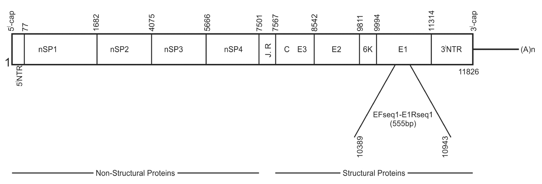
Chikungunya virus belongs to family togaviridae and genus alpha virus. It’s an enveloped RNA virus and RNA is linear, positive sense and single stranded. Genomic organisation of CHIKV is 5’cap-nsp1-nsp2-nsp3-nsp4-(junction region)-C-E3-E2-6K-E1-(poly A)3’cap. The length of RNA is 11805 bp excluding 5’ cap nucleotide, 3’ cap (I-poly A) tract and 3’ poly A tail. Two third of genomic RNA from 5’ end consists of non-structural proteins (nsP1, nsP2, nsP3 and nsP4) with length of 7425 nucleotides and one third towards 3’end consists of structural proteins (E1, E2 and E3, 6K and C) with length of 3735 nucleotides [Table/Fig-1]. The 5’ NTR has 76 nt, 3’ NTR has 526 nt and internal poly A region has 68 nucleotides. 3’ end has internal polyadenylation site and repeated sequence elements (RSEs). CHIKV genome consists of two open reading frames, one code for non-structural poly-proteins (2474 aa) and another for structural proteins (1244 aa) [5].
Structure of Chikungunya virus whole genome with arrangement of structural and non structural genes. The location primers marked in E1 gene.
Chikungunya virus has three genotypes namely East Central South African (ECSA), West african and Asian. The genotypes were named according to prior geographical distribution. The ECSA genotype was confined to East, Central and South Africa previously but in the year 2000 same genotype was first time isolated in India from mosquito samples collected from Yawat, Pune district, Maharastra, India [6]. The same genotype caused explosive outbreak in different regions of Indian ocean island, India, Europe and other parts of the world between 2005-2009 [7,8]. The Mutations in structural and non-structural coding region of viral genome in alpha virus affects infectivity and virulence [9,10].
Aim
In the present study, Molecular diagnosis and Molecular Characterisation of Chikungunya virus isolates, in detail novel mutations in E1 gene responsible for increased host adaptability, infectivity and virulence of Chikungunya virus have been studied.
Materials and Methods
Clinical Samples
Total of 33 serum samples with symptoms of fever and arthralgia, which were Chikungunya serodiagnosed (IgM antibody) included in the study. Blood samples were collected from BLDE’s hospital, Bijapur and Government PHC, CHC, taluk and district hospitals of Bijapur district, Karnataka from 2011 to 2014. Serum was separated and stored at -70°C. Informed consent was obtained from all cases before sample collection.
RNA Extraction
RNA was isolated from 33 serum samples using QIAamp viral RNA mini kit (Qiagen) according to manufacturer instructions [11].
Details of primers used for PCR amplification in the study.
| Primer Name | Sequences 5’-3’ | Genome position | Amplicon size bp |
|---|
| E1-F1 | GCTCCGCGTCCTTTAC | 10389bp–10943pb | 555 |
| E1-R1 | ATGGCGACGCCCCCAAAGTC |
RT-PCR was carried out with 5μl of isolated RNA using Amplisure® Chikungunya RTPCR kit on ABI7500 thermo cycler at RAS Lifesciences Pvt Ltd. The pathogen detection was based on amplification of specific regions in NSP gene. The steps of RT-PCR were: a reverse transcription step at 42°C for 15 min; followed by 40 cycles of thermo cycling which includes denaturation step at 95°C for 1 min, annealing step at 94°C for 15 sec and extension step at 60° for 1 min. Strict adherence to manufacturer’s instructions was followed for optimal results and to avoid PCR contamination. Kit supplied Internal control (IC) was used to identify possible PCR inhibition [12].
Sequencing
Sequencing was performed at RAS Lifesciences Pvt Ltd by using commercial facility. Nested PCR was performed and E1 partial gene sequencing was carried out by Sanger sequencing method for 9 samples. Approximately, 555 base pairs were amplified from samples using E1F1 & E1R1 primers [13,14]. Sequencing was done by using DNA Sequencer (ABI 3130 xl GA) instrument.
Phylogenetic Analysis
Chikungunya sequences were aligned using Clustal W2 software. The unrooted tree was constructed using Neighbour-Joining method [15]. The optimal tree with the sum of branch length = 0.2448 is shown. The percentage of replicate trees in which the associated taxa clustered together in the bootstrap test (10000 replicates) are shown next to the branches [16]. The tree is drawn to scale, with branch lengths in the same units as those of the evolutionary distances used to infer the Phylogenetic tree. The evolutionary distances were computed using the Maximum Composite Likelihood method [17] and are in the units of the number of base substitutions per site. The analysis involved 18 nucleotide sequences. All positions containing gaps and missing data were eliminated. There were a total of 502 positions in the final dataset. Evolutionary analyses were conducted in MEGA6 [18]. The accession numbers used for the study are given below along with the positions matching with the samples [Table/Fig-3].
Reference strains with nucleotide and protein accession number.
| Nucleotide Sequence Accession Numbers with position | Protein Sequence Accession Numbers (Uniprot) | Strain | Year of isolation |
|---|
| AF369024.2|:10427-10941 | Q8JUX5 | S27 (ECSA) | 1952 |
| DQ309336.1|:223-737 | A0SE38 | Reunion 223/05 (ECSA) | 2005 |
| AM258992.1|:10376-10890 | Q1W367 | Reunion (ECSA) | 2006 |
| EF027139.1|:10428-10942 | A6MH23 | INDIA-00-MH4 (Asian) | 2007 |
| HM045811.1|:10410-10924 | D7R978 | Tanzania (ECSA) | 1953 |
| HM045797.1|:10410-10916 | D7R952 | RSU1 (Asian) | 1985 |
| EU192143.1|:2861-3367 | B2BZX4 | Indonesia (Asian) | 2007 |
| HM045817.1|:10412-10926 | D7R990 | Senegal (West Africa) | 2005 |
| HM045816.1|:10412-10926 | D7R988 | Senegal (West Africa) | 1966 |
Mutational Analysis
It was carried out for 8 out of 9 samples because Sample Ck 403 couldn’t be translated.
Protein Modelling
Template search with Blast [19] and HHBlits [20] has been performed against the SWISS-MODEL template library. For each identified template, the template’s quality has been predicted from features of the target-template alignment. The templates with the highest quality have then been selected for model building i.e. 3n43 was used as template for model building. Models are built based on the target-template alignment using Promod-II. Coordinates which are conserved between the target and the template are copied from the template to the model. Insertions and deletions were remodelled using a fragment library. Side chains are then rebuilt. Finally, the geometry of the resulting model is regularized by using a force field. In case loop modelling with ProMod-II [21] does not give satisfactory results, an alternative model is built with MODELLER [22]. The global and per-residue model quality has been assessed using the QMEAN scoring function [23]. For improved performance, weights of the individual QMEAN terms have been trained specifically for SWISS-MODEL.
Mutation Mapping
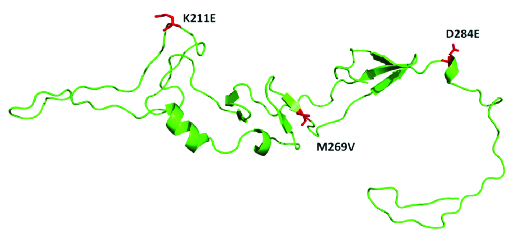
Positions of three observed mutations in E1 protein was mapped in current isolates by using PyMOL software. The sequence positions were labelled according to reference strain S-27.
Results
RT-PCR
Chikungunya RNA was detected in 31 (93.9%) samples. Two Sero-positive samples were found negative for Chikungunya RNA.
Phylogenetic analysis based on Partial E1 nucleotide sequences and relationship with reference strains. Gene bank accession number for reference strain labelled.
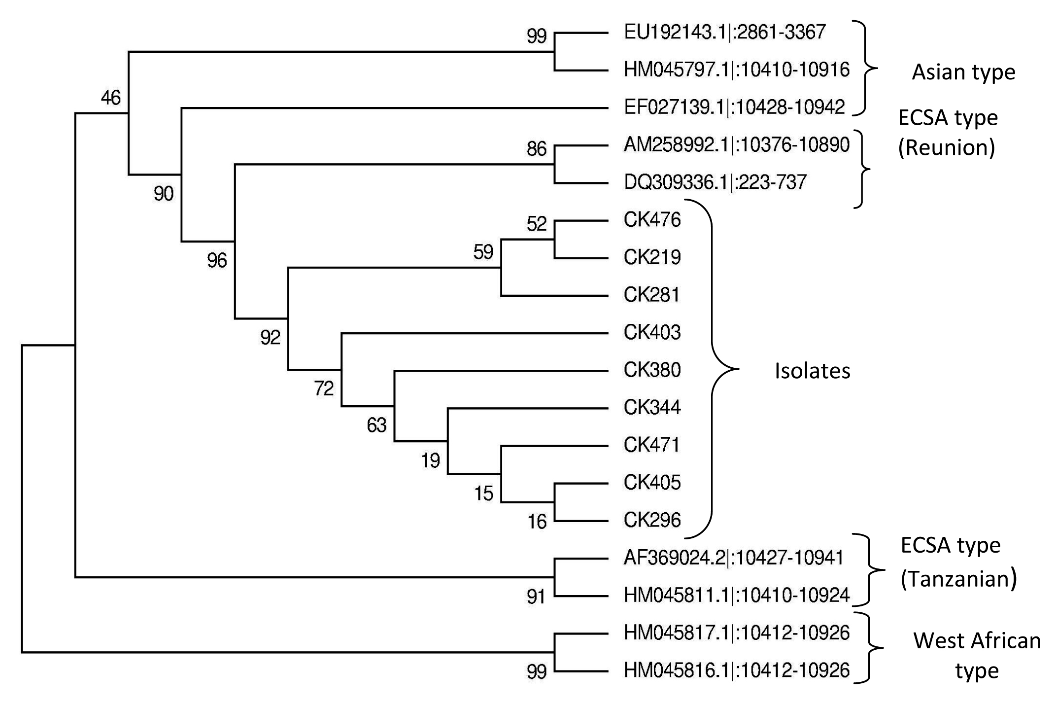
Unrooted Phylogenetic tree shows isolates are closely related to Reunion strains and distantly related to S27 Tanzanian strain and made sister group of ECSA. So, isolates belong to ECSA genotype. Isolates were even more divergent to Asian and West African genotypes. Comparative nucleotide and amino acid homology analysis reveals that isolates are 94.9±5.1% nucleotide homology and 97.1±2.9% at amino acid homology with S-27 strain.
Amino acid Mutational analysis of isolates with respect to positions.
| Position | Reference strains with uniprot ID | Isolates |
|---|
| Nucleotide | Polypeptide | Protein | Q8JUX5-S27 | A0SE38-Reunion | Q1W367 Reunion | A6MH23-INDIA-00-MH4 | D7R978-Tanzania | D7R952-RSU1 | B2BZX4-Indonesia | D7R990-Senegal | D7R988Senegal | 219 | 281 | 296 | 344 | 380 | 405 | 471 | 476 |
|---|
| A10624G | 1020 | 211 | K | . | . | . | . | E | E | . | . | E | E | E | E | E | E | E | E |
| A10798G | 1078 | 269 | M | V | V | V | . | . | . | I | V | V | V | V | V | V | V | V | V |
| T10845A | 1093 | 284 | D | E | E | . | . | . | . | . | . | E | E | E | E | E | E | E | E |
Few random nucleotide changes were observed in partial E1 region. Following amino acid mutations were observed. E1K211E, E1M269V and E1D284E in all isolates.
Molecular Modelling
Homology modelling with mutations has been projected to three dimensional structure [Table/Fig-6]. All the three observed mutations lies in area in major secondary structure. So it couldn’t affect local protein structure.
Mapping of mutations positions in E1 gene.
Discussion
South Indian states Karnataka and Kerala had major Chikungunya outbreaks during 2006-2009. In 2006 Karnataka state reported 7,62,026 number of Chikungunya suspected cases, Bijapur was one among the district experienced huge number of cases [24]. Statistically significant number of Chikungunya confirmed cases (sporadic and epidemic) are being reported in present years. In the present study phylogenetic analysis of Chikungunya virus shows close relation of isolates with Reunion strains than prototype (S 27). It indicates that these isolates may have evolved from Reunion ECSA genotype (subtype Indian ocean lineage-IOL).
Chikungunya RNA couldn’t be detected from two Sero-positive samples. The probable reason for molecular negativity may be due to very low viral load. The cases which produce strong antibody mediated and cell mediated immune response experience short period of viremia (median 6 days) [25]. These two samples were collected on day 7, so in the samples viremia may be short. Antigenic cross-reactivity was observed between Chikungunya and other alpha viruses like Ross River, Onyong nyong, Mayaro, and Sindbis viruses, causing similar clinical manifestations [26].
Niyas KP et al., reported two important mutations A226V and V291I in E1 gene [13]. Shrinet J et al., reported number of mutations in E1 gene namely, K211E, M269V, D284E, V179A, S234P, R196K and R247C [14]. But, in the present study we have found only three mutations K211E, M269V and D284E [Table/Fig-7].
Comparison of E1 gene amino acid substitution reported in Indian isolates.
| Author | Mutations |
|---|
| Isabelle Schuffenecker et al., [7] | A226V and D284E |
| Kudukkil P Niyas et al., [13] | A226V and V291I |
| Jatin Shrinet et al., [14] | K211E, M269V, D284E, V179A, S234P, R196K and R247C |
| Current study | K211E, M269V and D284E |
Previously, it has been demonstrated that E1A226V mutation increases the midgut infectivity and viral dissemination to secondary organs and in turn enhances Chikungunya virus transmissibility by Aedes albopictus mosquitoes. Similarly, it was postulated that mutation decreases cholesterol dependence in target cells and increases fitness of Chikungunya virus on Aedes albopictus mosquitoes [27]. This mutation was absent in the current isolates.
First time ECSA genotype was isolated in India from mosquito during 2000 (Yawat strain). Thereafter, ECSA and Asian genotypes have been circulating in India. The ECSA genotype has caused outbreaks in Indian ocean Island, India and presently causing sporadic and epidemic cases in India. A study did conduct to find out the origin and spread of ECSA genotype in India. It was concluded that Reunion ECSA genotype was not resulted from recombination of prototype ECSA (S27) and Asian genotypes. It’s under purifying selection and may evolved due to random neutral and non-synonymous mutations [28].
In a study, infection of C6/36 cell lines by E1226A and E1226V strains, resulted in higher titre than prototype (S27) [29]. It indicates that there are some un-identified mutations in ECSA lineage responsible for adaptation of ECSA to Aedes albopictus mosquitoes [30,31]. These mutations might affect the displacement of Asian lineage by ECSA lineage in India where both genotypes exist [32].
It was established that E1K211E mutation was positively selected (new non-synonymous advantageous mutations) site with a posterior probability of >75% [28,33]. In Bijapur district, Aedes aegypti mosquitoes were predominant species than Aedes albopictus. Mutations present in E2 gene might play a vital epistatic role in E1 gene for the adaptation of Chikungunya virus to Aedes aegypti and Aedes albopictus mosquitoes. However, further studies are required to elucidate on the effect of mutations in whole genome on viral infectivity, epidemiology and vector adaptability.
Limitations
Genetic characterization was carried out for 9 of 31 samples due to low viral load, as we couldn’t get sequences from remaining samples.
Conclusion
Comparative nucleotide and amino acid homology studies reveal that the current isolates may have evolved from Reunion island strains. The mutation A226V which claimed to increase the host adaptability, infectivity and virulence in Chikungunya virus was absent in present isolates. The identified unique mutations in E1 gene K211E, M269V and D284E were still maintained in current isolates. Homology modelling studies concluded that observed mutations have not altered the local protein structure.
[1]. Lumsden WHR, An epidemic of virus disease in Southern Province, Tanganyika territory, in 1952–1953 II. General description and epidemiologyTransactions of royal society of tropical medicine and hygiene 1955 49(1):28-32. [Google Scholar]
[2]. de Lamballerie X, Leroy E, Charrel RN, Ttsetsarkin K, Higgs S, Gould EA, Chikungunya virus adapts tiger mosquito via evolutionary convergence, a sign of things to come?Virology Journal 2008 5:33 [Google Scholar]
[3]. Guidelines of clinical management of Chikungunya fever. WHO Regional office for south East Asia. 2008; SEA-CD-180 [Google Scholar]
[4]. Kashyap RS, Morey SH, Chandak NH, Purohit HJ, Taori GM, Daginawala HF, Detection of viral antigen, IgM and IgG antibodies in cerebrospinal fluid of Chikungunya patients with neurological complicationsCerebrospinal Fluid Research 2010 7:12 [Google Scholar]
[5]. Khan AH, Morita K, Parquet Md Mdel C, Hasebe F, Mathenge EG, Igarashi A, Complete nucleotide sequence of Chikungunya virus and evidence for an internal polyadenylation siteJ Gen Virol 2002 83(Pt 12):3075-84. [Google Scholar]
[6]. Mourya DT, Thakare JR, Gokhale MD, Powers AM, Hundekar SL, Jayakumar PC, Isolation of Chikungunya Virus from Aedes aegypti mosquitoes collected in the town of Yawat, Pune district, Maharastra state, IndiaActa Virol 2001 45:305-09. [Google Scholar]
[7]. Schuffenecker I, Iteman I, Michault A, Frangeul L, Vaney M, Lavenir R, Genomic microevaluation of Chikungunya virus causing the Indian ocean outbreakPLOS medicine 2006 7:1058-70. [Google Scholar]
[8]. Rezza G, Nicoletti L, Angelini R, Romi R, Finarelli AC, Panning M, Infection with chikungunya virus in Italy: an outbreak in a temperate regionLancet 2007 370:1840-46. [Google Scholar]
[9]. Fazakerley JK, Boyd A, Mikkola ML, Kääriäinen L, A single amino acid change in the nuclear localization sequence on the nsp2 protein affects the neuro-virulence of semliki forest virusJournal of Virology 2002 76:392-96. [Google Scholar]
[10]. Mayuri Geders TW, Smith JL, Kuhn RJ, Role of conserved residues on Sindbis virus nsp2 methyl transferase-like domain in regulation of minus strand synthesis and development of cytopathic infectionJournal of virology 2008 82:7284-97. [Google Scholar]
[11]. Yergolkar PN, Tandale BV, Arankalle VA, Sathe PS, Sudeep A, Gandhe MD, Jacob GP, Hundekar SL, Mishra AC, Chikungunya outbreaks caused by african genotypes, IndiaEmerg Infect Dis 2006 12(10):1580-83. [Google Scholar]
[12]. Amplisue® Chikungunya RTPCR kit. Product insert. RAS Life sciences Pvt ltd [Google Scholar]
[13]. Niyas KP, Abraham R, Unnikrishnan RN, Mathew T, Nair S, Manakkadan A, Molecular characterisation of Chikungunya virus isolates from clinical samples and adult Aedes albopictus mosquitoes emerged from larvae from Kerala south IndiaVirology Journal 2010 7:189 [Google Scholar]
[14]. Shrinet J, Jain S, Sharma A, Shekhar SS, Mathur K, Rana V, Genetic characterization of Chikungunya virus from New Delhi reveal emergence of a new molecular signature in Indian isolatesVirology journal 2012 9:100 [Google Scholar]
[15]. Saitou N, Nei M, The neighbor-joining method: A new method for reconstructing phylogenetic treesMolecular Biology and Evolution 1987 4:406-25. [Google Scholar]
[16]. Felsenstein J, Confidence limits on phylogenies: An approach using the bootstrapEvolution 1985 39:783-91. [Google Scholar]
[17]. Tamura K, Nei M, Kumar S, Prospects for inferring very large phylogenies by using the neighbor-joining methodProceedings of the National Academy of Sciences (USA) 2004 101:11030-35. [Google Scholar]
[18]. Tamura K, Stecher G, Peterson D, Filipski A, Kumar S, MEGA6: Molecular Evolutionary Genetics Analysis version 6.0Molecular Biology and Evolution 2013 30:2725-29. [Google Scholar]
[19]. Altschul SF, Madden TL, Schaffer AA, Zhang J, Zhang Z, Miller W, Gapped BLAST and PSI-BLAST: a new generation of protein database search programsNucleic Acids Res 1997 25:3389-402. [Google Scholar]
[20]. Remmert M, Biegert A, Hauser A, Soding J, HH blits: lightning-fast iterative protein sequence searching by HMM-HMM alignmentNat Methods 2012 9:173-75. [Google Scholar]
[21]. Guex N, Peitsch MC, SWISS-MODEL and the Swiss-Pdb Viewer: an environment for comparative protein modelingElectrophoresis 1997 18:2714-23. [Google Scholar]
[22]. Sali A, Blundell TL, Comparative protein modelling by satisfaction of spatial restraintsJ Mol Biol 1993 234:779-815. [Google Scholar]
[23]. Benkert P, Biasini M, Schwede T, Toward the estimation of the absolute quality of individual protein structure modelsBioinformatics 2011 27:343-50. [Google Scholar]
[24]. Talwar AS, Pujar HS, An outbreak of Chikungunya epidemic in south India- KarnatakaIJRRAS 2012 5(3):229-34. [Google Scholar]
[25]. Poo YS, Rudd PA, Gardner J, Wilson JAC, Larcher T, Colle MA, Multiple immune factors are involved in controlling acute and chronic chikungunya virus infectionPLOS Neglected Tropical Diseases 2014 8(12):e3354 [Google Scholar]
[26]. Chanas AC, Johnson BK, Simpson DIH, Antigenic Relationships of Alphaviruses by a Simple Micro-culture Cross-neutralization Method. genVirol 1976 32:295-300. [Google Scholar]
[27]. Konstantin A, Tsetsarkin Dana L, Vanlandingham Charles E, McGee Stephen Higgs, A single mutation in Chikungunya virus affects vector specificity and epidemic potentialPLOS pathogens 2007 3:12 [Google Scholar]
[28]. Arankalle VA, Shrivastava S, Cherian S, Gunjikar RS, Walimbe AM, Jadhav SM, Genetic divergence of Chikungunya viruses in India (1963-2006) with special reference to the 2005-2006 explosive epidemicJournal of General Virology 2007 88:1967-76. [Google Scholar]
[29]. Wikan N, Sakoonwatanyo P, Ubol S, Yoksan S, Smith DR, Chikungunya virus infection of cell lines: analysis of East Central and South African lineagePLOS ONE 2012 7:e31102 [Google Scholar]
[30]. Raharimalala FN, Ravaomanarivo LH, Ravelonandro P, Rafarasoa LS, Zouche K, Biogeography of the two major arbovirus mosquito vectors, Aedes aegypti and Aedes albopictus (Diptera, Culicidae) in MadagascarParasit vectors 2012 5:56 [Google Scholar]
[31]. Delatte H, Bagny L, Brengue C, Bouetard A, Paupy C, The invaders: Phylogeography of dendue and Chikungunya viruses Aedes vectors on the south west islands of the Indian oceanInfect Genet Evol 2011 11:1769-81. [Google Scholar]
[32]. Sam IC, Loong SK, Micheal JC, Chua CL, Sulaiman WYW, Vythilingam I, Genotypic and phenotypic characterisation of Chikungnya virus of different genotypes from MalaysiaPLOS ONE 2012 7:e50476 [Google Scholar]
[33]. Sumathy K, Ella KM, Genetic diversity of Chikungunya virus India 2006-2010: Evolutionary dynamics and serotype analysesJ Med Virol 2012 84:462-70. [Google Scholar]