Analysis of the Genotoxic Effects of Mobile Phone Radiation using Buccal Micronucleus Assay: A Comparative Evaluation
Sumita Banerjee1, Narendra Nath Singh2, Gadiputi Sreedhar3, Saikat Mukherjee4
1 Assistant Professor, Department of Oral Pathology and Oral Microbiology, Dental College, Regional Institute of Medical Sciences, Lamphelpat, Imphal, Manipur, India.
2 Professor and Head of the Department, Department of Oral Pathology, Kothiwal Dental College and Research Center, Moradabad, Uttar Pradesh, India.
3 Professor and Head of the Department, Department of Oral and Maxillofacial Pathology and Microbiology, Babu Banarasi Das College of Dental Sciences, Lucknow, Uttar Pradesh, India.
4 DBT–Research Associate, Department of Biochemistry, Manipur University, Imphal, Manipur, India.
NAME, ADDRESS, E-MAIL ID OF THE CORRESPONDING AUTHOR: Dr. Sumita Banerjee, Assistant Professor, Department of Oral Pathology and Oral Microbiology, Dental College, Regional Institute of Medical Sciences, Lamphelpat, Imphal, Manipur-795004, India.
E-mail: banerjeesumi10@gmail.com
Introduction
Micronucleus (MN) is considered to be a reliable marker for genotoxic damage and it determines the presence and the extent of the chromosomal damage. The MN is formed due to DNA damage or chromosomal disarrangements. The MN has a close association with cancer incidences. In the new era, mobile phones are constantly gaining popularity specifically in the young generation, but this device uses radiofrequency radiation that may have a possible carcinogenic effect. The available reports related to the carcinogenic effect of mobile radiation on oral mucosa are contradictory.
Aim
To explore the effects of mobile phone radiation on the MN frequency in oral mucosal cells.
Materials and Methods
The subjects were divided into two major groups: low mobile phone users and high mobile phone users. Subjects who used their mobile phone since less than five years and less than three hours a week comprised of the first group and those who used their mobile since more than five years and more than 10 hours a week comprised of the second group. Net surfing and text messaging was not considered in this study. Exfoliated buccal mucosal cells were collected from both the groups and the cells were stained with DNA-specific stain acridine orange. Thousand exfoliated buccal mucosal cells were screened and the cells which were positive for micronuclei were counted. The micronucleus frequency was represented as mean±SD, and unpaired Student t-test was used for intergroup comparisons.
Results
The number of micronucleated cells/ 1000 exfoliated buccal mucosal cells was found to be significantly increased in high mobile phone users group than the low mobile phone users group. The use of mobile phone with the associated complaint of warmth around the ear showed a maximum increase in the number of micronucleated cells /1000 exfoliated buccal mucosal cells.
Conclusion
Mobile phone radiation even in the permissible range when used for longer duration causes significant genotoxicity. The genotoxicity can be avoided to some extent by the regular use of headphones.
Acridine orange, DNA damage, DNA specific stain, Micronucleus, Oral mucosa, Radiofrequency radiation
Introduction
In this emerging era of mobile phones, more than three billion people are using mobile phones in the world [1]. India has world’s second-largest population of mobile phone users [2] having the” teledensity” of 80% [3]. Mobile phones use microwave radiation in the carrier frequency range of 900 to 1800 MHz [4]. There are disagreements regarding the health hazards of mobile phone radiation. Several studies have confirmed the genotoxic effect of mobile phone radiation [5–9]. WHO has classified mobile phone radiation on the IARC scale as Group 2B – ‘possibly carcinogenic by increased risk of Glioma formation’ [10]. But here we have to consider that brain tissue is well protected in the skull and to have an effect, mobile radiation has to penetrate several layers of tissue like skin, muscle, bone and even the blood-brain barrier. As a matter of fact, the oral mucosa is the tissue that is present in the closest vicinity of the area of a mobile phone while in use and has chances to show possible genotoxic changes by the effect of mobile phone radiation. Some authors have confirmed the genotoxicity of mobile radiation on oral mucosa [4,11], but others have apparently denied the genotoxic effect [12–14]. Considering all these disagreements, reevaluation of the effect of mobile radiation on the oral epithelium is needed. This study was designed to evaluate this effect by using the micronuclei index in the buccal exfoliated cells, as a marker for genotoxicity. Micronucleus (MN) is presented as microscopically visible chromatin mass in the cytoplasm that is present near the nucleus with no direct communication with the nucleus. The MN represents the eccentric chromosomes or chromatin fragments formed due to abnormal mitosis [15]. The presence of an increased number of micronucleated cells indicates DNA damage [16]. An evaluation of the MN frequency in exfoliated oral mucosal cells and its comparison between high and low mobile users can solve the controversy related to the genotoxicity of mobile phone radiation.
Materials and Methods
Subject Selection
The study was done after getting Institutional Ethical Clearance. A total of 300 male subjects between the age group of 20-30 (150 high mobile users and 150 low mobile users) were selected from the OPD of Department of Oral Pathology and Microbiology, Kothiwal Dental College and Research Centre, Moradabad, Uttar Pradesh, from March 2010 to December 2010. The low mobile phone users (Group I), used mobile phone since less than five years and less than three hours a week. The high mobile phone users (Group II), used mobile since more than five years and more than 10 hours a week. Receiving or making calls was considered, while net surfing and text messaging was not included. In group II,95 subjects were CDMA users and 55 subjects were GSM users. These high mobile users were further divided into wired headphone users (70) and non head phone users (80). For all the subjects, exfoliated buccal mucosal cells were collected from the same side in which the subject used their mobile phones the most. Only in the case of group II, a comparative evaluation of micronuclei was done between both the sides of buccal mucosa (right and left).
Inclusion Criteria
Subjects were selected in the age limit of 20-30 years. Individuals who didn’t have any history of medication in last three months had a similar body mass index, no nutritional deficiency and didn’t have any deleterious oral habits were finally selected for the study.
Exclusion Criteria
Subjects having deleterious oral habits, specifically tobacco and having any visible oral mucosal lesions were excluded from the study.
Before sample collection informed written consent from each subject was collected. A detailed questionnaire was prepared to evaluate the lifestyle, dietary habit, previous history of medication, locality of residence, type of mobile used (CDMA or GSM), duration of mobile phone usage (number of years and numbers of hours a week), use of headsets, wired or not. Associated symptoms like headaches, tingling of skin, rashes over the skin, the warmth of the ear were also noted.
Sample collection and evaluation
The subjects were initially asked to rinse the mouth with 1% glacial acetic acid. Exfoliated cells from the buccal mucosa were collected using a moistened wooden spatula and the cells were spread evenly on clean microscopic glass slides and air dried. The samples were fixed using cytofixative (Bio Fix) and the slides were stained with acridine Orange (Loba Chemie) staining solutions. Each slide was observed by a single observer.
Scoring of micronuclei
The criteria for identifying and scoring of MNi were based on the proposed description by Tolbert [17].
The MN has rounded smooth perimeter suggestive of the membrane.
It stains in the same intensity as the nucleus.
It is located within the cytoplasm of the cell, and usually, the diameter is 1/3 to 1/6 of the nucleus.
It has texture similar to nucleus.
It is located in the same focal plane as nucleus.
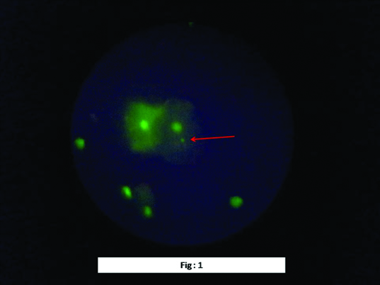
From each slide 1000 cells were observed under Fluorescence microscope (Kyowa, Japan) under 400X magnification for MNi identification and 1000X magnification MNi for scoring [Table/Fig-1].
Exfoliated Cytology cells of higher mobile users stained with acridine orange and observed under fluorescent microscope under 40x magnification (images were taken with sony cool pix camera under 2X zoom with total magnification 80X). Only one cell in the fields is showing single micronucleus. The micronucleus is the small intense green colored round structure highlighted by the arrow. The nucleus is the bigger intense green colored and situated in close approximation of the micronucleus.
Statistical Analysis
SPSS (Statistical package for Social Sciences) Version 15.0 Software was used for statistical analysis. The values were represented in Mean±SD. The comparative evaluations were done using unpaired Student t-test.
Results
There was a significant increase in the mean micronuclei count in group II (1.52±1.176) in comparison to the group I (0.77±0.815) [Table/Fig-2]. In group II, the micronuclei count in the side of mobile phone use was found to be statistically significantly elevated (1.52±1.176) in comparison to the opposite side (0.90±0.3992) [Table/Fig-3]. There was no significant difference in the mean micronuclei count of subjects using CDMA (0.64± 0.722) or GSM (0.90± 0.886) mobile phones [Table/Fig-4]. But the micronuclei mean count was found to be significantly increased in non-head phone users (2.08±1.291) in comparison to headphone users (0.96± 0.699) [Table/Fig-5]. It was also found that in group II, users without head phones, had complained about warmth around the ear, have showed the highest mean count for micronucleated cells (2.847± 0.341).
Mean micronucleus count in low mobile phone users and high mobile phone users showing their statistical differences.
| Mean | N | Std. Deviation | t-value | df | p-value* |
|---|
| Low mobile users | 0.77 | 150 | 0.815 | | | |
| High mobile users | 1.52 | 150 | 1.176 | 6.4199 | 298 | <.0001 |
Unpaired student t test:
*The two-tailed p-value is less than 0.0001. By conventional criteria this difference is considered to be extremely statistically significant.
Mean micronucleus count from the side of buccal mucosa of mobile phone used and from the opposite side of buccal mucosa of mobile phone used in case of high mobile users showing their statistical differences.
| Mean | N | Std. Deviation | t-value | df | p-value* |
|---|
| Opposite side of mobile use | 0.90 | 150 | 0.3992 | | | |
| Same side of mobile use | 1.52 | 150 | 1.176 | 14.1024 | 298 | <.0001 |
*The two-tailed p-value is less than 0.0001. By conventional criteria this difference is considered to be extremely statistically significant.
Mean micronucleus count in subjects using CDMA and GSM mobile users.
| Type of mobile phone | N | Mean | Std. Deviation | t-value | df | p-value |
|---|
| CDMA | 95 | 0.64 | .722 | | | |
| GSM | 55 | 0.90 | .886 | 1.9528 | 148 | 0.0527 |
*The two-tailed p-value equals 0.0527. By conventional criteria this difference is considered to be not quite statistically significant.
Mean micronucleus count in subjects using headphone and non using head phones.
| Head phone (wired) | N | Mean | Std. Deviation | t-value | df | p-value* |
|---|
| Users | 70 | 0.96 | 0.699 | | | |
| Non users | 60 | 2.08 | 1.291 | 6.2677 | 128 | <.0001 |
*The two-tailed p-value is less than 0.0001. By conventional criteria this difference is considered to be extremely statistically significant.
Discussion
The most conventional mobile communication system in India is GSM and CDMA. GSM (Global System for Mobile Communications) uses frequencies of around 900 MHz bandwidth and CDMA (Code Division Multiple Access) works on higher bandwidth i.e. 1800MHz [4]. So, CDMA handsets may have more genotoxicity. The International Commission on Non-ionizing radiation Protection (ICNIRP) has determined the permissible range on the level of SAR (Specific Absorption Rate i.e. the amount of energy absorbed per unit time per unit mass of tissue) [18]. The ICNIRP permissible range of SAR is 2.0W/Kg [19]. Now-a-days the most popular 3G network system works under the frequency range of 2100 MHz [20]. Mobile phones radiate an average of power of 0.2-0.6 Watt/Kg, 40% of which is absorbed in the head and neck region [11]. Though the radio frequency wave from emitting the mobile phones are considered to be low-grade emission in the range of 1.6 to 2W/kg i.e. within the permissible range it can be still harmful in case of long term use for the prolonged period. Microwave radiation in the range of 2.45 GHz indicates significant DNA damage in mice model [21]. Another study conducted by Kesari et al., reported an increase in MN count, caspase 3 levels and apoptosis rate in mice model due to 3G cell phone exposure for two hours a day for 60 days [22]. Long-term use of mobile radiation even in low range can affect the reproductive system also. Shahin et al., have shown decreased sperm count in male Swiss strain mice for a long time low radiofrequency exposure by mobile phones [23]. As the head and neck region are the most closely approximated area for mobile phone use, the maximum radiation effect can be expected here. In this study micronucleus count in exfoliated buccal mucosal cells were used to evaluate the genotoxic effect of mobile phone radiation. MN count in the exfoliated cells can be used as a marker for an abnormal cell cycle as it is formed as a result of aberrant mitosis when the whole chromosome or chromatid fragment fails to reach the spindle pole. It is one of the best indicators of mitotic interference and chromosomal mutations or breakage [24]. The MNi index is preferable for mass screening as it is rapid, simple, sensitive and cost-effective [25]. Counting of MNi is very technique sensitive, and different staining methods cause significant variations for the evaluation of it’s frequency [26]. For proper evaluation of MN, DNA specific stain should be used [27] and thereby acridine orange stain was used in this study. As stated in the result, MN count was found to be significantly higher in high mobile phone users in comparison to low users, and it directly indicates the genotoxic effect of prolonged mobile phone use for longer period. In our study, all probable causes for the increase in the MN count were excluded (tobacco, alcohol, recent medication, systemic factor etc.). Therefore mobile phone radiation was expected to be the immediate and possible cause for increased MN count in higher mobile phone users, and this finding was similar to various research groups [4,11]. On the contrary, other research groups have directly denied any significant increase of MN count in mobile phone users [12,28,29]. In this study, the higher mobile phone users were also evaluated by type of mobile phone used i.e., CDMA or GSM. Though the CDMA phones work under higher electromagnetic frequency (1800 MHz) in comparison to GSM mobiles (900MHz), no significant MN count variability in these two groups were observed. This result indicates that when used within the permissible range the strength of radiofrequency radiation is not a major factor for genotoxic damage. When the high mobile phone users were questioned for usage of wired headphones, a significantly dramatic decrease in MN count was observed in headphone users. When headphones are used with mobile phones, it helps to keep the mobile phone away from the body, and there is no direct contact of the radio-frequency receiver with the body.
The headphones also contribute to reducing the local temperature rise around the ear, and this is a commonly encountered problem by the long-term mobile phone users not using headphones. A significant finding of this study was that, people complaining of warmth around the ear were found to have the highest micronuclei count indicating that heat provides a synergistic effect on genotoxic damage, probably by the activation of heat shock proteins along with the radiofrequency radiation. It has been a proven fact that heat shock protein 70 level is increased as a radioadaptive response [30]. As the local rise of temperature is a direct effect of heating up of the battery of the phone or as a result of the long press of the mobile phone against the cheek, cellular response due to direct heat also can’t be ignored. Localized heat stress is proven to increase the vascular permeability making the tissue more susceptible to genotoxic stress [31]. The local rise of temperature may even facilitate heat stress-induced mitochondrial membrane damage, release of cytochrome c, and activation of caspase-9 and -3 [32] which in turn can be considered as a prerequisite for cytotoxicity. On the other hand, the increase micronuclei production can also be directly associated with localized hyperthermia [33]. So, the increased micronuclei count in subjects complaining of warmth around the ear may be synergistic effects of both mobile radiations induced genotoxicity and local thermal effects. In our study although any visible oral mucosal changes were not observed but it should be taken into consideration that mobile phones are being used extensively by the general population only last 10-12 years and increased micronucleus count indicate that in future days it may cause visible oral lesions. The complete avoidance of mobile technology is not possible but few precautionary steps such as: keeping the mobile phone away from the body when connecting; use of headphones; keeping the phone in switched off mode when possible, may help us to reduce the deleterious effects of mobile radiation.
Conclusion
Mobile phones when used for prolonged periods can cause genotoxicity. Although according to the SAR values most of the mobile phones emit radiofrequency radiation within safety limit, long-term use of mobile phones shows definite signs of DNA damage. The genotoxicity accentuates when associated symptom like warmth around the ear is related. Headphone use reduces the harmful effects of mobile phone radiation.
Unpaired student t test:
*The two-tailed p-value is less than 0.0001. By conventional criteria this difference is considered to be extremely statistically significant.
*The two-tailed p-value is less than 0.0001. By conventional criteria this difference is considered to be extremely statistically significant.
*The two-tailed p-value equals 0.0527. By conventional criteria this difference is considered to be not quite statistically significant.
*The two-tailed p-value is less than 0.0001. By conventional criteria this difference is considered to be extremely statistically significant.
[1]. Hoskote SS, Kapdi M, Joshi SR, An epidemiological review of mobile telephones and cancerJ Assoc Phys India 2008 56:980-84. [Google Scholar]
[2]. Shah C, Nair A, Naiku M, Bakshi S, Cell phone radiation and genomic damage: in vitro exposure and assessmentInternational Journal of Innovative Research in Science, Engineering and Technology 2015 4(2):401-05. [Google Scholar]
[3]. www.kas.de/wf/doc/2245-1442-2-30.pdf [Google Scholar]
[4]. Yadav AS, Sharma MK, Increased frequency of micronucleated exfoliated cells among humans exposed in vivo to mobile telephone radiationMutation Research 2008 650:175-80. [Google Scholar]
[5]. Foster KR, Repacholi MH, Biological effects of radiofrequency fields: does modulation matter?Radiation Research 2004 162(2):219-25. [Google Scholar]
[6]. Karaca E, Durmaz B, Aktug H, Yildiz T, Guducu C, Irgi M, The genotoxic effect of radiofrequency waves on mouse brainJ Neurooncol 2012 106(1):53-58. [Google Scholar]
[7]. Hardell L, Carlberg M, Hansson Mild K, Pooled analysis of two case-control studies on use of cellular and cordless telephones and the risk for malignant brain tumours diagnosed in 1997-2003Int Arch Occup Environ Health 2006 79:630-39. [Google Scholar]
[8]. Hardell L, Carlberg M, Mobile phones, cordless phones and the risk for brain tumoursInt J Oncol 2009 35:5-17. [Google Scholar]
[9]. Takebayashi T, Varsier N, Kikuchi Y, Wake K, Taki M, Watanabe S, Mobile phone use, exposure to radiofrequency electromagnetic field, and brain tumour: a case control studyBr J Cancer 2008 98:652-59. [Google Scholar]
[10]. www.iarc.fr/en/media-centre/pr/2011/pdfs/pr208_E.pdf [Google Scholar]
[11]. Gandhi G, Singh P, Cytogenetic damage of mobile phone users: preliminary dataInt J Hum Genet 2005 5(4):259-65. [Google Scholar]
[12]. Hintzsche H, Stopper H, Micronucleus frequency in buccal mucosa cells of mobile phone usersToxicology Letters 2010 193(1):124-30. [Google Scholar]
[13]. McNamee JP, DNA damage and micronucleus induction in human leukocytes after acute in vitro exposure to a 1.9 GHz continuous-wave radiofrequency fieldRadiat Res 2002 158:523-33. [Google Scholar]
[14]. Adelman G, International Encyclopedia of Neuroscience. Third Edition. New York: Elsevier. Antonopoulos A, Eisenbrandt H, Obe G. Effects of high-frequency electromagnetic fields on human lymphocytes in vitroMutat Res 1997 395:209-14. [Google Scholar]
[15]. Thomas P, Holland N, Bolognes C, Volders MK, Bonassi S, Zieiger E, Buccal micronucleus cytome sssayNature Protocols 2009 4(6):825-37. [Google Scholar]
[16]. Holland N, Bolognsei C, Volders KM, Bonassi S, Zeiger E, Knasmueller S, The micronucleus assay in human buccal cells as a tool for biomonitering DNA damage: The HUMN project perspective on current status and knowledge gapsMutation Research 2008 659(1-2):93-97. [Google Scholar]
[17]. Tolbert PE, Shy CM, Allen JW, Micronuclei and other nuclear anomalies in buccal smears: methods developmentMutation Research 1992 271:69-77. [Google Scholar]
[18]. International Commission on non ionizing radiation protection (ICNIRP) guidelines for limiting exposure to time-varying electric, magnetic, and electromagnetic fields (up to 300 ghz)Health phys 1998 74:494 [Google Scholar]
[19]. www.dot.gov.in/sites/default/files/Annexures/advertisement_0.pdf [Google Scholar]
[20]. www.telcoantennas.com.au/site/guide-to-mobile-networks [Google Scholar]
[21]. Chaturvedi CM, Singh VP, Singh P, Basu P, Singaravel M, 2.45 ghz (Cw) microwave irradiation alters circadian organization, spatial memoiy, dna structure in the brain cells and blood cell counts of male miceMus Musculus Electromagnetic Research B 2011 29:23-42. [Google Scholar]
[22]. Kesari KK, Meena R, Wirala J, Kumar J, Verma HN, Effect of 3G cell phone exposure with computer controlled 2-D stepper motor on non-thermal activation of the hsp27/p38MAPK stress pathway in rat brainCell Biochem Biophys 2014 68(2):347-58. [Google Scholar]
[23]. Shahin S, Mishra V, Singh SP, Chaturvedi CM, 2.45-GHz microwave irradiation adversely affects reproductive function in male mouse, Mus musculus by inducing oxidative and nitrosative stressFree Radic Res 2014 48(5):511-25. [Google Scholar]
[24]. Sivasankari NP, Kaur S, Reddy KS, Vivekanandam S, Ramchandra RK, Micronucleus Index: An early diagnosis in oral carcinomaJ Anat Soc India 2008 57:8-13. [Google Scholar]
[25]. Saran R, Tiwari RK, Reddy PP, Ahuja YR, Risk assessment of oral cancer in patients with precancerous states of the oral cavity using micronucleus test and challenge assayOral Oncology 2008 44(4):354-60. [Google Scholar]
[26]. Dias VM, Manelli-Oliveria R, Machado-Santelli GM. Using flurosence for the improvement of the quantititative analysis of MN in cell cultureMutation Research 2005 565:173-79. [Google Scholar]
[27]. Thomas P, Fenech M, Buccal micronucleus cytome assayNature Protocols 2009 4(6):825-37. [Google Scholar]
[28]. Ros-Llor I, Sanchez-Siles M, Camacho-Alonso F, Lopez-Jornet P, Effect of mobile phones on micronucleus frequency in human exfoliated oral mucosal cellsOral Diseases 2012 18(8):786-92. [Google Scholar]
[29]. Souza L da CM, Cerqueira E de MM, Meireles JRC, Assessment of nuclear abnormalities in exfoliated cells from the oral epithelium of mobile phone usersElectromagnetic Biology and Medicine 2014 33(2):98-102. [Google Scholar]
[30]. Park SH, Lee SJ, Chung HY, Kim TH, Cho CK, Yoo SY, Inducible heat-shock Protein 70 is involved in the radioadaptive responseRadiation Research 2000 153(3):318-26. [Google Scholar]
[31]. Suganuma T, Irie K, Fujii E, Yoshioka T, Muraki T, Effect of heat stress on lipopolysaccharide-induced vascular permeability change in miceJPE 2002 303:656-63. [Google Scholar]
[32]. Gu ZT, Li L, Wu F, Zhao P, Yang H, Liu YS, Heat stress induces apoptosis through transcription-independent p53-mediated mitochondrial pathways in human umbilical vein endothelial cellScientific Reports 2014 4:4469 [Google Scholar]
[33]. Hintzsche H, Riese T, Stopper H, Hyperthermia-induced micronucleus formation in a human keratinocyte cell lineMutat Res 2012 71(4):738-39. [Google Scholar]