A Rare Case of Multiple Myeloma Presenting As Lytic Lesion of the Rib
Dhruva Sharma1, Vivek Rawat2, Rajkumar Yadav3
1 Resident, Department of Cardiothoracic & Vascular Surgery, SMS Medical College & Attached Group of Hospitals, Jaipur, India.
2 Resident, Department of Cardiothoracic & Vascular Surgery, SMS Medical College & Attached Group of Hospitals, Jaipur, India.
3 Professor and Head, Department of Cardiothoracic & Vascular Surgery, SMS Medical College & Attached Group of Hospitals, Jaipur, India.
NAME, ADDRESS, E-MAIL ID OF THE CORRESPONDING AUTHOR: Dr. Dhruva Sharma, Resident, Department of Cardiothoracic & Vascular Surgery, SMS Medical College & Attached Group of Hospitals, Jaipur, India. E-mail : nsharma226@gmail.com
Multiple Myeloma (MM) is a disease which results from malignant proliferation of plasma cells. It is commonly encountered in elderly patients. Diffuse bony lesions are the most frequent thoracic involvement with MM. We report a case of 42-year-old male patient who came with pain and full swelling in the right chest wall since two years. On CT scan of thorax, heterogenously enhancing soft tissue density lesion with lytic sclerotic destruction of right 4th rib seen. En-bloc resection of third and fourth rib was done and plasmacytoma was confirmed on biopsy.
Haematologic cancers, Lytic lesion of rib, Multiple myeloma
Case Report
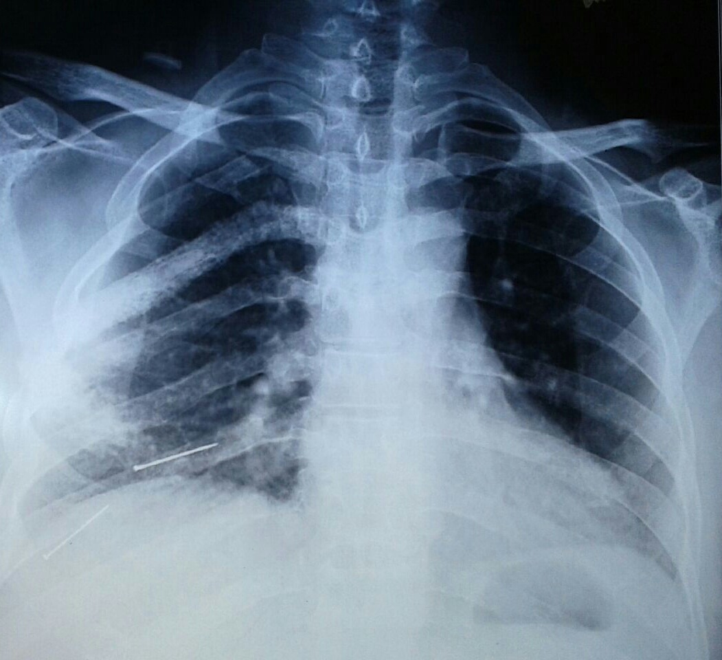
A 42-year-old male patient came to CTVS (CardioThoracic & Vascular Surgery) out-patient department with pain and full swelling in the right chest wall since two years. There was no history of cough, chest trauma or fever. There were no associated co-morbidities and no history of any addictions. There was negative history of herpes infection to rule out post zoster neuralgia. Family history was unremarkable. On examination there was palpable, mildly tender, swollen 4th rib present anteriorly at the level of right nipple. Baseline investigations of the patient were non- significant except for mild thrombocytopenia. On serum nephelometry, increased production of free monoclonal light chains was seen leading to a change in k/λ light chain which was suggestive of existence of monoclonal gammopathy. Cardiac examination was unremarkable. On chest X- ray right fourth rib was invisible with no evidence of any pleural effusion. Chest X-ray of the patient showed thickening of the pleura around 4th rib, as depicted in [Table/Fig-1].
Chest X-ray showing thickened pleura around 4th rib.
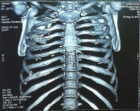
On CT (Computed Tomography) scan of thorax, heterogenously enhancing soft tissue density lesion of approximate size 34×26mm with lytic sclerotic destruction of right 4th rib seen as shown in [Table/Fig-2].
CT-scan of thorax showing heterogenously enhancing soft tissue density lesion with lytic sclerotic destruction of right 4th rib.
Intrathoracic extension was not present. But, multiple bilateral axillary lymph nodes were seen and largest was measuring 17mm. Bilateral lungs were clear, trachea was central and tracheal bifurcation was well defined. There was no evidence of pleural effusion or any thickening.
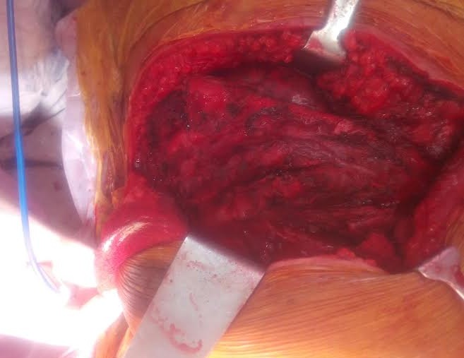
Patient was taken to operation theatre after clearance from the anaesthetist. Written informed consent was sought from the patient before operation. On exploration, third and fourth rib was found to be fused to each other as depicted in [Table/Fig-3].
Intraoperative picture showing fused third and fourth rib.
Excision of third and fourth rib was done and the specimen was sent for biopsy. Prolene mesh was used for reconstruction of the rib cage. On microscopic examination of the osteoytic lesion rib, sections showed fibro-collagen and adipose tissue showing congestion of blood vessels and infiltration by sheets of plasma cells. Sections from the bone showed marrow cavity with diffuse infiltration by plasma cells suggestive of multiple myeloma. While, biopsy of right pleura also showed fibrocollagenous tissue with congested blood vessels and marked infiltration by chronic non specific inflammatory cells. Bone marrow aspiration was done under strict aseptic conditions. It showed cellular bone marrow with M:E ratio of 20:1. Erythroid series was normoblastic and leucopoiesis was increased. It was negative for any haemoparasite and granuloma. Per’s stain for iron showed adequate iron stores.
The patient was referred for further treatment to Tata Memorial Centre, Mumbai for radiotherapy. He underwent whole body bone scan showing abnormal radiotracer uptake involving right 5th and 6th rib posteriorly. It was suggestive of metastases involving right 5th and 6th rib posteriorly. Patient was suggested radiotherapy to the primary site to a dose of 33 Gy for eleven cycles using appropriate energy, portals and technique.
Discussion
Multiple Myeloma (MM) is a disease which results from malignant proliferation of plasma cells. It is commonly encountered in elderly patients [1]. Diffuse bony lesions are the most frequent thoracic involvement with MM [2]. A pulmonary, bronchial and pleural involvement is reported to be rare [3,4]. It accounts for approximately 1% of neoplastic diseases and 13% of haematologic cancers. MM occurs as a result of monclonal gammopathy. Genetic abnormalities also play a major role in its pathogenesis [5].
A huge number of literatures are available addressing multiple myeloma cases but still there is paucity in the research work about it. It has been reported in previous studies that, pleural effusion occurs in nearly 6% of patients with MM [1,6]. Pleural effusion have also been reported in the literature by other studies which was predominantly left sided [2,3]. But in our case there was no evidence of pleural effusion.
Bone haemangiomas are one of the differential diagnosis of rib tumour that is commonly seen in middle aged females [7]. It accounts for approximately 1% of all osseous tumours [8]. Tuberculosis is another differential diagnosis of lytic rib lesion which was reported by Ozol D et al., [9].
A solitary plasmacytoma of the rib occurs as a result of destruction of the bone cortex which extends into the surrounding soft tissues [10]. Solitary plasmacytomas of bone are formed by clonal proliferations of plasma cells which produces localized osseous growth. Plasmacytomas can be classified as multiple, solitary osseous, and solitary extraosseous or extramedullary plasmacytomas and rare as compared with multiple myeloma [10,11]. As far as treatment of plasmacytoma is concerned, radiotherapy and surgical resection are sufficient to achieve long term survival [10,12]. In our case we did surgical resection and referred the patient for radiotherapy as mentioned in previous studies [10,13].
We also referred the patient for adjuvant chaemotherapy because it has been reported in previous studies that adjuvant chaemotherapy has increased the survival of the patient [10].
Conclusion
To conclude we suggest that en-bloc resection of the rib followed by subsequent radiotherapy and adjuvant chaemotherapy can increase the survival rate of solitary plasmacytoma cases.
This article might fulfill the gap in the original research work in this field.
Consent of the Patient : Written informed consent was obtained from the patient for publication of this case report and accompanying images.
[1]. AlShati MH, Kumar R, Kannan S, Dyspnea, massive effusion and lytic rib lesion as initial presentation of multiple myeloma in a young manCan Respir J 2013 20(4):253-55. [Google Scholar]
[2]. Kintzer JS, Rosenow EC, Kyle RA, Thoracic and pulmonary abnormalities in multiple myeloma. A review of 958 casesArch Intern Med 1978 138(5):727-30. [Google Scholar]
[3]. Fekih L, Fenniche S, Hassene H, Abdelghaffar H, Yaalaoui S, EL Mezni F, Multiple Myeloma Presenting with Multiple Thoracic ManifestationsIndian J Chest Dis Allied Sci 2010 52:47-49. [Google Scholar]
[4]. Ascari E, Merlini G, Riccardi A, Multiple myelomaAnn Ital 1990 5:312-21. [Google Scholar]
[5]. Palumbo A, Anderson K, Multiple MyelomaN Engl J Med 2011 364:1046-60. [Google Scholar]
[6]. Alexandrakis MG, Passam FH, Kyriakou DS, Peural effusions in haematologic malignanciesChest 2004 125(4):1546-55. [Google Scholar]
[7]. Jain SK, Songra M, Malhotra A, Kapoor N, Malik R, Shrivastava A, Rib Haemangioma: A Rare Differential for Rib TumoursIndian J Surg 2011 73(6):447-49. [Google Scholar]
[8]. Dorfman HD, Steiner GC, Jaffe HL, Vascular tumours of boneHum Pathol 1971 2(3):349-76. [Google Scholar]
[9]. Ozol D, Köktener A, Uyar ME, Active pulmonary tuberculosis with vertebra and rib involvement: case reportSouth Med J 2006 99(2):171-73. [Google Scholar]
[10]. Singal R, Dalal U, Dalal AK, Attri AK, Gupta S, Raina R, Solitary plasmacytoma of the rib: A rare caseLung India 2011 28(4):309-11. [Google Scholar]
[11]. Masood A, Hudhud KH, Hegazi A, Syed G, Mediastinal plasmacytoma with multiple myeloma presenting as a diagnostic dilemmaCases J 2008 1(1):116 [Google Scholar]
[12]. Ooi GC, Chim JC, Au WY, Khong PL, Radiologic manifestations of primary solitary extramedullary and multiple solitary plasmacytomasAJR Am J Roentgenol 2006 186(3):821-27. [Google Scholar]
[13]. Mendenhall CM, Thar TL, Million RR, Solitary plasmacytoma of bone and soft tissueInt J Radiat Oncol Biol Phys 1980 6(11):1497-501. [Google Scholar]