A Series of Congenital High Airway Obstruction Syndrome – Classic Imaging Findings
Rajaram Sharma1, Amit Kumar Dey2, Shah Alam3, Kartik Mittal4, Hemangini Thakkar5
1 Student, Deparment of Radiology, Seth GS Medical College and KEM Hospital, Mumbai, Maharashtra, India.
2 Student, Deparment of Obstetrics and Gynaecology, Seth GS Medical College and KEM Hospital, Mumbai, Maharashtra, India.
3 Student, Deparment of Radiology, Seth GS Medical College and KEM Hospital, Mumbai, Maharashtra, India.
4 Student, Deparment of Radiology, Seth GS Medical College and KEM Hospital, Mumbai, Maharashtra, India.
5 Additional Professor, Deparment of Radiology, Seth GS Medical College and KEM Hospital, Mumbai, Maharashtra, India.
NAME, ADDRESS, E-MAIL ID OF THE CORRESPONDING AUTHOR: Dr. Rajaram Sharma, Radiology, Seth GS Medical College and KEM Hospital, Acharya Donde Marg, Room no. 107, KEM Main Boy’s Hostel, Parel, Mumbai – 400012, Maharashtra, India.
E-mail: rajaramsharma12345@gmail.com
Congenital high airway obstruction syndrome (CHAOS) is a very rare entity with very poor prognosis in which upper airway is intrinsically obstructed, the most common reason being laryngeal atresia. In summary prenatal early diagnosis of patients with CHAOS is necessary so that perinatal management can be undertaken successfully or elective termination of pregnancy can be undertaken. The fetoscopic approach may be a life saving modality in a subset of CHAOS patients. Involving a multidisciplinary team comprising of paediatricians, radiologists, obstetricians and anaesthesiologists increases the efficiency of management.
CHAOS, EXIT, Ultrasonography
Case Report 1
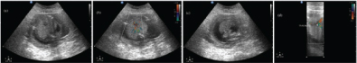
A 23-year-old primigravida came for routine obstetric malformation scan at 17 weeks of gestation to our hospital. Routine ultrasonography and colour Doppler was done which showed that the heart of the fetus was compressed by surrounding enlarged hyperechoic lungs, flattening of the diaphragm in the fetus, significant fetal ascites and severe Oligohydramnios in the patient [Table/Fig-1]. The mother had no significant family or personal history. Following this the case was diagnosed as CHAOS. Other differentials like cystic adenomatous malformation of the lung and Bronchopulmonary sequestration were ruled out. Associated abnormalities like digeorge and Fraser syndrome was evaluated for but was not found. A fetal echocardiogram was done which showed no signs of heart failure. Karyotyping was done which was normal. Following multiple discussions and counseling sessions with the patient and her husband elective termination of pregnancy was suggested. However, patient refused termination of pregnancy and on follow up it was found that the fetus was spontaneously aborted and expelled.
(a) Trans-abdominal antenatal ultrasound show bulky echogenic fetal lungs compressing the heart. Oligohydramnios and fetal ascites are also evident.
(b)Trans-abdominal antenatal ultrasound showing compressed heart and major fetal vessels between over expanded lungs. The heart is squeezed between the lungs and decrease in cardiothoracic circumference is noted. (c) Trans-abdominal antenatal ultrasound, at lower down section showing fetal ascites and oligohydramnios. (d) Trans-abdominal antenatal ultrasound in coronal orientation view showing dilated trachea with bulky echogenic lungs causing flattening of diaphragm. Dilated airways including trachea and bronchi are seen.
Case Report 2
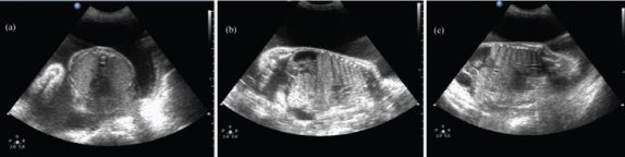
A 26-year-old primigravida came for routine obstetric malformation scan at 19 weeks of gestation to our hospital. Routine ultrasonography and colour Doppler was done which showed that the heart of the fetus was compressed by surrounding hyperplastic lungs, Inversion of the diaphragm of the fetus, significant ascites in the fetus and polyhydramnios in the mother [Table/Fig-2]. The mother had no significant family or personal history. Following this the case was diagnosed as CHAOS. Other differentials like cystic adenomatous malformation of the lung and Bronchopulmonary sequestration were ruled out. Associated abnormalities like digeorge and Fraser syndrome was evaluated for but was not found. A fetal echocardiogram was done which showed no signs of heart failure. Karyotyping was done which was normal. Following multiple discussions and counseling sessions with the patient and her husband elective termination of pregnancy was suggested and undertaken.
(a) Trans-abdominal antenatal ultrasound showing bulky echogenic fetal lungs compressing the heart. Oligohydramnios is not seen but mild polyhydramnios is seen. (b) Trans-abdominal antenatal ultrasound in coronal orientation view showing bulky echogenic lungs compressing the heart. Polyhydramnios is also present. Fetal diaphragm is inverted. (c) Trans-abdominal antenatal ultrasound in coronal orientation view showing dilated trachea with bulky echogenic lungs. Polyhydramnios is also evident.
Discussion
Congenital high airway obstruction syndrome is a very rare entity with very poor prognosis in which upper airway is intrinsically obstructed, the most common reason being laryngeal atresia [1].
The obstruction of upper airway leads to entrapment of fluids produced by the lungs as a result of which the lungs and the trachea are enlarged leading to flattening of the diaphragm which can be viewed on ultrasonography [2].
The term CHAOS was given by Hedrick MH et al., and when prenatal diagnosis of upper airway obstruction is diagnosed, it is very difficult to pin point the specific cause, thus giving rise to the broad term CHAOS [3]. Artunc Ulkumen B et al., reported two cases of CHAOS due to tracheal atresia diagnosed by antenatal ultrasonography and fetal MRI [4]. Another study pointed out the importance of MRI as an adjunctive role in demonstrating the level of obstruction which may not always be identified on ultrasound and in excluding extrinsic causes of obstruction [5]. Hamid-Sowinska A et al., pointed the importance of early diagnosis, detailed fetal assessment and an adequate postnatal intervention for establishing fetal airways in CHAOS [6]. CHAOS can be detected on routine ultrasonography due to secondary structural changes like lung enlargement etc. and some cases are detectable as early as 16 weeks [7].
The most common cause of CHAOS is usually laryngeal atresia. However other causes like laryngeal or tracheal webs, laryngeal cysts, tracheal atresia, subglottic stenosis or atresia and tracheal or laryngeal agenesis are also seen as possible causes of CHAOS [1].
Obstruction of the upper airway leads to accumulation of fluid in the tracheo – bronchial area leading to secondary pulmonary hyperplasia indicated by high echogenicity as compared to the kidneys on ultrasonography. Dilatation of the trachea and flattening of the diaphragm are associated findings. Because of enlargement of the lungs the heart appears small and squeezed in the middle of the thorax. Because of elevated intrathoracic pressure venous return is decreased eventually leading to heart failure which in turn leads to ascites, placentomegaly and hydrops fetalis [8]. Ascites is almost always associated with cases of CHAOS [3]. Ascites appears very early in CHAOS and is probably due to kinking of the aorta and inferior vena cava [9].
Sometimes pulmonary enlargement may resolve later in pregnancy indicating less severe course of the disease due to minor pharyngotracheal or laryngotracheal communications [10].
Early in pregnancy oligohydramnios may be present due to decreased amniotic volume because of obstruction. Oligohydramnios is also seen in Fraser syndrome (renal or ureteral agenesis, cryptophthalmous, syndactyly, ambiguous genitalia and laryngeal atresia). Later on in pregnancy polyhydramnios may develop because of oesophagus getting compressed and thus decreased fetal swallowing [1].
Other common fetal hyperechogenic lung lesions are congenital cystic adenomatoid malformation and pulmonary sequestration. To differentiate between CCAM, PS and CHAOS tracheal enlargement should be looked for as it is specific to CHAOS.
To determine the general level of obstruction Transvaginal sonography can be done where as some have used the breathing movements of the fetus as a way for finding out the level of obstruction. MRI is also beneficial but to determine the specific cause of obstruction is challenging [11].
Prenatal diagnosis of CHAOS is necessary so that perinatal management can be achieved. To create a surgical pathway a perinatal operation on placental support can be undertaken. Partial abdominal delivery of the head of the fetus with the fetus attached to the placenta followed by laryngoscopic examination and subsequent tracheostomy can be achieved. Only three cases have been reported [12]. De Cou JM et al., successfully used the EXIT ex utero intrapartum treatment procedure to treat a case of CHAOS in 1988 due to laryngeal atresia [13]. A percutaneous fetoscopic approach can also be undertaken which doesn’t affect the utero placental blood flow but premature rupture of membranes is still a complication [14]. Fetal laryngoscopy has been used in the past to achieve decompression of obstruction of larynx or trachea in CHAOS [11].
The prognosis of CHAOS is usually lethal because of the anomaly of the larynx by itself, or because of associated hydrops fetalis [8].
Conclusion
In summary prenatal early diagnosis of patients with CHAOS is necessary so that perinatal management can be undertaken successfully or elective termination of pregnancy can be undertaken. The fetoscopic approach may be a life saving modality in a subset of CHAOS patients. Involving a multidisciplinary team comprising of paediatricians, radiologists, obstetricians and anaesthesiologists increases the efficiency of management.
[1]. Vidaeff AC, Szmuk P, Mastrobattista JM, Rowe TF, Ghelber O, More or less CHAOS: case report and literature review suggesting the existence of a distinct subtype of congenital high airway obstruction syndromeUltrasound Obstet Gynecol 2007 30:114-17. [Google Scholar]
[2]. Gilboa Y, Achiron R, Katorza E, Bronshtein M, Early sonographic diagnosis of congenital high-airway obstruction syndromeUltrasound Obstet Gynecol 2009 33:730-34. [Google Scholar]
[3]. Hedrick MH, Ferro MM, Filly RA, Flake AW, Harrison MR, Adzick NS, Congenital high airway obstruction syndrome (CHAOS): a potential for perinatal interventionJ Pediatr Surg 1994 29:271-74. [Google Scholar]
[4]. Artunc Ulkumen B, Pala HG, Nese N, Tarhan S, Baytur Y, Prenatal Diagnosis of Congenital High Airway Obstruction Syndrome: Report of Two Cases and Brief Review of the LiteratureCase Reports in Obstetrics and Gynecology 2013 2013:728974 [Google Scholar]
[5]. Joshi P, Satija L, George R, Chatterjee S, D’Souza J, Raheem A, Congenital high airway obstruction syndrome—antenatal diagnosis of a rare case of airway obstruction using multimodality imagingMedical Journal, Armed Forces India 2012 68(1):78-80. [Google Scholar]
[6]. Hamid-Sowinska A, Ropacka-Lesiak M, Breborowicz GH, Congenital high airway obstruction syndromeNeuro Endocrinol Lett 2011 32(5):623-26. [Google Scholar]
[7]. Berg C, Geipel A, Germer U, Petersen-Hansen A, Koch- Dörfler M, Prenatal detection of Fraser syndrome without cryptophthalmos: case report and review of the literatureUltrasound Obstet Gynecol 2001 18:76-80. [Google Scholar]
[8]. Martínez JM, Castañón M, Gómez O, Prat J, Eixarch E, Bennasar M, Evaluation of Fetal Vocal Cords to Select Candidates for Successful Fetoscopic Treatment of Congenital High Airway Obstruction Syndrome: Preliminary Case SeriesFetal Diagn Ther 2013 34:77-84. [Google Scholar]
[9]. Tang PTM, Meagher SE, Kahan AA, Woodward CS, Laryngeal atresia: antenatal diagnosis in a twin pregnancyUltrasound Obstet Gynecol 1996 7:371-73. [Google Scholar]
[10]. Meagher SE, Fisk NM, Harvey JG, Watson GF, Boogert A, Disappearing lung echogenicity in fetal bronchopulmonary malformations: a reassuring sign?Prenat Diagn 1993 13:495-501. [Google Scholar]
[11]. Ruano R, Cass DL, Rieger M, Javadian P, Shamshirsaz AA, Olutoye OO, Fetal laryngoscopy to evaluate vocal folds in a fetus with congenital high airway obstruction syndrome (CHAOS)Ultrasound Obstet Gynecol 2014 43:102-05. [Google Scholar]
[12]. Oepkes D, Teunissen AKK, Van De Velde M, Devlieger H, Delaere P, Deprest J, Congenital high airway obstruction syndrome successfully managed with ex-utero intrapartum treatmentUltrasound Obstet Gynecol 2003 22:437-39. [Google Scholar]
[13]. De Cou JM, Jones DC, Jacobs HD, Touloukian RJ, Successful ex utero intrapartum treatment (EXIT) procedure for congenital high airway obstruction syndrome (CHAOS) owing to laryngeal atresiaJ Pediatr Surg 1998 33:1563-65. [Google Scholar]
[14]. Kohl T, Hering R, Bauriedel G, Van De Vondel P, Heep A, Keiner S, Fetoscopic and ultrasound-guided decompression of the fetal trachea in a human fetus with Fraser syndrome and congenital high airway obstruction syndrome (CHAOS) from laryngeal atresiaUltrasound Obstet Gynecol 2006 27:84-88. [Google Scholar]