The aesthetic principles have generated interest in guidelines and standards in designing the restorations [1,2]. The principles that delineate the aesthetics are subtle and they can be used with predictability [3]. The efforts have been made to establish standards and there exists little inconsistency in aesthetic perception [4]. Lack of directive makes it difficult to fabricate aesthetic restoration that is satisfying to both patients and clinicians [5].
The teeth within the confines of the gingival architecture have a tremendous impact on the aesthetics of the smile. Literature revealed greater number of the patients displayed gingiva during smile [6]. The aesthetic impact due to uneven gingival contour height can influence the axial inclination, emergence profile of the teeth affecting the smile aesthetics [7]. The height of the interdental papillae and its relationship to the gingival zenith is undefined. This location can be helpful in fabricating aesthetic restoration and smile designing.
The aim of this study was to evaluate the distance from the crest of gingival zenith position (HGZ) and tip of the interdental papilla (HIP) to the incisal edge of all maxillary anterior dentition and to determine for consistent ratio between the HIP and HGZ.
Materials and Methods
A random population of 100 volunteers reported to Department of Prosthodontics, SRM Dental College, Ramapuram, Chennai, over a period of six months with an average age group of 25 to 30 years were chosen for the study. A five member panel consisting of two prosthodontist, two restorative dentists and one periodontist was involved in selecting and standardisation of patient samples. The institutional ethical committee approved the study and informed consent was obtained from the participants.
Study participants: The subjects included in the study had all their natural teeth with no history of orthodontic treatment, no tooth size alterations, no spacing and no restorations. All subjects had class I molar and canine occlusion. The subject were excluded if they had periodontal diseases, gingival recession, hypertrophy, missing or altered interdental papilla, teeth spacing, crowding, restorations and attrition. Impacted, angulated, crowded dentitions were excluded from the study.
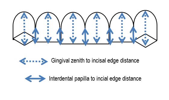
Procedure: Impressions were made with irreversible hydrocolloid impression (Algitex, DPI, India) material and the cast were made with type IV gypsum product. Face bow transfer was done on the subjects using Hanau spring face bow and the cast were oriented to the Hanau wide vue semi adjustable articulator. The mandibular cast was mounted to the articulator using the static centric relation record. The cast transfer to semi-adjustable articulator aided in exact positioning of the incisal edge position and the occlusal plane. This procedure reduced the errors in indirect evaluation of the casts. Digital caliper, (Series: ECO6, Japan) with minimum scale of 0.01mm was used. The tip of caliper was modified to sharper point for more accurate results. On the mounted study cast the long axis was drawn with a pencil from the gingival zenith point to the occlusal plane of all maxillary anterior dentition [Table/Fig-1]. Using the digital vernier caliper the measurement from gingival zenith to incisal edges and the interdental papilla tip to incisal edge distance was measured using 2.5X optical loupes for accurate visualisation. Control measurements were also performed by a second investigator. The distance was measured thrice and the mean of it was used for analysis. The caliper was calibrated prior to each measurement and the procedure was done on all six maxillary teeth.
Schematic representation of measurements.
Statistical Analysis
The data obtained was tabulated and analysed statistically using SPSS statistical package version 15. Descriptive statistics were calculated for the HIP and HGZ in terms of frequency. Chi-square analysis was used to determine the existence of proportion between the maxillary teeth and association between different quadrants. Alpha error was set at 5% and p-value less than 0.05 was considered statistically significant.
Results
The mean of HIP for the various maxillary anterior teeth were 5.6mm, 5.16mm, and 4.46mm for central incisor, lateral incisor and canine respectively. The mean of HGZ of central incisor was 9.9mm; lateral incisor 8.4mm, canine was 8.7mm [Table/Fig-2].
Independent samples T-Test to compare the mean values between gingival zenith and the interdental papillae.
| Variables | Measure to Incisal Edge | N | 1st Quadrant | 2nd Quadrant | p-value |
|---|
| Mean | Std. Dev | Mean | Std. Dev |
|---|
| Central Incisor | Gingival Zenith | 100 | 9.99 | 1.022 | 9.96 | 0.550 | <0.001 |
| Tip of Inter Dental Papillae | 100 | 5.66 | 0.833 | 5.63 | 0.965 |
| Lateral Incisor | Gingival Zenith | 100 | 8.46 | 1.010 | 8.46 | 1.352 | <0.001 |
| Tip of Inter Dental Papillae | 100 | 5.16 | 1.002 | 5.13 | 1.057 |
| Canine | Gingival Zenith | 100 | 8.71 | 1.257 | 8.42 | 1.496 | <0.001 |
| Tip of Inter Dental Papillae | 100 | 4.46 | 0.939 | 4.45 | 0.933 |
The mean ratio for central incisor was 1.8, for lateral incisor was 1.7 and 2.03 for canine. The inverse ratio between interdental papilla and gingival zenith was 0.5 for central incisor, 0.6 for lateral incisor and 0.5 for canine [Table/Fig-3]. The p-value was > 0.9 and there was no statistically significance between 1st and 2nd maxillary anterior quadrants.
Independent samples T-Test to compare the mean values of interdental papillae and gingival zenith between quadrants. Foot note:
| Variables | Side | N | Mean | Std. Dev | p-value |
|---|
| Central Incisor (GZ/TIDP) | Right Side | 100 | 1.80 | 0.288 | 0.367 |
| Left Side | 100 | 1.86 | 0.234 |
| Lateral Incisor (GZ/TIDP) | Right Side | 100 | 1.71 | 0.420 | 0.893 |
| Left Side | 100 | 1.70 | 0.356 |
| Canine (GZ/TIDP) | Right Side | 100 | 2.03 | 0.484 | 0.297 |
| Left Side | 100 | 2.10 | 0.486 |
| Central Incisor (TIDP/GZ) | Right Side | 100 | 0.57 | 0.101 | 0.604 |
| Left Side | 100 | 0.58 | 0.094 |
| Lateral Incisor (TIDP/GZ) | Right Side | 100 | 0.62 | 0.148 | 0.799 |
| Left Side | 100 | 0.61 | 0.131 |
| Canine (TIDP/GZ) | Right Side | 100 | 0.52 | 0.130 | 0.309 |
| Left Side | 100 | 0.50 | 0.128 |
GZ: Gingival Zenith
TIDP: Tip of Interdental Papilla
Right side: 1st Quadrant
Left side: 2nd Quadrant
Discussion
The dental and gingival aesthetics play important role in prosthodontic restorations [5]. Several studies have been published on the significance of interdental papilla and gingival zenith [6,7]. However, despite the imperative influence of interdental papilla and gingival zenith, small impact is provided on the proportions. This study evaluated for the existence of proportions between the interdental papilla and gingival zenith.
The study was done on 100 Indian patients of equal gender representation having healthy periodontal status for more idealistic evaluation. The impression was made with irreversible hydrocolloid material (Alginate, Algitex, DPI, India). Alginate impression material can record the best of surface details and economic advantage over other elastomeric impression materials [6,8]. Digital caliper was the simplest instrument in measuring the distances and the reading could be easily visualised from the display screen compared to the conventional calipers. The measurement for the study was made thrice with digital caliper and the average reading was considered to avoid inter operator errors [8].
The result of the study determined that the mean height from the gingival zenith to the incisal edge was 9.99 mm, 8.46 mm, 8.71mm for central, lateral incisors and canine on 1st quadrant and 9.96 mm, 8.46 mm, 8.42 mm similarly, on 2nd quadrant. Though, there are minor numerical differences, there was no significant statistical difference between the two quadrants. The results demonstrated no definitive proportion and the measurements were in accordance with previous studies [9,10] inferring central incisors displayed a distal GZP and no major differences existed between bilateral quadrants [11].
The mean height from the tip of the interdental papilla to the incisal edge were statistically significant and in accordance with previous studies on the height and symmetry of interdental papilla [9–13].
The mean ratios between the gingival zenith and the tip of interdental papilla to the incisal edge as listed in [Table/Fig-2] for 1st and 2nd quadrant were statistically significant with no major differences existing between bilateral quadrants. The ratio of gingival zenith and Interdental papilla to the incisal edges of maxillary anterior dentition was not in accordance with any proportions alike golden proportion, recurrent aesthetic dental proportion [14]. The study revealed that there was no definitive proportion between gingival zenith and Interdental papilla in the anterior teeth. The inverse ratio between gingival zenith and interdental papilla was not definitive and proved to have no proportion between the 1st and 2nd maxillary quadrants.
Limitations
The limitations of the study was possible occurrence of minor dimensional changes of the impression material, cast or on the evaluation data of the patient in measuring with occlusal plane. Care was taken to ensure and standardize the methodology to eliminate all operator and methodology bias during the course of the entire study. Further studies are required to determine the existence of similar proportion between mandibular anterior teeth and posterior teeth.
Conclusion
Within the limitations of the study it was concluded that the mean gingival zenith-incisal edge distance of the study population for Central incisor is 9.99mm, lateral incisor is 8.46mm and canine is 8.71mm. The mean papilla occlusal plane distance of the study population for central incisor is 5.66mm, Lateral incisor is 5.16mm and canine is 4.46mm.
The mean ratio between GZ and the tip of the interdental papillae to the incisal edges of all maxillary anteriors in 1st and 2nd quadrant is 1.80mm, 1.71mm, 2.03mm in central incisor, lateral incisor and canine respectively. No proportional ratio existed between gingival zenith and interdental papilla to the incisal edges of maxillary anterior in 1st and 2nd quadrants.
GZ: Gingival Zenith
TIDP: Tip of Interdental Papilla
Right side: 1st Quadrant
Left side: 2nd Quadrant