Iatrogenic Pseudo-aneurysm of Profunda Femoris Artery Following Fixation of Intertrochanteric Femur Fracture – A Case Report and Review of Literature
Kunal Dwijen Roy1, Rishi Anil Aggarwal2, Shaligram Purohit3, Gokul Bandagi4, Nandan Marathe5
1 Senior Registrar, Department of Orthopaedics, Seth GS Medical College and KEM Hospital, Mumbai, Maharashtra, India.
2 Senior Registrar, Department of Orthopaedics, Seth GS Medical College and KEM Hospital, Mumbai, Maharashtra, India.
3 Assistant Professor, Department of Orthopaedics, Seth GS Medical College and KEM Hospital, Mumbai, Maharashtra, India.
4 Final year Resident, Department of Orthopaedics, Seth GS Medical College and KEM Hospital, Mumbai, Maharashtra, India.
5 House Surgeon, Department of Orthopaedics, Seth GS Medical College and KEM Hospital, Mumbai, Maharashtra, India.
NAME, ADDRESS, E-MAIL ID OF THE CORRESPONDING AUTHOR: Dr. Kunal Dwijen Roy, 1, Rajiv Nagar, Roy Nursing home, Wardha Road, Nagpur - 440025, Maharashtra, India.
E-mail: kunaldroy@gmail.com
The Profunda Femoris is a common site for arterial pseudo-aneurysms and these have been described in literature following fractures as well as orthopaedic procedures of the femur and hip region. These are an uncommon complication and a high index of suspicion is required for correct diagnosis and prompt management. We present a case of pseudo-aneurysm of the Profunda Femoris in an operated case of Intertrochanteric femur fracture with acute presentation. The case was managed successfully by angiographic coil embolization.
Angiographic coil embolization, Angiography, Anaemia
Case Report
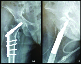
A 55-year-old female presented with a right-sided intertrochanteric femur fracture following a fall while walking. The patient was posted for fixation with dynamic hip screw and barrel plate [Table/Fig-1] on third day following trauma. While drilling hole for the second shaft screw, there was an inadvertent over-drilling of the medial cortex followed by immediate sudden bleeding from the medial soft tissues. Packing was done with sterile mops and the bleeding stopped spontaneously. Further procedure was uneventful. Postoperative vitals were stable. On day four postoperatively, sudden bleeding along suture line was noted with appearance of thigh swelling and pain. Blood tests revealed severe anaemia with a fall in haemoglobin from 10.2mg/dl preoperatively to 5.9mg/dl on the fourth Postoperative day. Local ultrasonography was performed which revealed a pseudo-aneurysm arising from Profunda Femoris Artery (PFA). Angiography confirmed the diagnosis of pseudo-aneurysm and the PFA was embolized using 2 coil wires in the same setting [Table/Fig-2]. The patient was given three pints of packed cells and closely monitored. At three weeks the patient showed no signs of active bleeding or distal vascular deficit with eventual uneventful recovery.
Four part Intertrochanteric femur fracture fixed with 135 DHS and fixed angle barrel plate. 1a: Postoperative AP view. 1b: Postoperative lateral view.
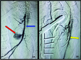
(a) Pre-embolization image showing a pseudoaneurysm arising from the Profunda Femoris artery. (Red arrow showing the Pseudoaneurysm & Blue arrow showing the Profunda Femoris) (b) Post-embolization image showing embolization of pseudoaneurysm using a coil (Yellow arrow showing the coil). The bleeding from pseudoaneurysm has stopped.
Discussion
Occurrence of pseudo-aneurysms of the proximal femoral artery after orthopaedic procedures in the peri-trochanteric region and the proximal femur is rarely described in literature. Many such cases might be sub-clinical and therefore might be missed. Diagnosis of this condition following either trauma or orthopaedic procedures needs high degree of clinical suspicion as well as sound awareness about the condition.
The triad of swelling, bleeding from the incision site and anaemia or decreasing trend of haemoglobin has been classically described to be associated with the occurrence of pseudo-aneurysms especially of the profunda femoris and it’s branches [1]. Our patient showed postoperative soakage and high operative site daily drain output pointing clinically towards the possibility of a pseudo-aneurysm.
The location of profunda femoris is such that it is protected by the vastus medialis from external trauma. It is vulnerable to injury in the subtrochanteric region [2]. Pseudo-aneurysms have been described by various authors to have occurred following blunt and penetrating trauma to the thigh and also following various orthopaedic procedures. Various procedures described in literature associated with the occurrence of pseudo-aneurysm are external fixation of femur, core decompression and internal fixation of the proximal femoral fractures. The mechanism of occurrence of pseudo-aneurysm is usually due to a spike of fractured bone, protruding cortical screw tip, Gamma-nail or over-penetration by a drill-bit leading to injury and disruption of the arterial wall [3]. Due to the force of the arterial blood flow the tissues of the damaged arteries get dissected and a sac or pseudo-aneurysm gets formed which has a luminal connection to the artery. This sac is surrounded by soft tissue structures present around the vessel or more often the media or adventitial layer of the vessel itself [4].
Pseudo-aneurysm may present early as in our case but even delayed presentation up to several weeks or years has been described. Chong C et al., described the occurrence of pseudo-aneurysm following traumatic proximal femur fracture and external fixation of femur which was managed by embolization and wire coiling of the false aneurysm [1]. A similar case was reported by Canbaz S et al., but this case was managed by ligation and excision of the aneurysmal sac via a medial approach [5]. Yang KH has reported a case of pseudo-aneurysm of the superficial femoral artery following closed nailing of a femur fracture possibly due to excess adduction and internal rotation of the limb [6]. Smejkal K et al., have described another case of pseudo-aneurysm of the profunda femoris following initial external fixation and subsequent un-reamed nailing of a femur fracture that was eventually managed by embolization [3]. Occurrence of pseudo-aneurysms has also been reported following core decompression of femoral head in separate instances by Lazarides MK et al., and Unay K et al., [7,8]. A case of pseudo-aneurysm of the profunda femoris has also been reported after total hip arthroplasty by Nozawa M et al., [9]. Patelis N, Cowley A, Entwistle JJ and Chandrasen J have individually reported the occurrence of pseudo-aneurysms after DHS fixation in cases of inter-trochanteric femur fracture but each due to different causes [10–13]. A strong clinical index of suspicion supported by appropriate radiological imaging such as ultra-sonography, computed tomography (CT) and angiography plays a major role in reaching a diagnosis. Experience and knowledge regarding treatment of pseudo-aneurysms of PFA is still limited. Patients with asymptomatic small (2-3 cm) pseudo-aneurysms may be observed as many will thrombose and obliterate spontaneously [5]. Treatment options for larger and symptomatic ones includes open surgical repair, ultrasound-guided compression, ultrasound-guided thrombin injection and endovascular repair using coil embolization or stent- graft insertion. The standard approach for most procedures of profunda femoris reconstruction is the medial approach to whole trunk. Alternatively the lateral approach to the profunda femoris as described by Naraynsingh V et al., may also be used with the advantage of better exposure up to the origin of the vessel [14]. These approaches are used for open surgical repair and/or ligation of the pseudo-aneurysm. Recent avenues of treatment of pseudo-aneurysm include embolization with coils or the use of endovascular stents. However selective embolization has been increasingly considered as the most effective treatment for such cases. The profunda femoris artery usually has a well-developed collateral supply, therefore, it is pertinent to embolize both proximal and distal to the pseudo-aneurysm to completely exclude it from the circulation by preventing backflow from the collateral circulation.
[Table/Fig-3] gives a comprehensive overview of cases reported in literature of pseudo-aneurysms of the profunda femoris artery related to orthopaedic procedures.
Review of Iatrogenic Pseudo-aneurysms of Profunda Femoris associated with Orthopaedic procedures.
| Sr. No. | Author | No. of cases | Initial diagnosis | Procedure performed | Cause of aneurysm | No. of days post-op/ post-trauma | Presentation of pseudo-aneurysm | Treatment |
|---|
| 1. | Nozawa M [9] | 1 | Osteoarthritis Hip | Total Hip Arthroplasty | Bone Spur/Acetabular reaming | 1 month | Thigh swelling and massive bleeding | Angiographic embolization using Tornade coils |
| 2. | Patelis N [10] | 1 case | Intertrochanteric femur fracture | Dynamic hip screw fixation | Protruding shaft screw tip | 2 months | Slow hemorrhage with gradually growing thigh swelling | Angiographic coil embolization |
| 3. | Hanna GB [2] | 2 case | A) Intertrochanteric femur fracture | Pugh nail | -------- | 1 week | Swelling with active bleeding | Angiographic coil embolization |
| B) Open comminuted femur fracture | AO intramedullary nail | Drill or Lower proximal locking screw | 2 weeks | Pulsatile swelling with audible bruit | Repair of the arterial defect |
| 4. | Cowley A [11] | 1 case | Intertrochanteric femur fracture | Dynamic hip screw fixation | Displaced fragment of lesser trochanter | 6 weeks | Drop in hemoglobin and bleeding | Angiographic coil embolization |
| 5. | Canbaz S [5] | 1 case | Open femur fracture | External fixation | Fracture fragment | --- | Thigh swelling, hemorrhage and fall in Hb | Surgical ligation of profunda femoris and excision of sac |
| 6. | Entwisle JJ [12] | 2 cases | A) Intertrochanteric femur fracture | Dynamic Hip Screw fixation | Drill bit/ Shaft screw | 24 hours | Painful and swollen Thigh | Angiographic coil embolization |
| B) Intertrochanteric femur fracture | Dynamic Hip Screw fixation | Drill bit / Shaft screw | Day 5 | Thigh swelling with fall in Hemoglobin | Angiographic coil embolization |
| 7. | Chong KC [1] | 2 cases | A) Open femur fracture | External fixation with fasciotomy | Not specified | 2 weeks | Tense and thigh swelling with fresh bleeding and fall in Hbhypotension(unstable) | Angiographic embolization |
| B) Closed comminuted femur fracture | Intramedullary nailing | Not specified | Day 13 | Thigh swelling with fresh bleeding and fall in Hb | Angiographic embolization |
| 8. | Unay K et al., [8] | 2 cases | A) Avascular necrosis | Core decompression | Deep insertion of lancet | Day 4 | Thigh swelling with fresh bleeding and rapid fall in Hb | Angiographic embolization with coil |
| B) Subtrochanteric Femur fracture | Closed Gamma nailing | Bone spike at fracture site | Day 7 | Profuse bleeding with thigh swelling and fall in Hb | Angiographice embolization with N-butyl-2-cyanoacrylate |
| 9. | Smejkal K et al., [3] | 1 case | Open Femurfrature | External fixation | Bone spike/ Fixator pin | 6 weeks | Long standing thigh swelling | Angiographic embolization |
| 10. | Chandrasen J [13] | 1 case | Intertrochanteric femur fracture | DHS fixation | Not specified | Day 9 | Pulsatile Swelling and fall in Hb | Angiographic embolization |
| 11. | Dillon JP [15] | 1 case | Intertrochanteric femur fracture | DHS fixation | Not specified | 7 months | Pulsatile thigh swelling and pain | Open surgical repair (failed embolization) |
Conclusion
Pseudo-aneurysms of the Profunda femoris artery following orthopaedic procedures are a rare occurrence. It is important to note that these patients usually present late and the presence of a pulsatile mass and distal ischemia is not always present. A high index of suspicion is required to correctly diagnose the condition. Ultrasonographic evaluation will usually establish diagnosis and an angiographic intervention should be considered early to reduce the morbidity associated with this condition.
[1]. Chong KC, Yap EC, Lam KS, Low BY, Profunda femoris artery pseudoaneurysm presenting with triad of thigh swelling, bleeding and anaemiaAnnals of the Academy of Medicine Singapore 2004 33(2):267-69. [Google Scholar]
[2]. Hanna GB, Holdsworth RJ, McCollum PT, Profunda femoris artery pseudoaneurysm following orthopaedic proceduresInjury 1994 25:477-79. [Google Scholar]
[3]. Smejkal K, Zva’k I, Trlica J, Raupach J, Neumann J, Traumatic pseudoaneurysm of arteria femoralis profunda— the case reportRozhledy v Chirurgii 2007 86(3):116-19. [Google Scholar]
[4]. Khoshnevis J, Sobhiyeh MR, Zavareh MF, Deep femoral artery branch pseudoaneurysm after orthopedic procedure requiring surgical treatment: a case reportTrauma Mon 2012 17(2):305-08. [Google Scholar]
[5]. Canbaz S, Acipayam M, Gurbuz H, Duran E, False aneurysm of perforating branch of the profunda femoris artery after external fixation for a complicated femur fractureJ Cardiovasc Surg 2002 43(4):519-22. [Google Scholar]
[6]. Yang KH, Park HW, Park SJ, Pseudoaneurysm of the superficial femoral artery after closed hip nailing with a Gamma nail: report of a caseJ Orthop Trauma 2002 16:124-27. [Google Scholar]
[7]. Lazarides MK, Arvanitis DP, Dayantas JN, Iatrogenic arterial trauma associated with hip joint surgery: an overviewEuropean Journal of Vascular Surgery 1991 5(5):549-56. [Google Scholar]
[8]. Unay K, Poyanli O, Akan K, Poyanli A, Profunda femoris artery pseudoaneurysm after surgery and traumaStrategies Trauma Limb Reconstr 2008 3:127-29. [Google Scholar]
[9]. Nozawa M, Irimoto M, Maezawa K, Hirose T, Shitoto K, Kurosawa H, False aneurysm of the profunda femoris artery after total hip arthroplastyJ Arthroplasty 2000 15:671-74. [Google Scholar]
[10]. Patelis N, Koutsoumpelis A, Papoutsis K, Kouvelos G, Vergadis C, Mourikis A, Iatrogenic injury of profunda femoris artery branches after intertro- chanteric hip screw fixation for intertrochanteric femoral fracture: a case report and literature reviewCase Rep Vasc Med 2014 2014:694235 [Google Scholar]
[11]. Cowley A, Williams D, Butler M, Edwards A, Parsons S, Pseudo-aneurysm of the profunda femoris artery as a late complication of hip fracture in a patient with myelodysplasiaAnn R Coll Surg Engl 2007 89:W4-W6. [Google Scholar]
[12]. Entwisle JJ, DeNunzio M, Hinwood D, Transcatheter emboli- zation of pseudoaneurysm of the profunda femoris artery complicating fracture of the femoral neckClin Radiol 2001 56:424-27. [Google Scholar]
[13]. Chandrasenan J, Garner JP, Meiring PD, Kumar K, Coil embolisation of an iatrogenic profunda femoris pseudoaneurysmInjury Extra 2006 37(7):249-52. [Google Scholar]
[14]. Naraynsingh V, Karmody AM, Leather RP, Corson JD, Lateral approach to the profunda femoris arteryAm J Surg 1984 147:813-14. [Google Scholar]
[15]. Dillon JP, O’Brien GC, Laing AJ, Adelowokan T, Dolan M, Pseudoaneurysm of the profunda femoris artery following an inter-trochanteric fracture of the femurInjury Extra 2004 35(3):30-2. [Google Scholar]