Duplication of inferior vena cava is an uncommon abnormality and is important in daily today practice for vascular surgeons, radiologist and urologist especially during retroperitoneal surgeries and treatment of thromboembolic disease. Radiologically, Duplicated IVC can be mistaken for lymphadenopathy or left pyeloureteric dilatation. Crossed fused kidney with a single ureter defy the embryological theory of ureteric bud crossing the opposite side and induce nephron formation associated anomaly of Duplication of inferior vena cava and malrotation of gut are not reported in a same patient. On meticulous search of literature no such combination of abnormalities has been reported. In this case report we bring forward this rare type of combination of three congenital malformations that is Duplication of IVC, crossed fused kidney and malrotation of gut.
Nulliparous, Ovarian cyst, Pain abdomen, Pyosalphix
Case Report
A 22-year-old nulliparous female patient visited the Outpatient Department of Krishna Institute of Medical Sciences, Karad with complain of Intermittent pain abdomen since 5 months which increased since last 5 days. Patient had history of fever for seven days, intermittent, low grade type, 1 month back. She had no bladder or bowel complaints. On clinical examination patient had tenderness in hypogastic region.
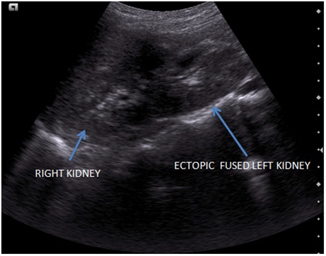
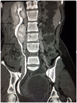
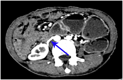
On investgations: Haemoglobin was 12gm/dl. Total leucocytes counts were raised i.e. 18,000. The renal function test was within normal limits. Ultrasonogram of abdomen was done which showed empty left renal fossa and left kidney was present on the right side, below the right kidney [Table/Fig-1] with a tubular cystic lesion and heteroechoic content in left adnexa seen separately from left ovary likely suggestive of pyosalphix. On contrast enhanced CT scan, a well defined peripherally enhancing tubular lesion was observed in left adnexa, likely to be Left Pyosalphinx, along with a cystic lesion not separated from the left ovary, likely to left ovarian serous cystadenoma. Also observed was, double Inferior vena cava each on either side of aorta with a preaortic segment crossing anterior to aorta at level of D12 vertebrae [Table/Fig-2,3 and 4].
Ultrasonogram showing right kidney and cross fused ectopic left kidney.
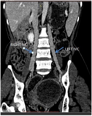
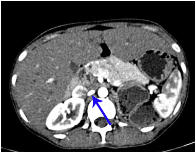
Coronal CECT image showing Duplication of Inferior vena cava (Right and Left IVC).
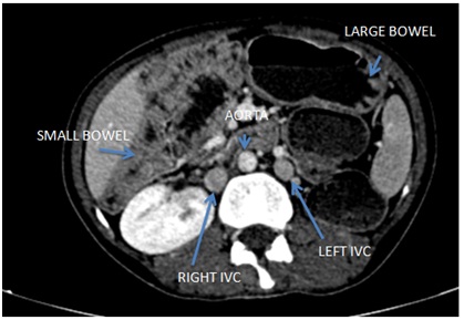
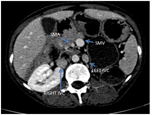
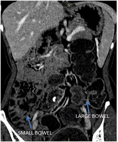
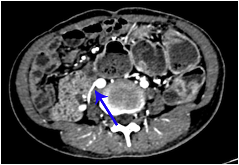
Axial CECT image showing Right and left inferior vena cava. Left IVC appears smaller than right IVC. Small bowel loops are on right side and large bowel loop on left side.
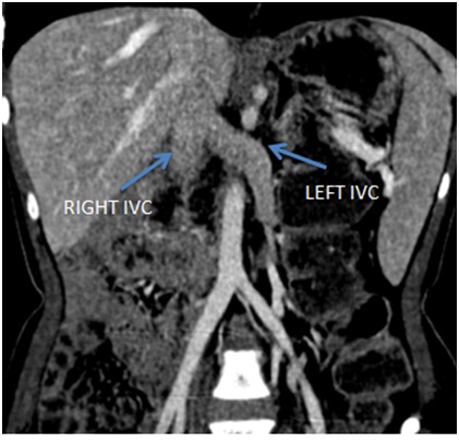
Coronal CT image showing Left IVC merges with right IVC at level of D12 vertebrae.
Left renal fossa was empty with abnormal rotation of left kidney and fusion to right kidney at midpole level likely suggestive of Crossed fused ectopia. Single ureter was noted on right side with normal excretion of contrast [Table/Fig-5,6 and 7].
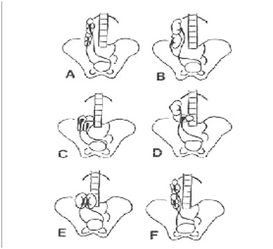
Types of crossed renal ectopia: a) Superior ectopia; b) Sigmoid shaped or S shaped ectopia; c) Lump kidney; d) L shaped kidney; e) Disk kidney; f) Inferior ectopia. Image taken from Crosed Fused Renal Ectopia with a Single Ureter.
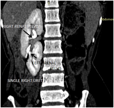
Coronal Reformat CECT image showing Right and left fused kidneys with Single ureter.
Coronal CECT image showing Fused right and left kidney with renal pelvis with single ureter.
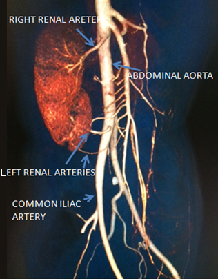
On CT angiogram: Right renal artery was seen arising from abdominal aorta and two left renal arteries arising from right common iliac artery [Table/Fig-8,9].
CT angiogram showing the right kidney being supplied by right renal artery and ectopic left kidney by 2 renal arteries arising from right common iliac artery.
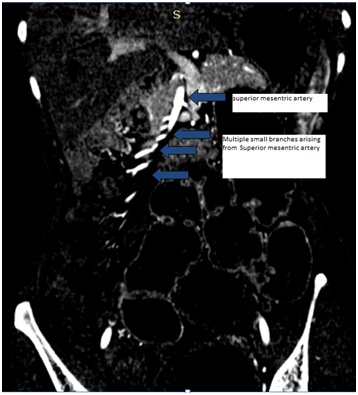
Multiple branches are seen entering the right kidney. These are the small arteries arising from superior mesenteric artery and are projected in front of kidney and appear to be entering the right kidney.
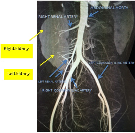
CT angiogram (VRT images): Right renal artery is arising from abdominal aorta and left renal arteries are arising from right common iliac artery.
Inverted relationship was observed between superior mesenteric vein (SMV) and artery (SMA) – the vein was lying to the left of the artery and small bowel was present almost entirely on the right side, the duodenojejunal junction was right-sided and the third and fourth duodenal portion did not cross the midline [Table/Fig-10,11].
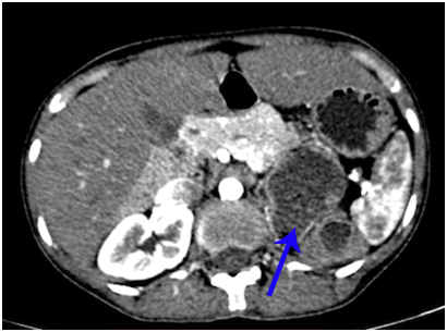
CECT axial images showing inversion of SMA- SMV relation (Superior mesenteric vein is ventral and to the left of superior mesenteric artery).
Coronal CECT image: Most of small bowel loops are on right side and large bowel loop on left side.
Patient was then referred to Surgery department for further management.
Discussion
Duplication of the IVC occurs because the left supracardinal vein fails to regress early in gestation resulting in large veins on both sides of the aorta that usually joins anterior to the level of the renal arteries to become the suprarenal IVC [1].
Three different variants of duplication of inferior vena cava are known i.e. Type I or Major duplication: Two bilaterally symmetrical trunks with a preaortic trunk of the same calibre. Type II or minor duplication with two bilaterally symmetrical trunks, but it is smaller than the preaortic trunk. In Type III or asymmetric duplication there is small left IVC, larger right IVC and even larger preaortic trunk. Genitourinary anomalies associated with Duplication of the IVC are Retro-aortic left renal vein, circum-aortic renal vein known as ‘venous collar’, cloacal exstrophy and horseshoe kidney [2]. In our patient right Inferior vena cava is larger in diameter compared to left inferior vena cava i.e. Type III [2]. Left inferior venacava merge with right at level of D12 vertebrae [Table/Fig-1,2 and 3].
Ayhan Coskun et al., reported a case of uterus didelphys with obstructed unilateral vagina and including ipsilateral renal agenesis, duplication of inferior vena cava, high-riding aortic bifurcation and intestinal malrotation [3].
Incidence rate of crossed fused kidney is 1:1000 with Male:Female – 2:1. Left to right ectopy is common then right to left, various theories are postulated to explain crossed fused ectopia i.e. abnormally situated umbilical artery prevents normal cephalic mirgration and developing kidney takes path of least resistance & crosses opposite side and cephalic migration occurs. According to another theory, cross fused anomaly occurs when the ureteral bud crosses the opposite side and induce nephron formation in contralateral metanephricblastema [4].
This case challenges the embryological theory that deviation of one of the ureteric buds to the opposite side results in crossed fused renal ectopia.
Six variations of crossed fusion Kidneys have been described (McDonald and McClellan classification) [Table/Fig-5] [5]. These are: type 1 – Superior crossed fused ectopia, type 2 - Sigmoid or S-shaped kidney, type 3 - unilateral lump kidney, type 4 –Lshaped kidney, type 5 –unilateral disk kidney and type 6 - Inferior crossed fused ectopia. According to this classification, the distinct feature is that ureters are present on either side of the urinary bladder. In our case of cross fused ectopia, there is a single ureter present which is draining both the kidneys and after comprehensive research we found that no such case is reported in the literature.
Mani et al., reported 2 cases with duplication of inferior vena cava is associated with polycystic kidney disease and congenital pelviureteric obstruction [6]. But in our case Duplication of inferior vena cava is associated with Crossed fused kidney with single ureter. Kaur et al., repoted a case of crossed fused renal ectopia with fused kidneys were present on the right side with a single ureter opening into the right side of the bladder [7]. These findings are similar to our case but there was no associated duplication of inferior vena and malrotation of gut noted.
According to Kulkarni et al., in an anatomic dissection the caecum, vermiform appendix, and ascending colon were found on the left side of the abdominal cavity along side of the descending colon [8]. The left renal fossa was found empty with crossed fused ectopic kidney and the kidneys were present as a lobulated masss near pelvic brim on right side and hila facing anteromedially.
In the present case left renal fossa was empty and the left kidney was present below the right kidney in the right lumbar region, and appeared to be fused with the right kidney [Table/Fig-1]. The right kidney was supplied by the right renal artery which was arising from the aorta and arterial supply for the ectopically placed inferior kidney (left kidney) was by two small branches from right common iliac artery [Table/Fig-8,9]. The crossed fused renal ectopia in this case was unusual as there was a single ureter [Table/Fig-5,6 and 7].
Various research works done on the duplication of inferior vena cava and crossed fused ectopia were done [Table/Fig-12].
Various similar research work done on the duplication of inferior vena cava and crossed fused ectopia.
| Author’s name | Clinical presentation | Imaging |
|---|
| Benoy Starly [2] | A 51-year-old lady presented with complaints of pedal oedema for 2 weeks. | Type III asymmetric duplication of the IVC. |
| Ayhan Coskun [3] | A 16-year-old patient with progressive dysmenorrhea and pelvic pain that had begun within the first years after menarche, and persistent vaginal spotting after menstruation. | Uterus didephys with obstructed unilateral vagina and including ipsilateral renal agenesis, duplication of inferior vena cava, high-riding aortic bifurcation, and intestinal malrotation. |
| Mani et al., [6] | 1. A 14-year-old boy who had right-sided congenital ureteropelvic junction obstruction with hydronephrosis.2. A 10-year-old boy, who was diagnosed to have polycystic disease of the kidney (autosomal recessive type). | 1. Small right kidney, which was hydronephrotic with a grossly dilated pelvis with duplication of IVC.2. Enlarged kidneys with multiple cysts in kidneys and liver with duplication of inferior vena cava. |
| Kaur et al., [7] | A 22-year-old female presented with intermittent episodes of colicky pain in the right flank for eight months. | Crossed fused renal ectopia with fused kidneys on the right side with a single ureter opening into the right side of the bladder. |
| Milani et al., [10] | A 23-year-old female patient with complain of acute shortness of breath, abdominal pain, and severe left lower extremity pain. | Thrombosis of left-sided iliac veins and left duplicated Inferior vena cava. |
| Anees Dudekula et al., [11] | A 49-year-old male patient presented with complaints of recurrent right loin pain since 3 year. | Mild Right hydronephrosis.with dilated upper ureter with retrocaval course of right ureter at the L3-L4. intervertebral level with Duplication of inferior vena cava. |
| Present case | A 22-year-old nulliparous female patient with complain of intermittent pain abdomen since 5 months which increased since last 5 days. | Pyosalphinx with left ovarian cystic lesion. Duplication of Inferior vena cava. with Crossed fused ectopia with single ureter and Malrotation of gut. |
Any deviation from the normal 270°counterclockwise rotation of the midgut during embryologic development is called Intestinal malrotation [9]. It can cause intermittent episodes of gastrointestinal obstruction or a midgut volvulus. Deviation from the normal relationship between the Superior Mesenteric Artery (SMA) and Superior Mesenteric Vein (SMV) is a useful indicator of malrotation. In addition to multiple spleens, other abdominal findings may include a left-sided inferior venacava, azygos or hemiazygos continuation of an interrupted inferior vena cava, a preduodenal portal vein, and a truncated appearance to the pancreas
In our case there is inversion of SMA – SMV relation and Superior mesenteric vein lies ventrally and to the left of Superior mesenteric artery [Table/Fig-10]. Duodenum is not crossing the lumbar spine and most of small bowel lies on right side and large bowel on the left side [Table/Fig-11].
More CT images are shown in [Table/Fig-13,14,15,16 and 17] to ascertain the crossed fused kidneys with a single ureter.
CECT axial image arterial phase: At level of right renal artery (arrow), no left renal artery at level of right renal artery.
CECT axial image arterial phase: First left renal artery arising from right common iliac artery (arrow).
CECT axial image arterial phase: Second left renal artery arising from right common iliac artery (arrow).
CECT axial arterial phase: Empty left renal fossa.
Coronal CECT (Arterial phase) showing Superior mesenteric arteries and multiple small arteries arising from it.
To the best of our knowledge, this is the first case that has all of the forementioned anomalies in a same patient.
Conclusion
Duplication of inferior vena cava could be due to incidental finding and helps the surgeons and radiologists in their diagnosis and planning of surgeries like portosystemic shunts, abdominal aortic aneurysm repair, ligation of IVC in thromboembolic disease, placement of IVC filter and preventing postoperative complications. Urine flow or renal vascular complications may occur in cases of ectopic kidney and are associated with malignancy like Renal cell carcinoma and Transitional cell carcinoma. In addition, the ectopic kidney is vulnerable to traumas because of its position. Irrespective of age of the patient, surgical treatment of quiescent malrotation should be considered because surgery remains the only real safeguard against complications. These anatomical and embryological variations could be incidental findings on routine investigation but need thorough search for other associated anomaly and complications.
[1]. Kandpal H, Sharma R, Gamangatti S, Srivastava DN, Vashisht S, Imaging the Inferior Vena Cava: A Road Less TraveledRadioGraphics 2008 28:669-89. [Google Scholar]
[2]. Starly B, A rare vascular anomaly - Type III asymmetric duplicated inferior vena cava; Case 12098European society of radiology 2014 [Google Scholar]
[3]. Coskun A, Okur N, Ozdemir O, Kiran G, Arykan DC, Uterus didelphys with an obstructed unilateral vagina by a transverse vaginal septum associated with ipsilateral renal agenesis, duplication of inferior vena cava, high-riding aortic bifurcation, and intestinal malrotation: a case reportFertil Steril 2008 90(5):2006.e9-11. [Google Scholar]
[4]. Textbook of uroradiology 4th edition, N. Reed Dumick [Google Scholar]
[5]. Türkvatan A, Ölçer T, Cumhur T, Multidetector CT urography of renal fusion anomaliesTurkish Society of Radiology 2009 15:127-34. [Google Scholar]
[6]. Mani N, Venkataramu NK, Singh P, Suri S, Duplication of IVC and associated renal anomaliesIndian Journal of Radiology and Imaging 2000 10(3):157-58. [Google Scholar]
[7]. Kaur N, Saha S, Mriglani R, Saini P, Gupta A, Crosed Fused Renal Ectopia with a Single Ureter: A Rare AnomalySaudi Journal of Kidney Diseases and Transplantation 2013 24(4):773-76. [Google Scholar]
[8]. Kulkarni R, Appaji AC, Kulkarni RN, Crossed renal ectopia associated with malrotation of Intestine- a rare case reportInternational Journal of Anatomy and Research 2013 02:53-56. [Google Scholar]
[9]. Pickhardt PJ, Bhalla S, Intestinal Malrotation in Adolescents and Adults: Spectrum of Clinical and Imaging FeaturesAmerican Journal of Roentgenology 2002 179:1429-35. [Google Scholar]
[10]. Milani C, Constantinou M, Berz D, ButeraJ N, Colvin AG, Left sided inferior vena cava duplication and venous thromboembolism: case report and review of literatureJ HematolOncol 2008 1:24 [Google Scholar]
[11]. Dudekula A, Prabhu SD, A Rare Case of Right Retrocaval Ureter with Duplication of Infrarenal IVCCase Reports in Radiology Hindawi Publishing corporation 2014 2014:1-4. [Google Scholar]